

Paper Menu >>
Journal Menu >>
 Journal of Cancer Therapy, 2012, 3, 372-378 http://dx.doi.org/10.4236/jct.2012.324049 Published Online September 2012 (http://www.SciRP.org/journal/jct) 18FDG-PET/CT Is a Useful Tool in Staging Procedure before Chemo-Radiotherapy in Patients with Limited Disease Small-Cell Lung Cancer. Pattern of Failure and Survival Is Analyzed Anne Winther Larsen, Azza A. Khalil, Peter Meldgaard, Marianne M. Knap Department of Oncology, Aarhus University Hospital, Aarhus, Denmark. Email: [email protected] Received July 29th, 2012; revised August 30th, 2012; accepted September 12th, 2012 ABSTRACT Background: Th e purpose of this study was to evaluate the use of 18FDG-PET/CT in staging procedure, the pattern of failure and survival in patients with small-cell lung cancer limited disease (LD-SCLC) undergoing chemo-radiotherapy. Methods: A total of 79 LD-SCLC patients were treated with a combination of chemotherapy and chest radiotherapy. Radiotherapy of the tumour and the pathological lymph nodes was performed either as 45 Gy twice-daily or 46 - 50 Gy once-daily. 18Fluro-2-deoxy-D-glucose (18FDG)-PET/CT was performed in 35 patients as part of the staging procedure. Results: With a median follow-up time of 17 months 6% developed isolated loco-regional failures while 57% deve- loped distant metastases. No isolated regional failures were seen. Median overall survival was 22 months. Patients staged with a 18FDG-PET/CT had a significantly lower incidence of distant failures and a significantly improved overall survival compared with patients only staged with a CT scan (p = 0.03) (median ov erall surv ival of 34 versu s 17 months, respectively). Conclusion: The pattern of failure showed a high risk of distant metastases but a low incidence of iso- lated loco-regional failures. Patients staged with an 18FDG-PET/CT had a significantly lower incidence of distant fail- ures and better overall survival, indicating that 18FDG-PET could be beneficial in patients with LD-SCLC before decid- ing on treatment regimen. Keywords: Small-Cell Lung Cancer; Limited Disease; Thoracic Radiotherapy; Positron Emission Tomography; Pattern of Failure 1. Introduction Small-cell lung cancer (SCLC) accounts for about 12% - 15% of all lung cancers. It is an aggressive disease with a rapid doubling time and a considerable potential for early development of metastases. Only 20% - 30% of SCLC patients have limited disease (LD) at diagnosis and hence considered potentially curable. SCLC has a high sensiti- vity to chemotherapy (CT) and radiotherapy (RT), and an improvement in overall survival was achieved when CT and RT were combined [1,2]. Optimal dose, timing and fractionation of RT have been investigated previously but results are conflicting [3-5]. Despite the high sensitivity to treatment, most patients experience a relapse of dis- ease, particularly with distant metastases, but also in the form of local and regional failures, and overall survival rate is always low. Optimal treatment strategies should partly be based on an understanding of patterns of failure in standard therapies. Moreover, accurate staging is of utmost importance in the management of SCLC. During the last decade, 18FDG-PET scans have been incorpo- rated in the staging procedure in some institutes, but the documentation for its usefulness is not as robust as in non-small-cell lung carcinoma (NSCLC) [6]. The aim of this study was to present the treatment re- sults of consecutively enrolled LD-SCLC patients un- dergoing radical CT and RT over a four-year period par- ticularly focusing on pattern of failu re and effect of PET/ CT. 2. Materials and Methods 2.1. Patient Population All consecutive LD-SCLC patients treated with chest RT at the Department of Oncology at Aarhus University Hospital, Denmark between 2007 and 2010 were in- cluded. The inclusion criteria were: a pathologically pro- ven diagnosis of SCLC, a staging confirming LD and Copyright © 2012 SciRes. JCT 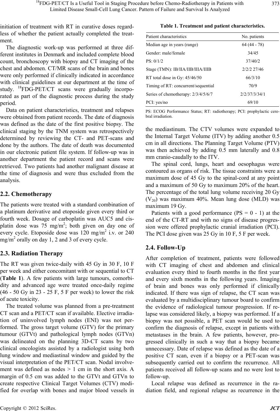 18FDG-PET/CT Is a Useful Tool in Staging Procedure before Chemo-Radiotherapy in Patients with Limited Disease Small-Cell Lung Cancer. Pattern of Failure and Survival Is Analyzed 373 initiation of treatment with RT in curative doses regard- less of whether the patient actually completed the treat- ment. The diagnostic work-up was performed at three dif- ferent institutes in Denmark and includ ed complete blood count, bronchoscopy with biopsy and CT imaging of the chest and abdomen. CT/MR scans of the br ain and bones were only performed if clinically indicated in accordance with clinical guidelines at our department at the time of study. 18FDG-PET/CT scans were gradually incorpo- rated as part of the diagnostic process during the study period. Data on patient characteristics, treatment and relapses were obtained from patient records. The date of diagnosis was defined as the date of the first positive biopsy. The clinical staging by the TNM system was retrospectively determined by reviewing the CT- and PET-scans and done by the authors. The date of death was documented in our electronic patient file system. If follow-up was in another department the patient record and scans were retrieved. Two patients had another malignant disease at the time of diagnosis and were thus excluded from the analysis. 2.2. Chemotherapy The patients were treated with a standard combinatio n of a platinum derivative and etoposide given every third or fourth week. Dosage of carboplatin was AUC5 and cis- platin dose was 75 mg/m2; both given on day one of every cycle. Etoposide dose was 120 mg/m2 i.v. or 240 mg/ m 2 orally on day 1, 2 and 3 of every cycle. 2.3. Radiation Therapy The RT was given twice-daily with 45 Gy in 30 F, 10 F per week and either concomitan t with or sequen tial to CT (Table 1). A few patients with large tumours, comorbi- dity and advanced age were treated once-daily regime (46 - 50 Gy in 23 - 25 F, 5 F per week) to lo wer the risk of acute toxicity. The treated volume was planned from a pre-treatment CT scan and a PET/CT scan if available. Elective irradia- tion of uninvolved lymph nodes (ENI) was not per- formed. The gross target volume (GTV) for the primary tumour (GTVt) and pathological lymph nodes (GTVn) was delineated on the planning 3D-CT scans by two clinical oncologists assisted by a radiologist using both lung window and mediastinal window and guided by the visual interpretation of the PET/CT scan. Nodal involve- ment was defined as nodes > 1 cm in the short axis. A margin of 0.5 cm was added to the GTVt and GTVn to create respective Clinical Target Volumes (CTV) modi- fied for overlap with bones and major blood vessels in Table 1. Treatment and patient characteristics. Patient characteristics No. patients Median age in year s (range) 64 (44 - 78) Gender: male/female 34/45 PS: 0/1/2 37/40/2 Stage (TMN): IB/IIA/IIB/IIIA/IIIB 2/2/2 27/46 RT total dose in Gy: 45/46/50 66/3/10 Timing of RT: concurrent/sequential 70/9 Series of chemotherapy: 2/3/4/5/6/7 2/2/37/3/34/1 PCI: yes/no 69/10 PS: ECOG Performance Status; RT: radiotherapy; PCI: prophylactic cere- bral irradiation. the mediastinum. The CTV volumes were expanded to the Internal Target Volume (ITV) by adding another 0.5 cm in all directions. The Plann ing Target Volume (PTV) was then achieved by adding 0.5 mm laterally and 0.8 mm cranio-caudally to th e ITV. The spinal cord, lungs, heart and oesophagus were contoured as organs of risk. The tissue constraints were a maximum dose of 45 Gy to the spinal-cord at any point and a maxi mum of 50 Gy t o maxi mum 2 0% of t he hear t. The percentage of the total lung vo lume receiving 20 Gy (V20) was maximum 40%. Mean lung dose (MLD) was maximum 19 Gy. Patients with a good performance (PS = 0 - 1) at the end of the CT-RT and with no signs of disease progres- sion were offered prophylactic cranial irradiation (PCI). The PCI dose given was 2 5 Gy in 1 0 F, 5 F pe r week . 2.4. Follow-Up After completion of treatment, patients were followed with CT imaging of chest and abdomen and clinical evaluation every third to fourth months in the first year and every sixth months in the following years. Imaging of brain and bones was only performed if clinically indicated. If there was sign of relapse, the CT scan was evaluated by a multidiscip linary tumour board to confirm the evidence of radiological tumour progression. If re- lapse was considered likely, a biopsy was performed. If a biopsy was not possible, a PET scan would be used to confirm the diagnosis of relapse, except in patients with metastases in the brain. A few patients, however, pro- gressed clinically in such a way that a biopsy became unnecessary. Date of relapse was defined as the date of a positive CT scan, even if a biopsy or a PET-scan was subsequently carried out to confirm the recurrence. All patients received all follow-up scans and no were lost to follow-up. Local relapse was defined as recurrence in the ra- diation field, and regional relapse as recurrence in the Copyright © 2012 SciRes. JCT 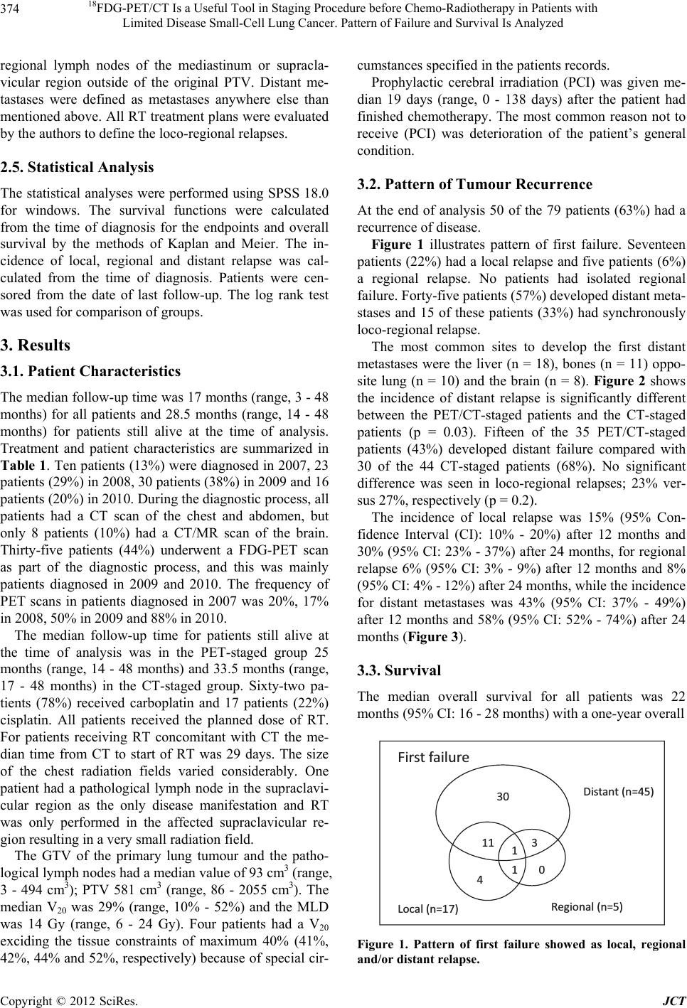 18FDG-PET/CT Is a Useful Tool in Staging Procedure before Chemo-Radiotherapy in Patients with Limited Disease Small-Cell Lung Cancer. Pattern of Failure and Survival Is Analyzed 374 regional lymph nodes of the mediastinum or supracla- vicular region outside of the original PTV. Distant me- tastases were defined as metastases anywhere else than mentioned above. All RT treatment plans were evaluated by the authors to define the loco-regional relapses. 2.5. Statistical Analysis The statistical analyses were performed using SPSS 18.0 for windows. The survival functions were calculated from the time of diagnosis for the endpoints and overall survival by the methods of Kaplan and Meier. The in- cidence of local, regional and distant relapse was cal- culated from the time of diagnosis. Patients were cen- sored from the date of last follow-up. The log rank test was used for comparison of groups. 3. Results 3.1. Patient Characteristics The median follow-up time was 17 months (range, 3 - 48 months) for all patients and 28.5 months (range, 14 - 48 months) for patients still alive at the time of analysis. Treatment and patient characteristics are summarized in Table 1. Ten patients (13%) were diagnosed in 2007, 23 patients (29%) in 2008, 30 patients (38%) in 2009 and 16 patients (20%) in 2010. During the diagnostic process, all patients had a CT scan of the chest and abdomen, but only 8 patients (10%) had a CT/MR scan of the brain. Thirty-five patients (44%) underwent a FDG-PET scan as part of the diagnostic process, and this was mainly patients diagnosed in 2009 and 2010. The frequency of PET scans in patients diagnosed in 2007 was 20%, 17% in 2008, 50% in 2009 and 88% in 2010 . The median follow-up time for patients still alive at the time of analysis was in the PET-staged group 25 months (range, 14 - 48 months) and 33.5 months (range, 17 - 48 months) in the CT-staged group. Sixty-two pa- tients (78%) received carboplatin and 17 patients (22%) cisplatin. All patients received the planned dose of RT. For patients receiving RT concomitant with CT the me- dian time from CT to start of RT was 29 days. The size of the chest radiation fields varied considerably. One patient had a pathological lymph node in the supraclavi- cular region as the only disease manifestation and RT was only performed in the affected supraclavicular re- gion resulting in a very small radiation field. The GTV of the primary lung tumour and the patho- logical lymph nodes had a median value of 93 cm3 (range, 3 - 494 cm3); PTV 581 cm3 (range, 86 - 2055 cm3). The median V20 was 29% (range, 10% - 52%) and the MLD was 14 Gy (range, 6 - 24 Gy). Four patients had a V20 exciding the tissue constraints of maximum 40% (41%, 42%, 44% and 52%, respectively) because of special cir- cumstances specified in the patients records. Prophylactic cerebral irradiation (PCI) was given me- dian 19 days (range, 0 - 138 days) after the patient had finished chemotherapy. The most common reason not to receive (PCI) was deterioration of the patient’s general condition. 3.2. Pattern of Tumour Recurrence At the end of analysis 50 of the 79 patients (63%) had a recurrence of disease. Figure 1 illustrates pattern of first failure. Seventeen patients (22%) had a local relapse and five patients (6%) a regional relapse. No patients had isolated regional failure. Forty-five patients (57%) developed distant meta- stases and 15 of these patients (33%) had synchronously loco-regional relapse. The most common sites to develop the first distant metastases were the liver (n = 18), bones (n = 11) oppo- site lung (n = 10) and the brain (n = 8). Figure 2 shows the incidence of distant relapse is significantly different between the PET/CT-staged patients and the CT-staged patients (p = 0.03). Fifteen of the 35 PET/CT-staged patients (43%) developed distant failure compared with 30 of the 44 CT-staged patients (68%). No significant difference was seen in loco-regional relapses; 23% ver- sus 27%, respectively (p = 0.2) . The incidence of local relapse was 15% (95% Con- fidence Interval (CI): 10% - 20%) after 12 months and 30% (95% CI: 23% - 37%) after 24 months, for regional relapse 6% (95% CI: 3% - 9%) after 12 months and 8% (95% CI: 4% - 12%) after 24 months, while the incidence for distant metastases was 43% (95% CI: 37% - 49%) after 12 months and 58% (95% CI: 52% - 74%) after 24 months (Figure 3). 3.3. Survival The median overall survival for all patients was 22 months (95% C I: 16 - 28 m ont hs) with a one-year overall 30 0 3 1 11 4 1 First failure Local (n=17) Regional (n=5) Distant (n=45) Figure 1. Pattern of first failure showed as local, regional and/or distant relapse. Copyright © 2012 SciRes. JCT 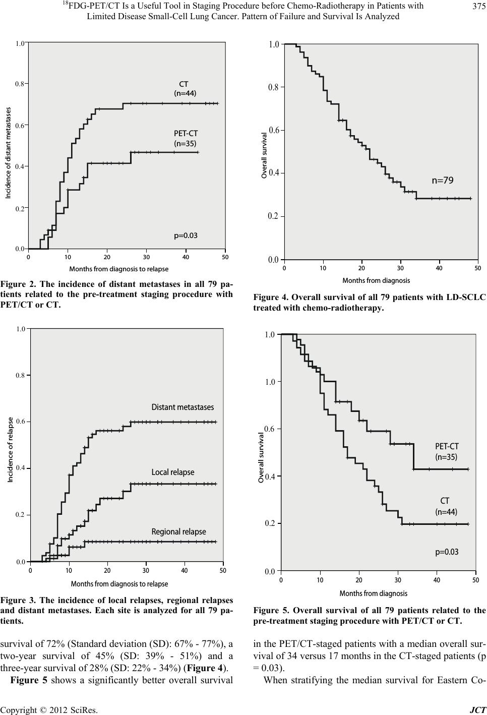 18FDG-PET/CT Is a Useful Tool in Staging Procedure before Chemo-Radiotherapy in Patients with Limited Disease Small-Cell Lung Cancer. Pattern of Failure and Survival Is Analyzed 375 Months from dia g nosis to rela p se 50403020100 Incidence of distant metastases 1,0 0,8 0,6 0,4 0,2 0,0 PET-CT (n=35) CT (n=44) p=0.03 1.0 0.8 0.6 0.4 0.2 0.0 Figure 2. The incidence of distant metastases in all 79 pa- tients related to the pre-treatment staging procedure with PET/CT or CT. Months from diagnosis to relapse 50403020100 Incidence of relapse 1,0 0,8 0,6 0,4 0,2 0,0 Distant metastases Local relapse Regional relapse 1.0 0.8 0.6 0.4 0.2 0.0 Figure 3. The incidence of local relapses, regional relapses and distant metastases. Each site is analyzed for all 79 pa- tients. survival of 72% (Standard deviation (SD): 67% - 77 %), a two-year survival of 45% (SD: 39% - 51%) and a three-year survival of 28% (SD: 22% - 34%) (Figure 4). Figure 5 shows a significantly better overall survival Months from diagnosis 50403020100 Overall survival 1,0 0,8 0,6 0,4 0,2 0,0 n=79 1.0 0.8 0.6 0.4 0.2 0.0 Figure 4. Overall survival of all 79 patients with LD-SCLC treated with chemo-radiotherapy. Months from diagnosis 50403020100 Overall survival 1,0 0,8 0,6 0,4 0,2 0,0 PET-C T (n=35) CT (n=44) p=0.03 1.0 1.0 0.6 0.4 0.2 0.0 Figure 5. Overall survival of all 79 patients related to the pre-treatment staging procedure with PET/CT or CT. in the PET/CT-staged patients with a median overall sur- vival of 34 versus 17 months in the CT-staged patients (p = 0.03). When stratifying the median survival for Eastern Co- Copyright © 2012 SciRes. JCT  18FDG-PET/CT Is a Useful Tool in Staging Procedure before Chemo-Radiotherapy in Patients with Limited Disease Small-Cell Lung Cancer. Pattern of Failure and Survival Is Analyzed 376 operative Oncology Group status (0 versus 1), stage, gen- der, RT schedule (twice-daily RT versus once-daily), concomitant CT-RT versus sequential, cisplatin versus carboplatin and the number of cycles of CT; none showed any significant difference in overall survival. 3.4. Treatment Time The median overall treatment time (from start of any treatment to the end of radiotherapy) was 57 days (range; 25 - 246 days). We stratified the patients in three groups of equal size according to their overall treatment time and compared the surv ival between the gro ups, but found no significant difference in the overall survival (p = 0.9). 3.5. Deaths At the time of analysis, 49 patients (62%) had died. Seven patients died without a diagnosed relapse; one pa- tient died of febrile neutropenia during chemotherapy and the death was related to the treatment. One patient died 2.5 months after end of treatment with symptoms of radiation pneumonitis. The V20 for this patient was 34% and the MLD 18 Gy. The last five patients died of causes not related to the treatment or to lung cancer. 4. Discussion These data describe the outcome of a consecutive patient population over a period of four years. The survival data are comparable with other retrospective consecutive stu- dies of non-selected patients [7,8]. We stratified for differences in patient characteristics and treatment and found no significant difference in overall survival, which was expected due to the low number of patients. However, did we observe a sig- nificantly improved overall survival for the 44% of the patients staged with 18FDG-PET/CT scans. Additionally, these patients had a lower incidence of distant failure; 43% versus 68%. This difference most likely indicates a more exact staging in the PET/CT group with some patients with extended disease (ED) erroneously staged with limited disease in the CT-staged group. However, as patients staged with a PET/CT scan were primarily diagnosed in 2009 and 2010, the median follow-up time for these patients was shorter than for the CT-staged group (25 months versus 33.5 months, respectively for surviving patients) and this could influence the result. A PET scan was found to be superior to a CT scan in the detection of lymph nodes and distant metastases in NSCLC and is now established in th e staging of NSCLC patients [9]. However, the documentation for the use of PET in SCLC is not as robust as in NSCLC [6], and its usefulness is still debated. Studies have been made to evaluate the stage migration phenomenon in SCLC pa- tients staged using a PET/CT scan, and these seem to up- stage 0% - 33% of the patients from LD seen on con- ventional imaging to ED after PET scan [6,10,11]. These results, however, have to be interpreted with caution since the studies were small and half of them retro- spective. However, the use of 18FDG-PET in the staging of SCLC has increased in recent years. Azad A. et al. [11] have evaluated the difference in overall survival in a re- trospective group of 46 consecutive patients undergoing staging by PET scans and found a significantly longer overall survival in patients with LD on PET staging compared with patients upstaged to ED on the PET scan (median 18.6 months versus 5.9 months). However, this result could easily be confounded by different subsequent treatments. One important question remains: Is the right staging in patients with a low burden of metastatic disease clini- cally important? These patients might benefit from a combined treatment with CT and RT. In a randomized study by Jeremic et al. [12], the effects of RT in patients with SCLC-ED were evaluated. A total of 206 patients with a complete response at distant sites and a complete or partial response in the thorax after three cycles of CT were randomized to either CT alone or accelerated hy- perfractionated RT (54 Gy; 1.5 Gy/F). The median over- all survival was found to be significantly higher in the RT-group (17 months versus 11 months). Two other trials are now ongoing to confirm this result (REST (NTR 1527) and RTOG-0937) [13,14]. Until this matter is properly clarified, it is of a great importance that pa- tients are protected from potentially toxic thoracic RT as a standard procedure. During follow-up, only few isolated loco-regional re- lapses (no = 5; 6%) and no isolated regional relapses were observed. The RT was planned so only the primary tumour and the pathological lymph nodes were encom- passed with omission of ENI resulting in a smaller ra- diation field. This strategy is, however, debated due to the potential risk of increasing the isolated nodal failure outside the radiation field. In 2006 two phase II [15,16] studies was preformed to evaluate concurrent CT and involved-field RT (45 Gy, 1.5 Gy/F) in 27 and 37 pa- tients, respectively. Baas et al. [15] observed an isolated regional relapse rate of 6% and De Ruysscher et al. [16] a higher rate of 11%. In both studies the RT was based solely on the pre-treatment CT imaging, and it was stated that a more precise staging with an 18FDG-PET scan might improve the rate of isolated regional nodal failures. A prospective study [17], by the same Dutch research group including De Ruysscher, in 2009 used 18FDG-PET scans to make omission of ENI in 60 LD-SCLC patients receiving concurrent CT (carboplatin and etoposide) to- gether with twice-daily RT (45 Gy, 1.5 Gy/F) and Copyright © 2012 SciRes. JCT  18FDG-PET/CT Is a Useful Tool in Staging Procedure before Chemo-Radiotherapy in Patients with Limited Disease Small-Cell Lung Cancer. Pattern of Failure and Survival Is Analyzed 377 observed a low rate of isolated nodal failures (3%). How- ever, it should be noticed that a difference in definition of isolated nodal failure was used in this study compared to the phase II trial by De Ruysscher et al. [16] making comparison difficult. In 2010 18FDG-PET guided omi- ssion of ENI was evaluated in a retrospective study by Shirvani et al. [18], where data on 60 patients treated with concurrent CT and RT resulting in a low rate of isolated elective nodal failure (1.7%). In contrast to the pr eviou sly mentioned study, intensity-modulated radiation therapy (IMRT) was used and the authors concluded that omis- sion of ENI was safe using a combination of 18FDG-PET and IMRT. Recently, two studies [19,20] again evaluated the omission of ENI guided by only a CT scan and found low rates of loco-regional relapses in 108 and 38 patients respectively, treated with RT concomitant with CT. In our study, omission of ENI was guided by 18FDG-PET in 44% of the patients and by CT scan in the remaining, and we observed no difference in locoregional relapse rate between the two groups. Further studies are needed to clarify the safety of omission of ENI. Results from two ongoing studies [13] evaluating RT regimes are expected. In the UK-led phase III CONVERT trial comparing the twice-daily RT regime (45 Gy in 30F) with a once-daily RT regime (66 Gy in 33 F), the use of ENI is not allowed. This differs from the U S-led CALG B 30610 /RTOG 05 38 trial, evaluating three different RT regimes where medi- astinal lymph nodes are irradiated electively. Another question is timing of the RT. Several authors support an early start of RT concomitant with CT [3,4, 21]. In a meta-analysis comparing phase III trials in LD-SCLC, De Ruysscher [22] showed that the most important predictor of five-year survival is the time from the start of any treatment until the end of RT (SER). The significantly higher five-year survival rate (>20%) was found in patients with a short SER of less than 30 days. In our study we had a median SER of 57 days with only one patient having a SER shorter than 30 days. We acknowledge that the limi tation of this stud y is the small number of patients included and the retrospective design. There were variations in the diagnostic work-up and the treatment, resulting in the possibility of selection bias for both the use of PET scans, imaging of the brain and for the choice of treatment. PET/CT was incorpo- rated in the diagnostic process during this time period and there might be a selection bias due to the availability of PET scanners. Patients were staged at three different institutes and only one of the institutes had a specific PET center. This could have influenced the patient selection. Carboplatin was the primary choice of platinum deri- vative. This is in contrast to most other studies where cisplatin is the treatment of choice and considered su- perior to carboplatin. Whether this could have affected the frequency of relapse and the overall survival is un- known. Despite the retrospective design of this study, the population is consecutive and no patients were lost to follow-up. Our data on relapse is therefore validated. Time and course of death are validated as well due to a complete registration of all patients in Denmark. 5. Conclusions The pattern of failure in patients with LD-SCLC under- going CT-RT showed that the challenge is still a very high risk of distant metastases. The incidence of isolated loco-regional failure was low, and this supports the safety of a treatment strategy of selective irradiation of pathological lymph nodes. Patients staged with an 18FDG-PET/CT scan had a sig- nificantly lower incidence of distant failure and a sig- nificantly better overall survival compared to patients staged with a CT scan only. This probably indicates the value of including an 18PET-CT scan in the staging of patients with SCLC before deciding on a treatment regi- men. REFERENCES [1] P. Warde and D. Payne, “Does Thoracic Irradiation Im- prove Survival and Local Control in Limited-Stage Small- Cell Carcinoma of the Lung? A Meta-Analysis,” Journal of Clinical Oncology, Vol. 10, 1992, pp. 890- 895. [2] J. P. Pignon, R. Arriagada, D. C. Ihde, D. H. Johnson, M. C. Perry, et al., “A Meta-Analysis of Thoracic Radio- therapy for Small-Cell Lung Cancer,” New England Jour- nal of Medicine, Vol. 327, No. 23, 1992, pp. 1618-1624. doi:10.1056/NEJM199212033272302 [3] D. B. Fried, D. E. Morris, C. Poole, J. G. Rosenman, J. S. Halle, et al., “Systematic Review Evaluating the Timing of Thoracic Radiation Therapy in Combined Modality Therapy for Limited-Stage Small-Cell Lung Cancer,” Journal of Clinical Oncology, Vol. 22, No. 23, 2004, pp. 4837-4845. doi:10.1200/JCO.2004.01.178 [4] M. Takada, M. Fukuoka, M. Kawahara, T. Sugiura, A. Yokoyama, et al., “Phase III Study of Concurrent versus Sequential Thoracic Radiotherapy in Combination with Cisplatin and Etoposide for Limited-Stage Small-Cell Lung Cancer: Results of the Japan Clinical Oncology Group Study 9104,” Journal of Clinical Oncology, Vol. 20, No. 14, 2002, pp. 3054-3060. doi:10.1200/JCO.2002.12.071 [5] A. T. Turrisi III, K. Kim, R. Blum, W. T. Sause, R. B. Livingston, et al., “Twice-Daily Compared with Once- Daily Thoracic Radiotherapy in Limited Small-Cell Lung Cancer Treated Concurrently with Cisplatin and Eto- poside,” New England Journal of Medicine, Vol. 340, 1999, pp. 265-271. doi:10.1056/NEJM199901283400403 [6] D. Thomson, P. Hulse, P. Lorigan and C. Faivre-Finn, Copyright © 2012 SciRes. JCT  18FDG-PET/CT Is a Useful Tool in Staging Procedure before Chemo-Radiotherapy in Patients with Limited Disease Small-Cell Lung Cancer. Pattern of Failure and Survival Is Analyzed Copyright © 2012 SciRes. JCT 378 “The Role of Positron Emission Tomography in Man- agement of Small Cell Lung Cancer,” Lung Cancer, Vol. 73, No. 2, 2011, pp. 121-126. doi:10.1016/j.lungcan.2011.03.013 [7] J. M. Watkins, A. E. Wahlquist, A. J. Zauls, K. Shirai, E. Garrett-Mayer, et al., “Involved-Field Radiotherapy with Concurrent Chemotherapy for Limited-Stage Small-Cell Lung Cancer: Disease Control, Patterns of Failure and Survival,” Journal of Medical Imaging and Radiation Oncology, Vol. 54, No. 5, 2010, pp. 483-489. doi:10.1111/j.1754-9485.2010.02201.x [8] A. Hallqvist, H. Rylander, T. Bjork-Eriksson and J. Ny- man, “Accelerated Hyperfractionated Radiotherapy and Concomitant Chemotherapy in Small Cell Lung Cancer Limited-Disease. Dose Response, Feasibility and Out- come for Patients Treated in Western Sweden, 1998- 2004,” Acta Oncologica, Vol. 46, 2007, pp. 969-974. doi:10.1080/02841860701316065 [9] Y. C. Ung, D. E. Maziak, J. A. Vanderveen, C. A. Smith, K. Gulenchyn, et al., “18Fluorodeoxyglucose Positron Emission Tomography in the Diagnosis and Staging of Lung Cancer: A Systematic Review,” Journal of the Na- tional Cancer Institute, Vol. 99, No. 23, 2007, pp. 1753- 1767. doi:10.1093/jnci/djm232 [10] N. Arslan, M. Tuncel, O. Kuzhan, E. Alagoz, B. Buda- koglu, et al., “Evaluation of Outcome Prediction and Disease Extension by Quantitative 2-Deoxy-2-[ 18F] Fluoro- D:-Glucose with Positron Emission Tomography in Pa- tients with Small Cell Lung Cancer,” Annals of Nuclear Medicine, Vol. 25, No. 6, 2011, pp. 406-413. doi:10.1007/s12149-011-0478-y [11] A. Azad, F. Chionh, A. M. Scott, S. T. Lee, S. U. Ber- langieri, et al., “High Impact of 18F-FDG-PET on Man- agement and Prognostic Stratification of Newly Diag- nosed Small Cell Lung Cancer,” Molecular Imaging and Biology, Vol. 12, No. 4, 2010, pp. 443-451. doi:10.1007/s11307-009-0295-z [12] B. Jeremic, Y. Shibamoto, N. Nikolic, B. Milicic, S. Mi- lisavljevic, et al., “Role of Radiation Therapy in the Combined-Modality Treatment of Patients with Extensive Disease Small-Cell Lung Cancer: A Randomized Study,” Journal of Clinical Oncology, Vol. 17, No. 7, 1999, pp. 2092-2099. [13] C. Faivre-Finn, F. Blackhall, M. Snee, S. Harsen, P. Hulse, et al., “Improving Survival with Thoracic Radio- therapy in Patients with Small Cell Lung Cancer. The CONVERT and the REST Trials,” Clinical Oncology (Royal College of Radiologists), Vol. 22, No. 7, 2010, pp. 547-549. doi:10.1016/j.clon.2010.05.008 [14] B. J. Slotman, “Radiotherapy for Extensive Stage Small Cell Lung Cancer,” Frontiers of Radiation Therapy and Oncology, Vol. 42, 2010, pp. 187-192. doi:10.1159/000262475 [15] P. Baas, J. S. Belderbos, S. Senan, H. B. Kwa, A. van Bochove, et al., “Concurrent Chemotherapy (Carboplatin, Paclitaxel, Etoposide) and Involved-Field Radiotherapy in Limited Stage Small Cell Lung Cancer: A Dutch Multi- center Phase II Study,” British Journal of Cancer, Vol. 94, No. 5, 2006, pp. 625-630. [16] D. De Ruysscher, R. H. Bremer, F. Koppe, S. Wanders, E. van Haren, et al., “Omission of Elective Node Irradiation on Basis of CT-Scans in Patients with Limited Disease Small Cell Lung Cancer: A Phase II Trial,” Radiotherapy & Oncology, Vol. 80, No. 3, 2006, pp. 307-312. doi:10.1016/j.radonc.2006.07.029 [17] J. van Loon, D. De Ruy ssc her, R. Wanders, L. Boersma , J. Simons, et al., “Selective Nodal Irradiation on Basis of (18)FDG-PET Scans in Limited-Disease Small-Cell Lung Cancer: A Prospective Study,” International Journal of Radiation Oncology*Biology*Physics, Vol. 77, No. 5, 2010, pp. 329-336. [18] S. M. Shirvani, R. Komaki, J. V. Hey mach, F. V. Fossella, J. Y. Chang, et al., “Positron Emission Tomography/ Computed Tomography-Guided Intensity-Modulated Ra- diotherapy for Limited-Stage Small-Cell Lung Cancer,” International Journal of Radiation Oncology*Biology* Physics, Vol. 82, No. 1, 2011, pp. 91-97. [19] R. Colaco, H. Sheikh, P. Lorigan, F. Blackhall, P. Hulse, et al., “Omitting Elective Nodal Irradiation during Tho- racic Irradiation in Limited-Stage Small Cell Lung Can- cer—Evidence from a Phase II Trial,” Lung Cancer, Vol. 74, 2011, pp. 75-79. [20] B. Xia, G. Y. Chen, X. W. Cai, J. D. Zhao, H. J. Yang, et al., “Is Involved-Field Radiotherapy Based on CT Safe for Patients with Limited-Stage Small-Cell Lung Ca ncer? ” Radiotherapy & Oncology, Vol. 6, No. 50, 2011. [21] D. De Ruysscher, M. Pijls-Johannesma, J. Vansteenkiste, A. Kester, I. Rutten, et al., “Systematic Review and Meta-Analysis of Randomised, Controlled Trials of the Timing of Chest Radiotherapy in Patients with Lim- ited-Stage, Small-Cell Lung Cancer,” Annals of Oncology, Vol. 17, No. 4, 2006, pp. 543-552. doi:10.1093/annonc/mdj094 [22] D. De Ruysscher, M. Pijls-Johannesma, S. M. Bentzen, A. Minken, R. Wanders, et al., “Time between the First Day of Chemotherapy and the Last Day of Chest Radiation Is the Most Important Predictor of Survival in Limited- Disease Small-Cell Lung Cancer,” Journal of Clinical Oncology, Vol. 24, No. 24, 2006, pp. 1057-1063. doi:10.1200/JCO.2005.02.9793 |