

Paper Menu >>
Journal Menu >>
 Open Journal of Obstetrics and Gynecology, 2012, 2, 298-303 OJOG http://dx.doi.org/10.4236/ojog.2012.23063 Published Online September 2012 (http://www.SciRP.org/journal/ojog/) The diagnostic accuracy of the usage of the Fetal Medicine Foundation’s (FMF) on-line risk calculator with first-trimester ultrasound for screening for pre-eclampsia in high-risk pregnant Brazilian population Ernesto Antonio Figueiró-Filho*, Maithe Vendas Galhardo, Bruno Areco de Souza Faculty of Medicine, University Hospital, Federal University of Mato Grosso do Sul, Campo Grande, Brazil Email: *[email protected] Received 28 July 2012; revised 25 August 2012; accepted 9 September 2012 ABSTRACT To evaluate the sensitivity and specificity of the usage of the FMF On-Line Risk Calculator with first-tri- mester ultrasound, in screening assessment for pre- eclampsia (PE), without serum markers. To define the best risk cut-off values for early, intermediate and late pre-eclampsia. Diagnostic accuracy study of preg- nant women who had first-trimester ultrasounds be- tween 11 and 13 weeks. The index test was the first- trimester ultrasound scan plus the FMF On-Line Risk Calculator to assess the risk for PE. The refer- ence standard was the confirmation of actual deve- lopment of early, intermediate or late PE. For calcu- lations of sensitivity and specificity to determine the best cut-off values for early, intermediate and late PE, all the information were processed into ROC curves. The assessment of pre-eclampsia risk in the first tri- mester using an ultrasound plus the FMF On-Line Risk Calculator demonstrated a significant (p < 0.05) area under the ROC curve for early, intermediate and late pre-eclampsia. The best risk cut-off values were defined as 2.1% for early, 2.5% for intermediate and 3.5% for late pre-eclampsia. The first trimester US plus the FMF On-Line Risk Calculator tool was useful and applicable when assessing the risk for pre-eclampsia in a specific pregnant Brazilian popu- lation. Keywords: Accuracy; Sensitivity; Specificity; Pre-Eclampsia; First Trimester; Risk 1. INTRODUCTION Pre-Eclampsia (PE) is a multisystem disorder exclusive to pregnancy characterized by hypertension and protein- uria that develops after 20 weeks of gestation [1-4]. PE affects 2% - 10% of all pregnancies and is an important cause of morbidity and maternal and perinatal mortality [1-3,5,6]. Early screening of women with a potential risk of developing pre-eclampsia is justified because it allows the possibility of early intensiv e maternal and fetal moni- toring, thereby avoiding adverse outcomes. Furthermore, there is evidence that prophylactic usage of aspirin may reduce the incidence of PE in 50% of women when the treatment is starte d bef o re 16 week s [7, 8] . Advances in fetal medicine and ultrasound imaging techniques have allowed extensive research on the role of first-trimester screening for several maternal and fetal conditions [4,9-14]. This new assessment of risk factors using fetal, maternal and biochemical markers suggests new approaches for prenatal care that are more focused on the first rather than third trimester [14,15]. Many current studies have demonstrated the possibi- lity of pre-eclampsia screening be tween 11 and 13 weeks with promising results [4,13,16-22]. This screening fo- cuses on the multivariate analysis of risk associatin g ma- ternal data, clinical, obstetric, ultrasound and serum ma rker measurements [19,20]. It was recently demonstrated that first-trimester ultra- sound scanning can predict early PE by identifying wo- men who will require delivery before 34 weeks’ gesta- tion, intermediate PE by identifying women requiring delivery at 34 - 37 weeks and late PE by identifying women requiring delivery after 37 weeks. This study conclude d t hat effe ct ive prediction of PE can be achieved at 11 - 13 weeks’ gestation [4]. Based on these results, the Fetal Medicine Foundation (FMF) provides an on- line risk calculator for the assessment of risk for early, inter-mediate and late PE using information from the ultrasound scan as well as clinical and serum biochemi- cal markers. Considering the paucity of studies using ultrasound *Corresponding author. OPEN ACCESS  E. A. Figueiró-Filho et al. / Open Journal of Obstetrics and Gynecology 2 (2012) 298-303 299 screening to assess the risk of pre-eclampsia in pregnant women in Brazil, the objective of this study was to evaluate the sensitiv ity and specificity o f th e usage of the FMF On-Line Risk Calculator and first trimester ultra- sound scan in assessment of risk for pre-eclampsia, but not serum markers. The actual development of pre- eclampsia was the reference standard. This study also aimed to define the best risk cut-off values for early, in- termediate and late pre-eclampsia, using the FMF on-line risk calculator. 2. METHODS We followed the STARD (Standards for Reporting of Diagnostic Accuracy) recommendations for reporting the accuracy of a diagnostic procedure [23]. This involved completing a checklist of items to ensure that all perti- nent information was present in the text, thus allowing the reader to detect the potential for bias and to judge the applicability of the results. This was a study of diagnostic accuracy among preg- nant women who had first-trimester ultrasound between 11 and 13 weeks at the High Risk Pregnancy Service, University Hospital (UH), School of Medicine (FAMED), Federal University of Mato Grosso do Sul (UFMS) be- tween October 2010 and May 2011. All pregnant women who had first-trimester ultrasound examinations during the study period and whose obstetric and perinatal clini- cal information were available in the medical records were includ ed. We excluded patients w ho were under 18 years of age or who had indigenous heritage and twin pregnancies. All patients included in study signed an informed consent. The study and its informed consent were approved by the Ethics Committee on Human Re- search, Protocol 1859, on September 30, 2010. The index test considered in this series was the first- trimester ultrasound scan plus the FMF On-Line Risk Calculator (https://courses.fetalmedicine.com/calculator/pe?locale=en). Together, they were used to assess the risk for early, in- termediate and late PE. The theoretical 3.0% cut-off risk for pre-eclampsia was defined for the index test (the ul- trasound scan + the FMF On-Line Risk Calculator), based upon previous pre-eclampsia prevalence studies in different populations [1-3], before confirmation using the reference standard. The reference standard was the confirmation of actual development of early, intermediate or late PE after pa- tient delivery. This study was planned after the patients had performed the index test and after they had delivered; therefore, this was a retrospective analysis of diagnostic accuracy. For the confirmation of maternal pre-eclampsia deve- lopment (reference standard), we considered women who presented with the diagnostic criteria of two or more blood pressure measurements exceeding 140/100 mmHg and proteinuria exceeding 300 mg/24 h [2,3], as reco rded in the medical record during prenatal care. If PE deve- loped and required delivery before 34 weeks, this was defined as early PE. If PE developed and required termi- nation of pregnancy between 34 - 37 weeks, we consid- ered this intermediate PE. Finally, patients that deve- loped late PE were those whose diagnosis required de- livery after 37 weeks [4]. To calculate the risk of pre-eclampsia, we used the FMF On-Line Risk Calculator for Pre-eclampsia, avail- able on the FMF web site. We also considered maternal clinical variables (e.g., age, ethnicity, weight, height, body mass index, mode of conception, smoking history, illicit drug us e, history of hypertension, histor y of diabe- tes and history of systemic lupus erythematous), obstet- rical variables (e.g., parity, history of PE in the woman’s mother, history of previous PE, history of miscarriages and stillbirths), ultrasound data (e.g., fetal crown-rump length and pulsatility ind ex of uterine arteries) and blood pressure (BP) measured at the time of examination [13, 17-22]. No maternal serum biochemical markers were used in the risk assessment for pre-eclampsia; only the clinical information, obstetric ultrasound and BP mea- surements were used. Blood pressure was measured twice in each arm, and the mean BP was calculated ac- cording to the criteria establishe d for the implementation of this examination by FMF [21,22]. All the ultrasound scanning data were collected by FMF-certified sonogra- phers. The Ultrasound used to acquire the images were Nemio 17, TOSHIBA, with preset of first trimester scan default. Clinical and obstetrical information, as well as ultra- sound data and blood pressure values, were included in the respective fields requested on the online risk calcula- tor homepage from the FMF website. The risks were registered on the patient’s record in the same format in which they were presented as percentages values for the following: risk for early, intermediate and late pre- eclampsia. The assessment risks from the index test were acquired blindly, without knowledge of the patient’s ac- tual development of pre-eclampsia; th e latter information was only available after delivery. Because each patient could have more than one risk factor according to the screening test, indicating their elevated risk for early, intermediate or late PE, they were grouped by the earliest possibility of developing PE. For example, if a patient was assessed to have a risk higher than 3.0% for early, intermediate and late PE, this patient was considered only in the early PE group. The design of the study is outlined in a flowchart (Figure 1). For calculations of sensitivity and specificity nd to determine the best cut-off values for early, inter- a Copyright © 2012 SciRes. OPEN ACCESS 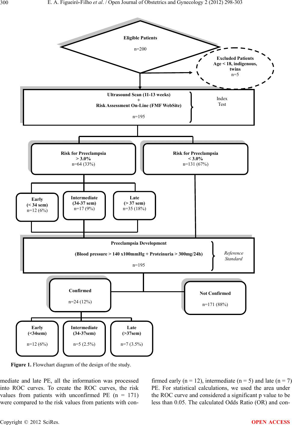 E. A. Figueiró-Filho et al. / Open Journal of Obstetrics and Gynecology 2 (2012) 298-303 Copyright © 2012 SciRes. 300 Eligible Patients n=200 Ultrasound Scan (11-13 weeks) + Risk Assessment On-Line (FMF Web Site) n=195 Excluded Patients Age < 18, indigenous, twins n=5 Risk for Preeclampsia > 3.0% n=64 (33%) Risk for Preeclampsia < 3.0% n=131 (67%) Early (< 34 sem) n=12 (6%) Intermediate (34-37 sem) n=17 (9%) Late (> 37 sem) n=35 (18%) Early (<34sem) n=12 (6%) Preeclampsia Development (Blood press ure > 140 x10 0mmHg + Proteinuria > 300mg/24h) n=195 Intermediate (34-37sem) n=5 (2.5%) Late (>37sem) n=7 (3.5%) Not Co nfir me d n=171 (88%) Confirmed n=24 (12%) Index Test Reference Standard Figure 1. Flowchart diagram of the design of the study. mediate and late PE, all the information was processed into ROC curves. To create the ROC curves, the risk values from patients with unconfirmed PE (n = 171) were compared to the risk values from patients with con- firmed early (n = 12), intermediate (n = 5) and late (n = 7) PE. For statistical calculations, we used the area under the ROC curve and considered a significant p value to be less than 0.05. The calculated Odds Ratio (OR) an d con- OPEN ACCESS 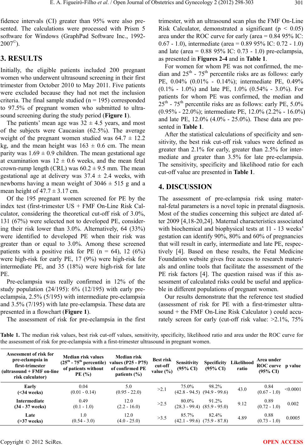 E. A. Figueiró-Filho et al. / Open Journal of Obstetrics and Gynecology 2 (2012) 298-303 301 fidence intervals (CI) greater than 95% were also pre- sented. The calculations were processed with Prism 5 software for Windows (GraphPad Software Inc., 1992- 2007©). 3. RESULTS Initially, the eligible patients included 200 pregnant women who underwent ultrasound screen ing in their first trimester from October 2010 to May 2011. Five patients were excluded because they had not met the inclusion criteria. The final sample studied (n = 195) corresponded to 97.5% of pregnant women who submitted to ultra- sound screening during the study period (Figure 1). The patients’ mean age was 32 ± 4.5 years, and most of the subjects were Caucasian (62.5%). The average weight of the pregnant women studied was 64.7 ± 12.2 kg, and the mean height was 163 ± 0.6 cm. The mean parity was 1.69 ± 0.9 ch ildren. The mean gestational age at examination was 12 ± 0.6 weeks, and the mean fetal crown-rump length (CRL) was 60.2 ± 9.5 mm. The mean gestational age at delivery was 37.4 ± 2.4 weeks, with newborns having a mean weight of 3046 ± 515 g and a mean height of 47. 7 ± 3. 17 c m. Of the 195 pregnant women screened for PE by the index test (first-trimester US + FMF On-Line Risk Cal- culator, considering the theoretical cut-off risk of 3.0%, 131 (67%) were selected not to developed PE, consider- ing their risk lower than 3.0%. Alternatively, 64 (33%) were identified to developed PE when their risk was greater than or equal to 3.0%. Among these screened patients with a positive risk for PE (n = 64), 12 (6%) were high-risk for early PE, 17 (9%) were high-risk for intermediate PE, and 35 (18%) were high-risk for late PE. Pre-eclampsia was really confirmed in 12% of the study population (24/195): 6% (12/195) with early pre- eclampsia, 2.5% (5/195) with intermediate pre-eclampsia and 3.5% (7/195) with late pre-eclampsia. These data are presented in a flow chart (Figure 1). The assessment of risk for pre-eclampsia in the first trimester, with an ultrasound scan plus the FMF On-Line Risk Calculator, demonstrated a significant (p < 0.05) area under the ROC curve for early (area = 0.84 95% IC: 0.67 - 1.0), intermediate (area = 0.89 95% IC: 0.72 - 1.0) and late (area = 0.88 95% IC: 0.73 - 1.0) pre-eclampsia, as presented in Figures 2-4 and in Table 1. For women for whom PE was not confirmed, the me- dian and 25th - 75th percentile risks are as follows: early PE, 0.04% (0.01% - 0.14%); intermediate PE, 0.49% (0.1% - 1.0%) and late PE, 1.0% (0.54% - 3.0%). For patients for whom PE was confirmed, the median and 25th - 75th percentile risks are as follows: early PE, 5.0% (0.95% - 22.0%); intermediate PE, 12.0% (2.2% - 16.0%) and late PE, 12.0% (4.0% - 25.0%). These data are pre- sented in Table 1. After the statistical calculations of specificity and sen- sitivity, the best risk cut-off risk values were defined as greater than 2.1% for early, greater than 2.5% for inter- mediate and greater than 3.5% for late pre-eclampsia. The sensitivity, specificity and likelihood ratio for each cut-off value a re p r es ented in Table 1. 4. DISCUSSION The assessment of pre-eclampsia risk using mater- nal-fetal parameters is a novel topic in prenatal diagnosis. Most of the studies concerning this subject are dated af- ter 2009 [4,18-20,24]. Maternal characteristics associated with biochemical and biophysical tests at 11 - 13 weeks’ gestation can iden tify 90%, 80% and 60% of pregn ancies that will result in early, intermed iate and late PE, respec- tively [4]. Based on these results, the Fetal Medicine Foundation website gives free access to research materi- als and online tools that facilitate the assessment of the PE risk factors [4]. The question raised was if this as- sessment of calculated risks could be useful and applica- ble in different populations of pregnant women. Our results demonstrate that the reference test studied (assessment of risk for PE with a first-trimester ultra- sound + the FMF On-Line Risk Calculator ) could accu- rately screen for early (cut-off risk value: >2.1%, 75% Ta ble 1. The median risk values, best risk cut-off values, sensitivity, specificity, likelihood ratio and area under the ROC curve for the assessment of risk for pre-eclampsia with a first-trimester ultrasound in pregnant women. Assessment of risk for pre-eclampsia in first-trimester (ultrasound + FMF on-line risk calculator) Median risk values (25th - 75th percentile) of patients without PE (%) Median risk values (P25 - P75) of confirmed PE patients (%) Best risk cut-off value (%) Sensitivity (95% CI)Specificity (95% CI)Likelihood ratio Area under ROC curve (95% CI)p value Early (<34 weeks) 0.04 (0.01 - 0.14) 5.0 (0.95 - 22.0) >2.1 75.0% (42.8 - 94.5)98.2% (94.9 - 99.6)43.0 0.84 (0.67 - 1.0)<0.0001 Intermediate (34 - 37 weeks) 0.49 (0.1 - 1.0) 12.0 (2.2 - 16.0) >2.5 80.0% (28.3 - 99.4)91.2% (85.9 - 95.0)9.12 0.89 (0.72 - 1.0)0.002 Late (>37 weeks) 1.0 (0.54 - 3.0) 12.0 (4.0 - 25.0) >3.5 85.7% (42.1 - 99.6)82.4% (75.9 - 87.8)4.89 0.88 (0.73 - 1.0)0.0005 Copyright © 2012 SciRes. OPEN ACCESS 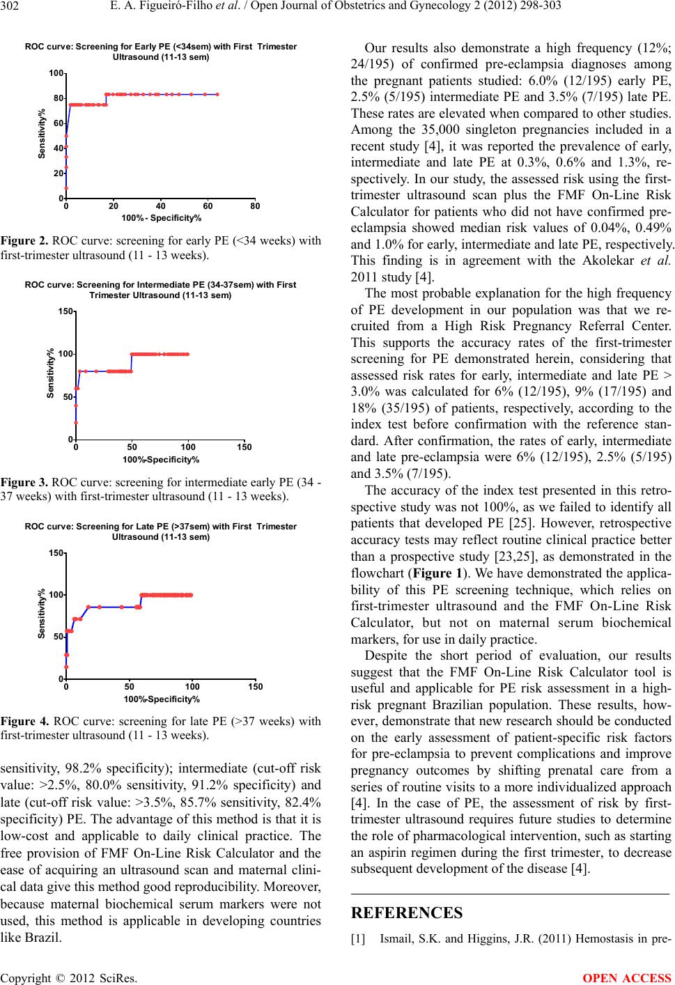 E. A. Figueiró-Filho et al. / Open Journal of Obstetrics and Gynecology 2 (2012) 298-303 302 ROC curv e: Screening for Early PE (<34sem) with First Trimester Ultrasound (11-13 sem) 020 40 60 80 0 20 40 60 80 100 100% - Specificity% Sensitivity% Figure 2. ROC curve: screening for early PE (<34 weeks) with first-trimester ultrasound (11 - 13 weeks). ROC curve: Screening for Intermediate PE (34-37sem) with First Trimester Ultrasound (11-13 sem) 050 100 150 0 50 100 150 100%-Specificity% Sensitivity% Figure 3. ROC curve: screening for intermediate early PE (34 - 37 weeks) with first-trimester ultrasound (11 - 13 weeks). ROC curve: Screening for Late PE (>37sem) with First Trimester Ultras ound (11-13 s em) 050100 150 0 50 100 150 100%-Specificity% Sensitivity% Figure 4. ROC curve: screening for late PE (>37 weeks) with first-trimester ultrasound (11 - 13 weeks). sensitivity, 98.2% specificity); intermediate (cut-off risk value: >2.5%, 80.0% sensitivity, 91.2% specificity) and late (cut-off risk value: >3.5%, 85.7% sensitivity, 82.4% specificity) PE. The advantage of this method is th at it is low-cost and applicable to daily clinical practice. The free provision of FMF On-Line Risk Calculator and the ease of acquiring an ultrasound scan and maternal clini- cal data give this method good reproducibility. Moreover, because maternal biochemical serum markers were not used, this method is applicable in developing countries like Brazil. Our results also demonstrate a high frequency (12%; 24/195) of confirmed pre-eclampsia diagnoses among the pregnant patients studied: 6.0% (12/195) early PE, 2.5% (5/195) intermediate PE and 3.5% (7/195) late PE. These rates are elevated when compared to other studies. Among the 35,000 singleton pregnancies included in a recent study [4], it was reported the prevalence of early, intermediate and late PE at 0.3%, 0.6% and 1.3%, re- spectively. In our study, the assessed risk using the first- trimester ultrasound scan plus the FMF On-Line Risk Calculator for patients who did not have confirmed pre- eclampsia showed median risk values of 0.04%, 0.49% and 1.0% for early, intermediate and late PE, respectively. This finding is in agreement with the Akolekar et al. 2011 study [4]. The most probable explanation for the high frequency of PE development in our population was that we re- cruited from a High Risk Pregnancy Referral Center. This supports the accuracy rates of the first-trimester screening for PE demonstrated herein, considering that assessed risk rates for early, intermediate and late PE > 3.0% was calculated for 6% (12/195), 9% (17/195) and 18% (35/195) of patients, respectively, according to the index test before confirmation with the reference stan- dard. After confirmation, the rates of early, intermediate and late pre-eclampsia were 6% (12/195), 2.5% (5/195) and 3.5% (7/195). The accuracy of the index test presented in this retro- spective study was not 100%, as we failed to identify all patients that developed PE [25]. However, retrospective accuracy tests may reflect routine clinical practice better than a prospective study [23,25], as demonstrated in the flowchart (Figure 1). We have demonstrated the applica- bility of this PE screening technique, which relies on first-trimester ultrasound and the FMF On-Line Risk Calculator, but not on maternal serum biochemical markers, for use in daily practice. Despite the short period of evaluation, our results suggest that the FMF On-Line Risk Calculator tool is useful and applicable for PE risk assessment in a high- risk pregnant Brazilian population. These results, how- ever, demonstrate that new research should be conducted on the early assessment of patient-specific risk factors for pre-eclampsia to prevent complications and improve pregnancy outcomes by shifting prenatal care from a series of routine visits to a more individualized approach [4]. In the case of PE, the assessment of risk by first- trimester ultrasound requires future studies to determine the role of pharmacological intervention, such as starting an aspirin regimen during the first trimester, to decrease subsequent development of the disease [4]. REFERENCES [1] Ismail, S.K. and Higgins, J.R. (2011) Hemostasis in pre- Copyright © 2012 SciRes. OPEN ACCESS 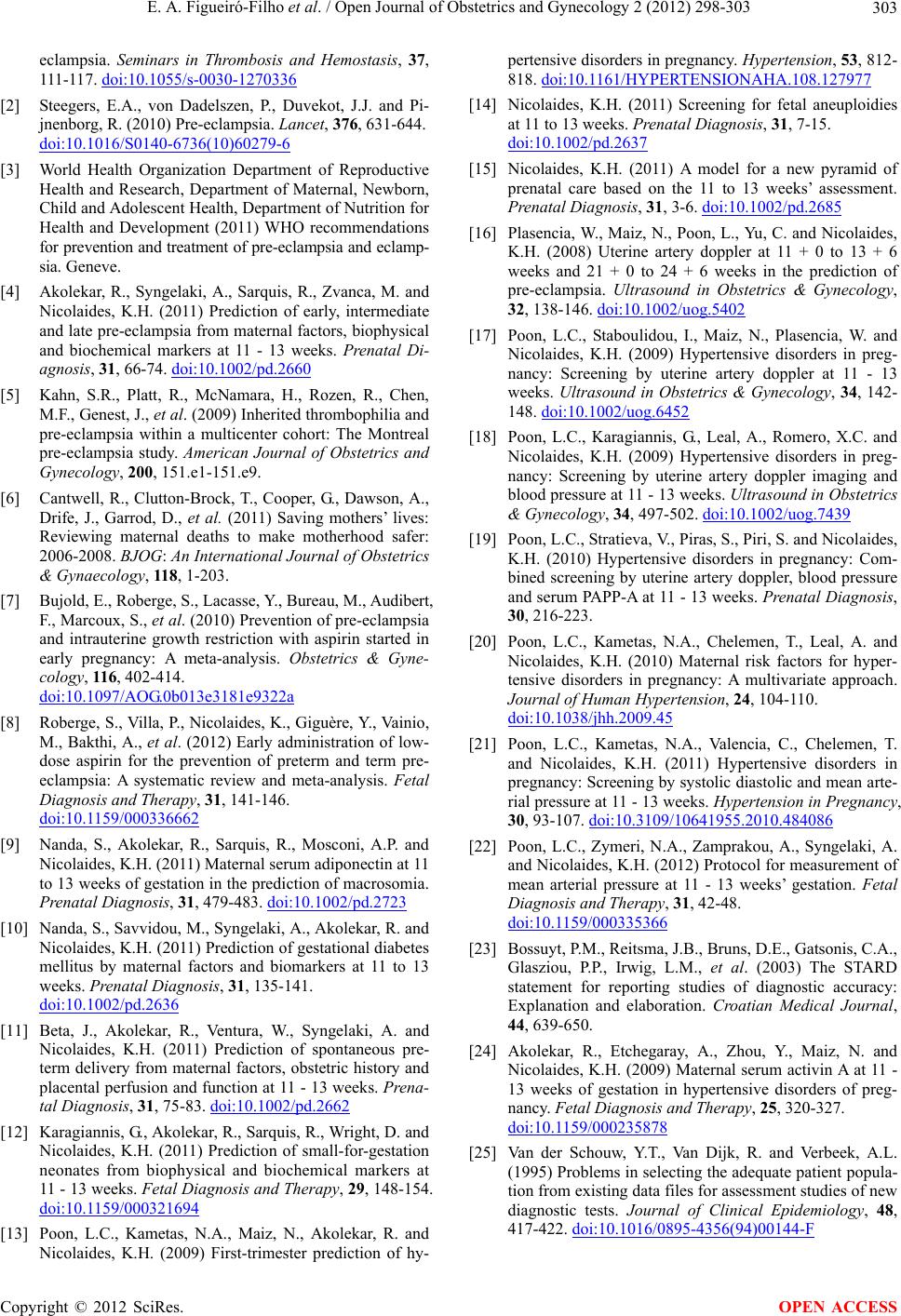 E. A. Figueiró-Filho et al. / Open Journal of Obstetrics and Gynecology 2 (2012) 298-303 303 eclampsia. Seminars in Thrombosis and Hemostasis, 37, 111-117. doi:10.1055/s-0030-1270336 [2] Steegers, E.A., von Dadelszen, P., Duvekot, J.J. and Pi- jnenborg, R. (2010) Pre-eclampsia. Lancet, 376, 631-644. doi:10.1016/S0140-6736(10)60279-6 [3] World Health Organization Department of Reproductive Health and Research, Department of Maternal, Newborn, Child and Adolescent Health, Department of Nutrition for Health and Development (2011) WHO recommendations for prevention and treatment of pre-eclampsia and eclamp- sia. Geneve. [4] Akolekar, R., Syngelaki, A., Sarquis, R., Zvanca, M. and Nicolaides, K.H. (2011) Prediction of early, intermediate and late pre-eclampsia from maternal factors, biophysical and biochemical markers at 11 - 13 weeks. Prenatal Di- agnosis, 31, 66-74. doi:10.1002/pd.2660 [5] Kahn, S.R., Platt, R., McNamara, H., Rozen, R., Chen, M.F., Genest, J., et al. (2009) Inherited thrombophilia and pre-eclampsia within a multicenter cohort: The Montreal pre-eclampsia study. American Journal of Obstetrics and Gynecology, 200, 151.e1-151.e9. [6] Cantwell, R., Clutton-Brock, T., Cooper, G., Dawson, A., Drife, J., Garrod, D., et al. (2011) Saving mothers’ lives: Reviewing maternal deaths to make motherhood safer: 2006-2008. BJOG: An International Journal of Obstetrics & Gynaecology, 118, 1-203. [7] Bujold, E., Roberge, S., Lac a sse, Y., Bure au, M., Audibert, F., Marcoux, S., et al. (2010) Prevention of pre-eclampsia and intrauterine growth restriction with aspirin started in early pregnancy: A meta-analysis. Obstetrics & Gyne- cology, 116, 402-414. doi:10.1097/AOG.0b013e3181e9322a [8] Roberge, S., Villa, P., Nicolaides, K., Giguère, Y., Vainio, M., Bakthi, A., et al. (2012) Early administration of low- dose aspirin for the prevention of preterm and term pre- eclampsia: A systematic review and meta-analysis. Fetal Diagnosis and Therapy, 31, 1 4 1-146. doi:10.1159/000336662 [9] Nanda, S., Akolekar, R., Sarquis, R., Mosconi, A.P. and Nicolaides, K.H. (2011) Maternal serum adiponectin at 11 to 13 weeks of gestation in the prediction of macrosomia. Prenatal Diagnosis, 31, 479-483. doi:10.1002/pd.2723 [10] Nanda, S., Savvidou, M., Syngelaki, A., Akolekar, R. and Nicolaides, K.H. (2011) Prediction of gestational diabetes mellitus by maternal factors and biomarkers at 11 to 13 weeks. Prenatal Diagnosi s, 31, 135-141. doi:10.1002/pd.2636 [11] Beta, J., Akolekar, R., Ventura, W., Syngelaki, A. and Nicolaides, K.H. (2011) Prediction of spontaneous pre- term delivery from maternal factors, obstetric history and placental perfusion and function at 11 - 13 weeks. Prena- tal Diagnosis, 31, 75-83. doi:10.1002/pd.2662 [12] Karagiannis, G., Akolekar, R., Sarquis, R., Wright, D. and Nicolaides, K.H. (2011) Prediction of small-for-gestation neonates from biophysical and biochemical markers at 11 - 13 weeks. Fetal Diagnosis and Therapy, 29, 148-154. doi:10.1159/000321694 [13] Poon, L.C., Kametas, N.A., Maiz, N., Akolekar, R. and Nicolaides, K.H. (2009) First-trimester prediction of hy- pertensive disorders in pregnancy. Hypertension, 53, 812- 818. doi:10.1161/HYPERTENSIONAHA.108.127977 [14] Nicolaides, K.H. (2011) Screening for fetal aneuploidies at 11 to 13 weeks. Prenatal Diagnosis, 31, 7-15. doi:10.1002/pd.2637 [15] Nicolaides, K.H. (2011) A model for a new pyramid of prenatal care based on the 11 to 13 weeks’ assessment. Prenatal Diagnosis, 31, 3-6. doi:10.1002/pd.2685 [16] Plasencia, W., Maiz, N., Poon, L., Yu, C. and Nicolaides, K.H. (2008) Uterine artery doppler at 11 + 0 to 13 + 6 weeks and 21 + 0 to 24 + 6 weeks in the prediction of pre-eclampsia. Ultrasound in Obstetrics & Gynecology, 32, 138-146. doi:10.1002/uog.5402 [17] Poon, L.C., Staboulidou, I., Maiz, N., Plasencia, W. and Nicolaides, K.H. (2009) Hypertensive disorders in preg- nancy: Screening by uterine artery doppler at 11 - 13 weeks. Ultrasound in Obstetrics & Gynecology, 34, 142- 148. doi:10.1002/uog.6452 [18] Poon, L.C., Karagiannis, G., Leal, A., Romero, X.C. and Nicolaides, K.H. (2009) Hypertensive disorders in preg- nancy: Screening by uterine artery doppler imaging and blood pressure at 11 - 13 weeks. Ultrasound in Obstetrics & Gynecology, 34, 497-502. doi:10.1002/uog.7439 [19] Poon, L.C., Stratieva, V., Piras, S., Piri, S. and Nicolaides, K.H. (2010) Hypertensive disorders in pregnancy: Com- bined screening by uterine artery doppler, blood pressure and serum PAPP-A at 11 - 13 weeks. Prenatal Diagnosis, 30, 216-223. [20] Poon, L.C., Kametas, N.A., Chelemen, T., Leal, A. and Nicolaides, K.H. (2010) Maternal risk factors for hyper- tensive disorders in pregnancy: A multivariate approach. Journal of Human Hypertension, 24, 104-110. doi:10.1038/jhh.2009.45 [21] Poon, L.C., Kametas, N.A., Valencia, C., Chelemen, T. and Nicolaides, K.H. (2011) Hypertensive disorders in pregnancy: Screening by systolic diastolic and mean arte- rial pressure at 11 - 13 weeks. Hypertension in Pregnancy, 30, 93-107. doi:10.3109/10641955.2010.484086 [22] Poon, L.C., Zymeri, N.A., Zamprakou, A., Syngelaki, A. and Nicolaides, K.H. (2012) Protocol for measurement of mean arterial pressure at 11 - 13 weeks’ gestation. Fetal Diagnosis and Therapy, 31, 42-48. doi:10.1159/000335366 [23] Bossuyt, P.M., Reitsma, J. B., Bruns, D.E., Gatsonis, C.A., Glasziou, P.P., Irwig, L.M., et al. (2003) The STARD statement for reporting studies of diagnostic accuracy: Explanation and elaboration. Croatian Medical Journal, 44, 639-650. [24] Akolekar, R., Etchegaray, A., Zhou, Y., Maiz, N. and Nicolaides, K.H. (2009) Maternal serum activin A at 11 - 13 weeks of gestation in hypertensive disorders of preg- nancy. Fetal Diagnosis and Therapy, 25, 320-327. doi:10.1159/000235878 [25] Van der Schouw, Y.T., Van Dijk, R. and Verbeek, A.L. (1995) Problems in selecting the adequate patient popula- tion from existing data files for assessment studies of new diagnostic tests. Journal of Clinical Epidemiology, 48, 417-422. doi:10.1016/0895-4356(94)00144-F Copyright © 2012 SciRes. OPEN ACCESS |