

Paper Menu >>
Journal Menu >>
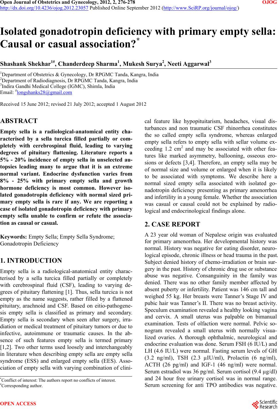 Open Journal of Obstetrics and Gynecology, 2012, 2, 276-278 OJOG http://dx.doi.org/10.4236/ojog.2012.23057 Published Online September 2012 (http://www.SciRP.org/journal/ojog/) Isolated gonadotropin deficiency with primary empty sella: Causal or casual association?* Shashank Shekhar1#, Chanderdeep Sharma1, Mukesh Surya2, Neeti Aggarwal3 1Department of Obs t e t r ics & Gynecology, Dr RPGMC Tanda, Kangra, India 2Department of Radi o d i a g n o s i s, Dr RPGMC Tanda, Kangra, India 3Indira Gandhi Medical College (IGMC), Shimla, India Email: #[email protected] Received 15 June 2012; revised 21 July 2012; accepted 1 August 2012 ABSTRACT Empty sella is a radiological-anatomical entity cha- racterised by a sella turcica filled partially or com- pletely with cerebrospinal fluid, leading to varying degrees of pituitary flattening. Literature reports a 5% - 20% incidence of empty sella in unselected au- topsies leading many to argue that it is an extreme normal variant. Endocrine dysfunction varies from 8% - 25% with primary empty sella and growth hormone deficiency is most common. However iso- lated gonadotropin deficiency with normal sized pri- mary empty sella is rare if any. We are reporting a case of isolated gonadotropin deficiency with primary empty sella unable to confirm or refute the associa- tion as causal or casual. Keywords: Empty Sella; Empty Sella Syndrome; Gonadotr o pi n Defi ci e ncy 1. INTRODUCTION Empty sella is a radiological-anatomical entity charac- terised by a sella turcica filled partially or completely with cerebrospinal fluid (CSF), leading to varying de- grees of pituitary flattening [1]. Thus, sella turcica is no t empty as the name suggests, rather filled by a flattened pituitary, arachnoid and CSF. Based on eitio-pathogene- sis empty sella is classified as primary and secondary. Empty sella is secondary when seen after surgery, irra- diation or medical treatment of pituitary tumors or due to infective, autoimmune or traumatic causes. In the ab- sence of such features empty sella is termed primary [1,2]. Two other terms used loosely and interchangeably in literature when describing empty sella are empty sella syndrome (ESS) and enlarged empty sella (EES). Asso- ciation of empty sella with varying combination of clini- cal feature like hypopituitarism, headaches, visual dis- turbances and non traumatic CSF rhinorrhea constitutes the so called empty sella syndrome, whereas enlarged empty sella refers to empty sella with sellar volume ex- ceeding 1.2 cm2 and may be associated with other fea- tures like marked asymmetry, ballooning, osseous ero- sions or defects [3,4]. Therefore, an empty sella may be of normal size and volume or enlarged when it is likely to be associated with symptoms. We describe here a normal sized empty sella associated with isolated go- nadotropin deficiency presenting as primary amenorrhea and infertility in a young female. Whether the association was casual or causal could not be explained by radio- logical and endocrinological findings alone. 2. CASE REPORT A 23 year old woman of Nepalese origin was evaluated for primary amenorrhea. Her developmental history was normal. History was negative for eating disorder, neuro- logical episode, ch ron ic illness or head trauma in th e past. Subject denied history of chemo-irradiation or brain sur- gery in the past. History of chronic drug use or substance abuse was negative. Consanguinity in the family was denied. There was no other family member affected by absent puberty or infertility. Patient was 146 cm tall and weighed 55 kg. Her breasts were Tanner’s Stage IV and pubic hair was Tanner’s II. There was no breast activity. Speculum examination revealed a healthy looking vagina and cervix. A small uterus was palpable on bimanual examination. Tests of olfaction were normal. Pelvic so- nogram revealed a small uterus with normally visua- lized ovaries. A thorough ophthalmic, neurological and endocrine evaluation w as done. Ser um FSH (6 IU/L) an d LH (4.6 IU/L) were normal. Fasting serum levels of GH (3.2 ng/ml), TSH (2.3 µIU/ml), Prolactin (6 ng/ml), ACTH (26 pg/ml) and IGF-1 (46 ng/ml) were normal. Serum estradiol was 36 pg/ml. Serum cortisol (9.4 µg/d l) and 24 hour free urinary cortisol was in normal range. Serum screening for anti TPO antibodies was negative. *Conflict of interest: The authors rep ort no conflicts of interest. #Corresponding author. OPEN ACCESS 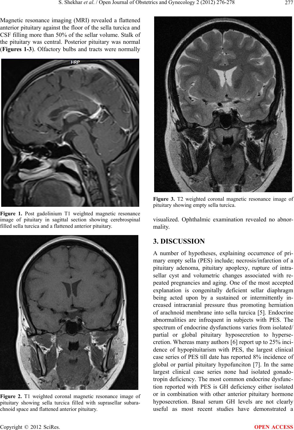 S. Shekhar et al. / Open Journal of Obstetrics and Gynecology 2 (2012) 276-278 277 Magnetic resonance imaging (MRI) revealed a flattened anterior pituitary against th e floor of the sella turcica and CSF filling more than 50% of the sellar volume. Stalk of the pituitary was central. Posterior pituitary was normal (Figures 1-3). Olfactory bulbs and tracts were normally Figure 1. Post gadolinium T1 weighted magnetic resonance image of pituitary in sagittal section showing cerebrospinal filled sella turcica and a flattened anterior pituitary. Figure 2. T1 weighted coronal magnetic resonance image of pituitary showing sella turcica filled with suprasellar subara- chnoid space and flattened anterior pituitary. Figure 3. T2 weighted coronal magnetic resonance image of pituitary showing empty sella turcica. visualized. Ophthalmic examination revealed no abnor- mality. 3. DISCUSSION A number of hypotheses, explaining occurrence of pri- mary empty sella (PES) include; necrosis/infarction of a pituitary adenoma, pituitary apoplexy, rupture of intra- sellar cyst and volumetric changes associated with re- peated pregnancies and aging. One of the most accepted explanation is congenitally deficient sellar diaphragm being acted upon by a sustained or intermittently in- creased intracranial pressure thus promoting herniation of arachnoid membrane into sella turcica [5]. Endocrine abnormalities are infrequent in subjects with PES. The spectrum of endocrine dysfunctions varies from isolated/ partial or global pituitary hyposecretion to hyperse- cretion. Whereas many authors [6] report up to 25% inci- dence of hypopituitarism with PES, the largest clinical case series of PES till date has reported 8% incidence of global or partial pituitary hypofunciton [7]. In the same largest clinical case series none had isolated gonado- tropin deficiency. The most common endocrine dysfunc- tion reported with PES is GH deficiency either isolated or in combination with other anterior pituitary hormone hyposecretion. Basal serum GH levels are not clearly useful as most recent studies have demonstrated a Copyright © 2012 SciRes. OPEN ACCESS 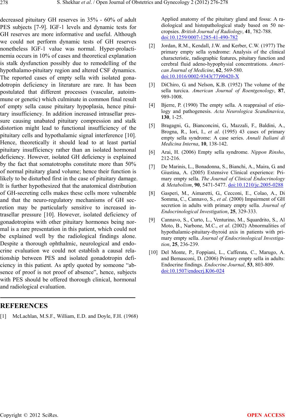 S. Shekhar et al. / Open Journal of Obstetrics and Gynecology 2 (2012) 276-278 Copyright © 2012 SciRes. OPEN ACCESS 278 decreased pituitary GH reserves in 35% - 60% of adult PES subjects [7-9]. IGF-1 levels and dynamic tests for GH reserves are more informative and useful. Although we could not perform dynamic tests of GH reserves nonetheless IGF-1 value was normal. Hyper-prolacti- nemia occurs in 10% of cases and theoretical explanation is stalk dysfunction possibly due to remodelling of the hypothalamo-pituitary region and altered CSF dynamics. The reported cases of empty sella with isolated gona- dotropin deficiency in literature are rare. It has been postulated that different processes (vascular, autoim- mune or genetic) which culminate in common final result of empty sella cause pituitary hypoplasia, hence pitui- tary insufficiency. In addition increased intrasellar pres- sure causing unabated pituitary compression and stalk distortion might lead to functional insufficiency of the pituitary cells and hypothalamic signal interference [10]. Hence, theoretically it should lead to at least partial pituitary insufficiency rather than an isolated hormonal deficiency. However, isolated GH deficiency is explained by the fact that somatotrophs constitute more than 50% of normal pituitary gland volume; hence their function is likely to be disturbed first in the case of pituitary damage. It is further hypothesized that the anatomical distribution of GH-secreting cells makes these cells more vulnerable and that the neuro-regulatory mechanisms of GH sec- retion may be particularly sensitive to increased in- trasellar pressure [10]. However, isolated deficiency of gonadotropins with other pituitary hormones being nor- mal is a rare presentation in this patient, which could not be explained well by the radiological findings alone. Despite a thorough ophthalmic, neurological and endo- crine evaluation we could not establish a causal rela- tionship between PES and isolated gonadotropin defi- ciency in this patient. As aptly quoted by someone “ab- sence of proof is not proof of absence”, hence, subjects with PES should be offered thorough clinical, hormonal and radiological evaluation. REFERENCES [1] McLachlan, M.S.F., William, E.D. and Doyle, F.H. (1968) Applied anatomy of the pituitary gland and fossa: A ra- diological and histopathological study based on 50 ne- cropsies. British Journal of Radiology, 41, 782-788. doi:10.1259/0007-1285-41-490-782 [2] Jordan, R.M., Kendall, J.W. and Kerber, C.W. (1977) The primary empty sella syndrome: Analysis of the clinical cha racteristic, radiographic features, pituitary functi on and cerebral fluid adeno-hypophysial concentrations. Ameri- can Journal of Medicine, 62, 569-580. doi:10.1016/0002-9343(77)90420-X [3] DiChiro, G. and Nelson, K.B. (1952) The volume of the sella turcica. American Journal of Roentgenology, 87, 989-1008. [4] Bjerre, P. (1990) The empty sella. A reappraisal of etio- logy and pathogenesis. Acta Neurologica Scandinavica, 130, 1-25. [5] Bragagni, G., Bianconcini, G., Mazzali, F., Baldini, A., Brogna, R., Iori, I., et al. (1995) 43 cases of primary empty sella syndrome: A case series. Annali Italiani di Medicina Interna, 10, 138-142. [6] Arai, H. (2006) Empty sella syndrome. Nippon Rinsho, 212-216. [7] De Marinis, L., Bonadonna, S., Bianchi, A., Maira, G. and Giustina, A. (2005) Extensive Clinical experience: Pri- mary empty sella. The Journal of Clinical Endocrinology & Metabolism, 90, 5471-5477. doi:10.1210/jc.2005-0288 [8] Gasperi, M., Aimaretti, G., Cecconi, E., Colao, A., Di Somma, C., Cannavo, S., et al. (2000) Impairment of GH secretion in adults with primary empty sella. Journal of Endocrinological Investigation, 25, 329-333. [9] Cannavo, S., Curto, L., Venturino, M., Squardrito, S., Al Moto, B., Narbone, M.C., et al. (2002) Abnormalities of hypothalamic-pituitary-thyroid axis in patients with pri- mary e mpty sella. Journal of Endocrinological Investiga- tion, 25, 236-239. [10] Del Monte, P., Foppiani, L., Cafferata, C., Marugo, A. and Bernasconi, D. (2006) Primary empty sella in adults: Endocrine findings. Endocrine Jour n al, 53, 803-809. doi:10.1507/endocrj.K06-024 |