

Paper Menu >>
Journal Menu >>
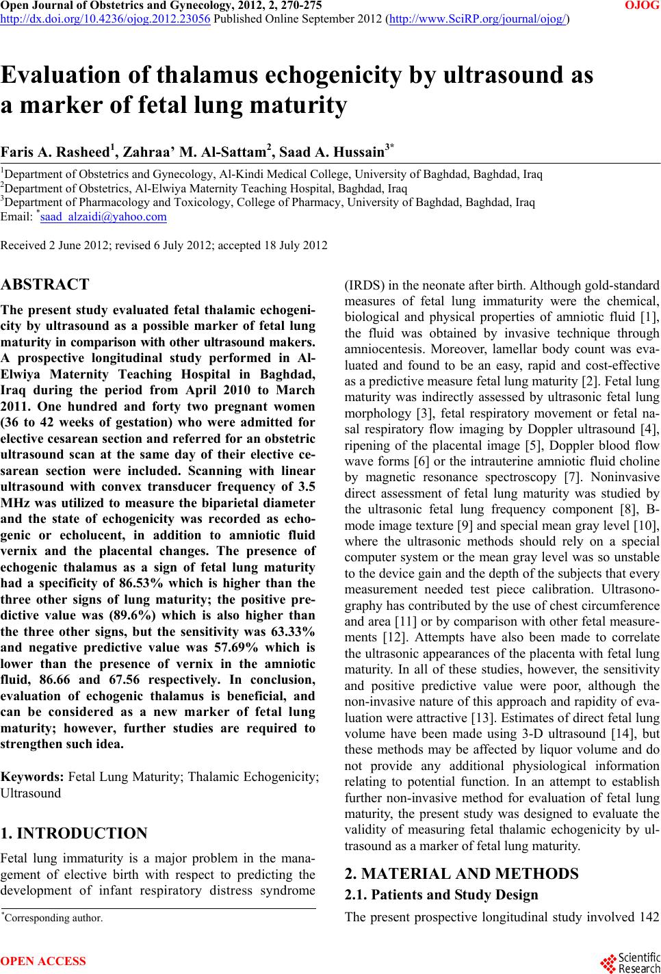 Open Journal of Obstetrics and Gynecology, 2012, 2, 270-275 OJOG http://dx.doi.org/10.4236/ojog.2012.23056 Published Online September 2012 (http://www.SciRP.org/journal/ojog/) Evaluation of thalamus echogenicity by ultrasound as a marker of fetal lung maturity Faris A. Rasheed1, Zahraa’ M. Al-Sattam2, Saad A. Hussain3* 1Department of Obstetrics and Gynecology, Al-Kindi Medical College, University of Baghdad, Baghdad, Iraq 2Department of Ob s tetrics, Al-Elwiya Maternity Teaching Hospital, Baghdad, Iraq 3Department of Pharmacology and Toxicology, College of Pharmacy, University of Baghdad, Baghdad, Iraq Email: *[email protected] Received 2 June 2012; revised 6 July 2012; accepted 18 July 2012 ABSTRACT The present study evaluated fetal thalamic echogeni- city by ultrasound as a possible marker of fetal lung ma tur ity in comparison with other ultrasound maker s. A prospective longitudinal study performed in Al- Elwiya Maternity Teaching Hospital in Baghdad, Iraq during the period from April 2010 to March 2011. One hundred and forty two pregnant women (36 to 42 weeks of gestation) who were admitted for elective cesarean section and referred for an obstetric ultrasound scan at the same day of their elective ce- sarean section were included. Scanning with linear ultrasound with convex transducer frequency of 3.5 MHz was utilized to measure the biparietal diameter and the state of echogenicity was recorded as echo- genic or echolucent, in addition to amniotic fluid vernix and the placental changes. The presence of echogenic thalamus as a sign of fetal lung maturity had a specificity of 86.53% which is higher than the three other signs of lung maturity; the positive pre- dictive value was (89.6%) which is also higher than the three other signs, but the sensitivity was 63.33% and negative predictive value was 57.69% which is lower than the presence of vernix in the amniotic fluid, 86.66 and 67.56 respectively. In conclusion, evaluation of echogenic thalamus is beneficial, and can be considered as a new marker of fetal lung maturity; however, further studies are required to strengthen such idea. Keywords: Fetal Lung Maturity; Thalamic Echogenicity; Ultrasound 1. INTRODUCTION Fetal lung immaturity is a major problem in the mana- gement of elective birth with respect to predicting the development of infant respiratory distress syndrome (IRDS) in the neonate after birth. Although gold-standard measures of fetal lung immaturity were the chemical, biological and physical properties of amniotic fluid [1], the fluid was obtained by invasive technique through amniocentesis. Moreover, lamellar body count was eva- luated and found to be an easy, rapid and cost-effective as a predictive measure fetal lung maturity [2]. Fetal lung maturity was indirectly assessed by ultrasonic fetal lung morphology [3], fetal respiratory movement or fetal na- sal respiratory flow imaging by Doppler ultrasound [4], ripening of the placental image [5], Doppler blood flow wave forms [6] or the intrauterine amniotic fluid choline by magnetic resonance spectroscopy [7]. Noninvasive direct assessment of fetal lung maturity was studied by the ultrasonic fetal lung frequency component [8], B- mode image texture [9] and special mean gray level [10], where the ultrasonic methods should rely on a special computer system or the mean gray level was so unstable to the device gain and the depth of the subjects th at every measurement needed test piece calibration. Ultrasono- graphy has contributed by the use of chest circumference and area [11] or by comparison with other fetal measure- ments [12]. Attempts have also been made to correlate the ultrasonic appearances of the placenta with fetal lung maturity. In all of these studies, however, the sensitivity and positive predictive value were poor, although the non-invasive nature of this approach and rapidity of eva- luation were attractive [13]. Estimates of direct fetal lung volume have been made using 3-D ultrasound [14], but these methods may be affected by liquor volume and do not provide any additional physiological information relating to potential function. In an attempt to establish further non-invasive method for evaluation of fetal lung maturity, the present study was designed to evaluate the validity of measuring fetal thalamic echogenicity by ul- trasound as a marker of fetal lung maturity. 2. MATERIAL AND METHODS 2.1. Patients and Study Design The present prospective longitudinal study involved 142 *Corresponding a uthor. OPEN ACCESS 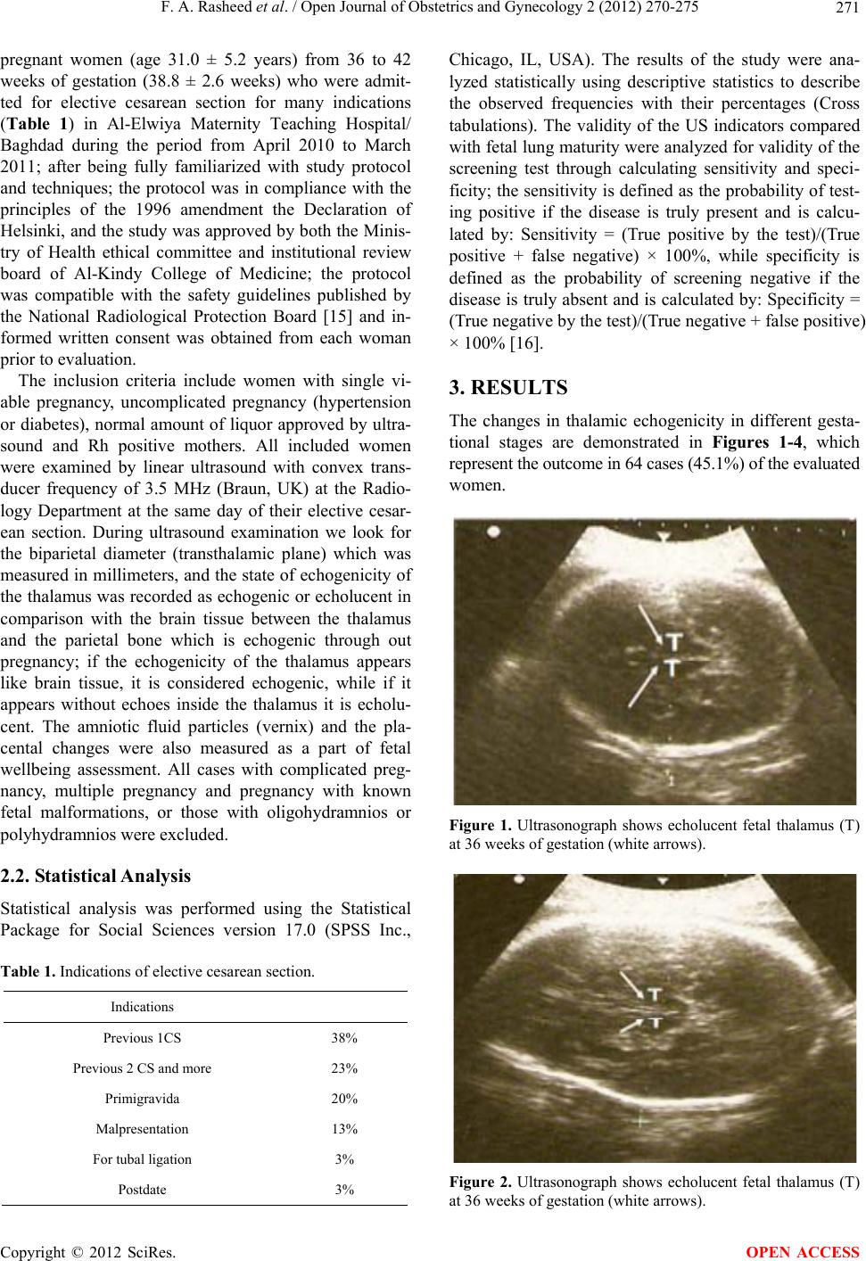 F. A. Rasheed et al. / Open Journal of Obstetrics and Gynecology 2 (2012) 270-275 271 pregnant women (age 31.0 ± 5.2 years) from 36 to 42 weeks of gestation (38.8 ± 2.6 weeks) who were admit- ted for elective cesarean section for many indications (Table 1) in Al-Elwiya Maternity Teaching Hospital/ Baghdad during the period from April 2010 to March 2011; after being fully familiarized with study protocol and techniques; the protocol was in compliance with the principles of the 1996 amendment the Declaration of Helsinki, and the study was approved by both the Minis- try of Health ethical committee and institutional review board of Al-Kindy College of Medicine; the protocol was compatible with the safety guidelines published by the National Radiological Protection Board [15] and in- formed written consent was obtained from each woman prior to evaluation. The inclusion criteria include women with single vi- able pregnancy, uncomplicated pregnancy (hypertension or diabetes), normal amount of liquor approved by ultra- sound and Rh positive mothers. All included women were examined by linear ultrasound with convex trans- ducer frequency of 3.5 MHz (Braun, UK) at the Radio- logy Department at the same day of their elective cesar- ean section. During ultrasound examination we look for the biparietal diameter (transthalamic plane) which was measured in millimeters, and the state of echogenicity of the thalamus was recorded as echogenic or echolucent in comparison with the brain tissue between the thalamus and the parietal bone which is echogenic through out pregnancy; if the echogenicity of the thalamus appears like brain tissue, it is considered echogenic, while if it appears without echoes inside the thalamus it is echolu- cent. The amniotic fluid particles (vernix) and the pla- cental changes were also measured as a part of fetal wellbeing assessment. All cases with complicated preg- nancy, multiple pregnancy and pregnancy with known fetal malformations, or those with oligohydramnios or polyhydramnios were excluded. 2.2. Statistical Analysis Statistical analysis was performed using the Statistical Package for Social Sciences version 17.0 (SPSS Inc., Table 1. Indications of elective cesarean section. Indications Previous 1CS 38% Previous 2 CS and more 23% Primigravida 20% Malpresentation 13% For tubal ligation 3% Postdate 3% Chicago, IL, USA). The results of the study were ana- lyzed statistically using descriptive statistics to describe the observed frequencies with their percentages (Cross tabulations). The validity of the US indicators compared with fetal lung maturity were analyzed fo r validity of the screening test through calculating sensitivity and speci- ficity; the sensitivity is de fined as the probability of test- ing positive if the disease is truly present and is calcu- lated by: Sensitivity = (True positive by the test)/(True positive + false negative) × 100%, while specificity is defined as the probability of screening negative if the disease is truly absent and is calculated by: Specificity = (True negative by the test)/(True negative + false positive) × 100% [16]. 3. RESULTS The changes in thalamic echogenicity in different gesta- tional stages are demonstrated in Figures 1-4, which represent the outcome in 64 cases (45.1%) of the evaluated women. Figure 1. Ultrasonograph shows echolucent fetal thalamus (T) at 36 weeks of gestation (white arrows). Figure 2. Ultrasonograph shows echolucent fetal thalamus (T) at 36 weeks of gestation (white arrows). Copyright © 2012 SciRes. OPEN ACCESS 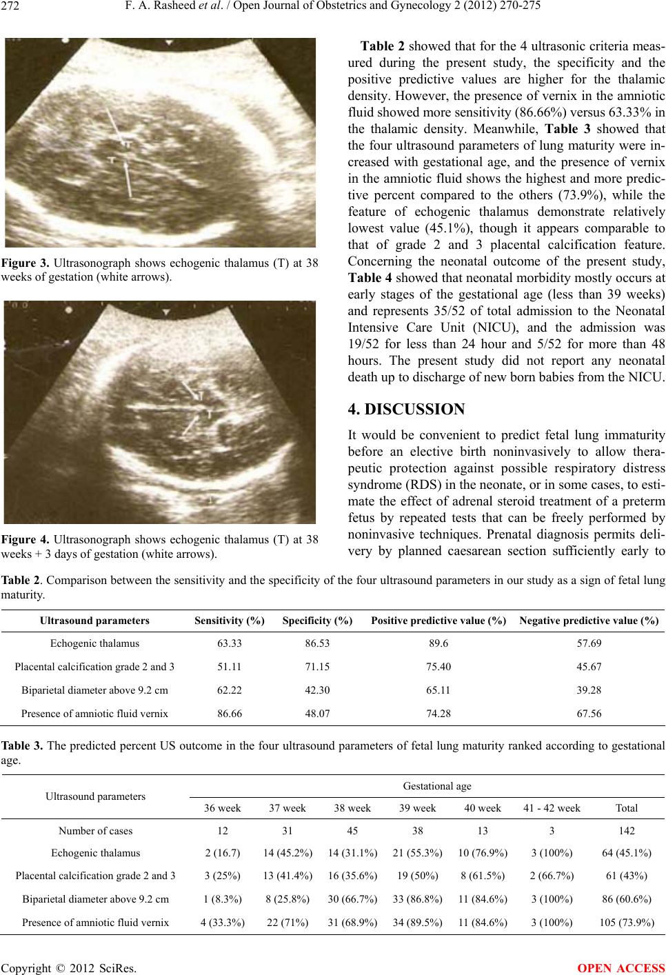 F. A. Rasheed et al. / Open Journal of Obstetrics and Gynecology 2 (2012) 270-275 Copyright © 2012 SciRes. 272 Table 2 showed that for the 4 ultrasonic criteria meas- ured during the present study, the specificity and the positive predictive values are higher for the thalamic density. However, the pr esence of vernix in the amniotic fluid showed more sensitivity (86.66 %) versus 63.33% in the thalamic density. Meanwhile, Table 3 showed that the four ultrasound parameters of lung maturity were in- creased with gestational age, and the presence of vernix in the amniotic fluid shows the highest and more predic- tive percent compared to the others (73.9%), while the feature of echogenic thalamus demonstrate relatively lowest value (45.1%), though it appears comparable to that of grade 2 and 3 placental calcification feature. Concerning the neonatal outcome of the present study, Table 4 showed that neonatal morbidity mostly occu rs at early stages of the gestational age (less than 39 weeks) and represents 35/52 of total admission to the Neonatal Intensive Care Unit (NICU), and the admission was 19/52 for less than 24 hour and 5/52 for more than 48 hours. The present study did not report any neonatal death up to discharge of ne w bo r n babi es fr o m the NICU. Figure 3. Ultrasonograph shows echogenic thalamus (T) at 38 weeks of gestation (white arrows). 4. DISCUSSION It would be convenient to predict fetal lung immaturity before an elective birth noninvasively to allow thera- peutic protection against possible respiratory distress syndrome (RDS) in the neonate, or in some cases, to esti- mate the effect of adrenal steroid treatment of a preterm fetus by repeated tests that can be freely performed by noninvasive techniques. Prenatal diagnosis permits deli- very by planned caesarean section sufficiently early to Figure 4. Ultrasonograph shows echogenic thalamus (T) at 38 weeks + 3 days of gestation (white arrows). Table 2. Comparison between the sensitivity and the specificity of the four ultrasound parameters in our study as a sign of fetal lung maturity. Ultrasound parameters Sensitivity (%)Specificity (%) Positive predictive value (%) Negative predictive value (%) Echogenic thalamus 63.33 86.53 89.6 57.69 Placental calcification grade 2 and 3 51.11 71.15 75.40 45.67 Biparietal diameter above 9.2 cm 62.22 42.30 65.11 39.28 Presence of amnio t ic fl u id ve rnix 86.66 48.07 74.28 67.56 Table 3. The predicted percent US outcome in the four ultrasound parameters of fetal lung maturity ranked according to gestational age. Gestational age Ultrasound parameters 36 week 37 week 38 week 39 week 40 week 41 - 42 week Total Number of cases 12 31 45 38 13 3 142 Echogenic thalamus 2 (16.7) 14 (45.2%)14 (31.1%)21 (55.3%)10 (76.9%) 3 (100%) 64 (45.1%) Placental calcification grade 2 and 3 3 (25% ) 13 (41.4%)16 (35.6%)19 (50%) 8 (61.5%) 2 (66.7%) 61 (43%) Biparietal diameter above 9.2 cm 1 (8.3%) 8 (25.8%)30 (66.7%)33 (86.8%)11 (84.6%) 3 (100%) 86 (60.6%) Presence of amniotic fluid vernix 4 (33.3%) 22 (71%) 31 (68.9%)34 (89.5%)11 (84.6%) 3 (100%) 105 (73.9%) OPEN ACCESS 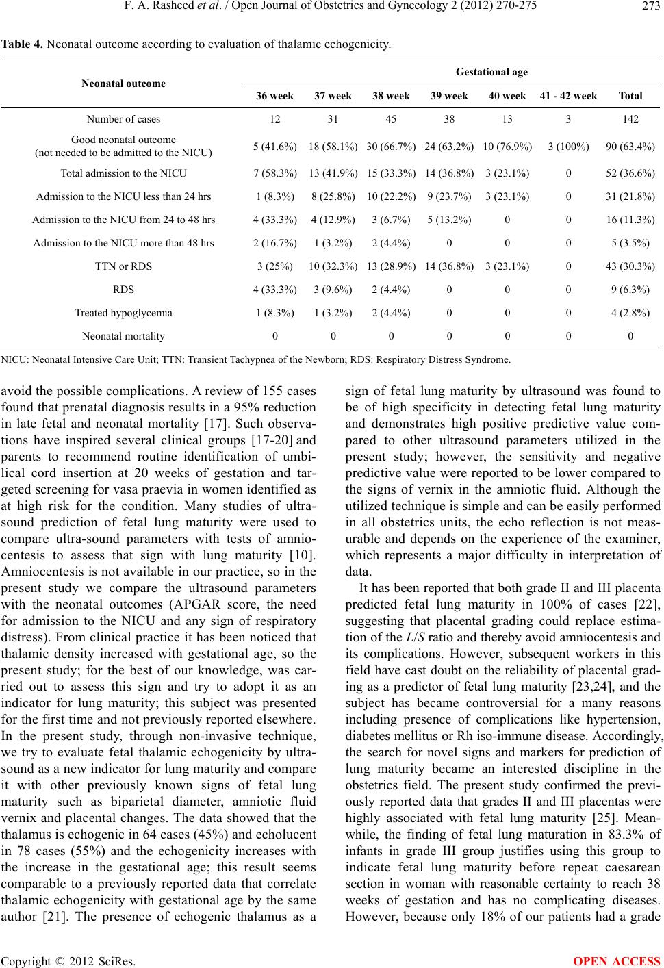 F. A. Rasheed et al. / Open Journal of Obstetrics and Gynecology 2 (2012) 270-275 273 Table 4. Neonatal outcome according to evaluation of thalamic echogenicity. Gestational age Neonatal outcome 36 week37 week38 week39 week40 week 41 - 42 weekTotal Number of cases 12 31 45 38 13 3 142 Good neonatal outcome (not needed to be admitted to the NICU) 5 (41.6%)18 (58.1%)30 (66.7%)24 (63.2%)10 (76.9%) 3 (100%) 90 (63.4%) Total admission to the NICU 7 (58.3%)13 (41.9%)15 (33.3%)14 (36.8%)3 (23.1%) 0 52 (36.6%) Admission to the NICU less than 24 hrs 1 (8.3%)8 (25.8%)10 (22.2%)9 (23.7%)3 (23.1%) 0 31 (21.8%) Admission to the NI CU from 24 to 48 hrs 4 (33.3%)4 (12.9%)3 (6.7%)5 (13.2%)0 0 16 (1 1.3%) Admission to the NICU more than 48 hrs 2 (16.7%)1 (3.2%)2 (4.4%)0 0 0 5 (3.5%) TTN or RDS 3 (25%) 10 (32.3%)13 (28.9%)14 (36.8%)3 (23.1%) 0 43 (30.3%) RDS 4 (33.3%)3 (9.6%)2 (4.4%)0 0 0 9 (6.3%) Treated hypoglycemia 1 (8.3%)1 (3.2%)2 (4.4%)0 0 0 4 (2.8%) Neonatal mortality 0 0 0 0 0 0 0 NICU: Neonatal Intensive Car e Unit; TTN: Transient Tachypnea of the Newborn; RDS: Respir atory Distress Syndrome. avoid the possible complications. A review of 155 cases found that prenatal diagnosis results in a 95% reduction in late fetal and neonatal mortality [17]. Such observa- tions have inspired several clinical groups [17-20] and parents to recommend routine identification of umbi- lical cord insertion at 20 weeks of gestation and tar- geted screening for vasa praevia in women identified as at high risk for the condition. Many studies of ultra- sound prediction of fetal lung maturity were used to compare ultra-sound parameters with tests of amnio- centesis to assess that sign with lung maturity [10]. Amnio centesis is not ava ilable in our prac tice, so in the present study we compare the ultrasound parameters with the neonatal outcomes (APGAR score, the need for admission to the NICU and any sign of respiratory distress). From clinical practice it has been noticed that thalamic density increased with gestational age, so the present study; for the best of our knowledge, was car- ried out to assess this sign and try to adopt it as an indicator for lung maturity; this subject was presented for the first time and not previously reported elsewhere. In the present study, through non-invasive technique, we try to evaluate fetal thalamic echogenicity by ultra- sound as a new indicator for lung maturity and compare it with other previously known signs of fetal lung maturity such as biparietal diameter, amniotic fluid vernix and placental changes. The data showed that the thalamus is echogenic in 64 cases (45%) and echolucent in 78 cases (55%) and the echogenicity increases with the increase in the gestational age; this result seems comparable to a previously reported data that correlate thalamic echogenicity with gestational age by the same author [21]. The presence of echogenic thalamus as a sign of fetal lung maturity by ultrasound was found to be of high specificity in detecting fetal lung maturity and demonstrates high positive predictive value com- pared to other ultrasound parameters utilized in the present study; however, the sensitivity and negative predic tive value w ere reported to be lower compared to the signs of vernix in the amniotic fluid. Although the utilized technique is simple and can be easily performed in all obstetrics units, the echo reflection is not meas- urable and depends on the experience of the examiner, which represents a major difficulty in interpretation of data. It has been reported that both grade II and III placenta predicted fetal lung maturity in 100% of cases [22], suggesting that placental grading could replace estima- tion of the L/S ratio and thereby avoid amniocentesis and its complications. However, subsequent workers in this field have cast doubt on the reliability of placental grad- ing as a predictor of fetal lung maturity [23,24], and the subject has became controversial for a many reasons including presence of complications like hypertension, diabetes mellitus or Rh iso-immune disease. Accordingly, the search for novel signs and markers for prediction of lung maturity became an interested discipline in the obstetrics field. The present study confirmed the previ- ously reported data that grades II and III placentas were highly associated with fetal lung maturity [25]. Mean- while, the finding of fetal lung maturation in 83.3% of infants in grade III group justifies using this group to indicate fetal lung maturity before repeat caesarean section in woman with reasonable certainty to reach 38 weeks of gestation and has no complicating diseases. However, because only 18% of our patients had a grade Copyright © 2012 SciRes. OPEN ACCESS  F. A. Rasheed et al. / Open Journal of Obstetrics and Gynecology 2 (2012) 270-275 274 III placenta, those with grade II placenta satisfying the provisos mentioned above should be managed in the same way as those in grade III on the basis of our find- ings, and even with this group 75.4% of fetuses had achieved lung maturity, such finding seems compatible with that reported previously [22]. Hadlock et al. (1985) utilized amniocentesis for determination of fetal lung maturity and ultrasonographic (US) evaluation of the biparietal diameter (BPD) and placental grade were per- formed simultaneously and concluded that the ability of the sonograph ic parameters to predict fetal lung maturity was closely related to menstrual age and the best use of US for predicting fetal lung maturity is in establishing menstrual age early in pregnancy [26]; the results ob- tained in the present study support the previously men- tioned idea. Concerning evaluation of the presence of FFPs in the amniotic fluid, Gross et al. (1985) correlate this ultrasound finding with fetal lung maturity and suggest that presence of FFPs on real-time ultrasound could be used to confirm fetal lung maturity [27]. The data presented in this study support the previously men- tioned one an d more than on e u ltr asound mar k er s may be better correlated for prediction of fetal lung maturity. Moreover, the present study demonstrates that a BPD greater than or equal to 92 mm predicted 56 of 90 (62.22%) cases with mature lung, while the presence of FFPs predicted 78 of 90 (86.66%); when utilized toge- ther, a BPD equal to or greater than 92mm and/or the presence of FFPs predicted 83 of 90 (92.22%) of cases with mature fetal lung, and such finding complies with data reported by others, where BPD greater than or equal to 92 mm and the presence of free-floating particles are independent, and considered as complimentary predic- tors of fetal lung maturity [28]. Subjective approach during measurement of echogenicity and lack of a me- thod to measure the gain of echoes are the limitations of the present study, in hope th at we can overco me in future studies. In conclusion, evaluation of echogenic thalamus by ultrasound at the lev el of the BPD is of value, and can be considered as a new marker of fetal lung maturity; however, further studies are required to strengthen such idea. 5. ACKNOWLEDGEMENTS The authors would like to thank University of Baghdad for support; also they gratefully acknowledge the ultrasonography staff of Al- Elwiya Maternity Teaching Hospital for technical assistance. REFERENCES [1] American College of Obstetricians and Gynecologists. (2008) ACOG practice bulletin No. 97. Fetal lung ma- turity. Obstetrics and Gynecology, 11 2, 717-726. doi:10.1097/AOG.0b013e318188d1c2 [2] Bahadsari, S. and Changizi, N. (2005) Association be- tween lamellar body count and respiratory distress in neonates. Saudi M ed ic al Journal, 26, 1414-1146. [3] Gerads, F.A., Twisk, J.W., Fetter, W.P., Wijnaendts, L.C. and van Vugt, J.M. (2008) Predicting pulmonary hy- poplasia with 2- or 3-dimensional ultrasonography in complicated pregnancies. American Journal of Obstetrics and Gyneology, 198, 140. [4] La Torre, R., Cosmi, E., Anceschi, M.H., Piazze, J.J., Piga, M.D. and Cosmi, E.V. (2003) Preliminary report on a new and noninvasive method for the assessment of fetal lung maturity. Journal of Perinatal Medicine, 31, 431- 434. doi:10.1515/JPM.2003.067 [5] Shan, A.G. and Graham, D. (1986) Relationship of pla- cental grade to fetal pulmonary maturity and respiratory distress syndrome. American Journal of Perinatology, 3, 53-55. doi:10.1055/s-2007-999827 [6] Piazze, J.J., Ancesc hi, M.M., Picone, G., Cereskja, A., La Torre, R. and Cosmi, E.V. (2003) Association between maternal-fetal Doppler velocimetry and fetal lung ma- turity. Journal of Perinatal Medicine, 31, 484-488. doi:10.1515/JPM.2003.074 [7] Clifton, M.S. , Joe, B.N., Zekt ozer, A.S., Kurhanewicz , J., Vigneron, D.B., Coakley, F.V., et al. (2006) Feasibility of magnetic resonance spectroscopy for evaluating fetal lung maturity. Journal of Pediatric Surgery, 41, 768-773. doi:10.1016/j.jpedsurg.2006.02.020 [8] Sohn, C., Stolz, W. and Bastert, G. (1991) Diagnosis of fetal lung maturity by ultrasound: A new method and first results. Ultrasound in Obstetrics and Gynecology, 1, 345- 348. doi:10.1046/j.1469-0705.1991.01050345.x [9] Prakash, K.N., Ramarkrishnan, A.G., Suresh, S. and Chow, T.W. (2001) An investigation into the feasibility of fetal lung maturity prediction using statistical textural features. Ultrasonography and Imaging, 23, 39-54. [10] Takesin, I., Anderer, G., Hellmeyer, L., Stein, W., Kunert, M. and Schmidt, S. (2004) Assessment of fetal lung de- velopment by quantitative ultrasonic tissue characteriza- tion: A methodical study. Prenatal Diagnnosis, 24, 671- 676. doi:10.1002/pd.951 [11] Nimrod, C., Davies, D., Iwanicki, S., Harder, J., Prsaud, D. and Nicholson, S. (1986) Ultrasound prediction of pulmonary hypoplasia. Obstetrics and Gynecology, 68, 495-498. [12] Johnson, A., Callan, N., Bhutan, V.K., Colmorgen, G.H.C., Weiner, S. and Bolognese, R.J. (1987) Ultrasonic ratio of fetal thoracic to abdominal circumference: An association with pulmonary hypoplasia. American Journal of Obstet- rics and Gynecology, 157, 764-769. [13] Kazzi, G.M., Gross, T.L., Sokol, R.J. and Kazzi, S.N. (1985) Non-invasive prediction of HMD: An optimised classification of sonographic placental maturation. Ame- rican Journal of Obstetrics and Gynecology, 152, 213. [14] Lee, A., Kratochwil, A., Stumpflen, I., Deutinger, J. and Bernaschek, G. (1996) Fetal lung volume determination by three-dimensional ultrasonography. American Journal of Obstetrics and Gynecology, 175, 588-592. Copyright © 2012 SciRes. OPEN ACCESS  F. A. Rasheed et al. / Open Journal of Obstetrics and Gynecology 2 (2012) 270-275 Copyright © 2012 SciRes. 275 OPEN ACCESS doi:10.1053/ob.1996.v175.a72478 [15] Mole, R.H. (1993) The biology and radiobiology of in utero development in relation to radiological protection. British Journal of Radiology, 66, 833-841. doi:10.1259/0007-1285-66-792-1095 [16] Beth, D. and Robert, G. (2004) Basic and clinical biosta- tistics, 4th Editon, McGraw-Hill, New York. [17] Oyelese, Y., Catanzarite, V., Prefumo, F., Lashley, S., Schachter, M., Tovbin, Y., et al. (2004) Vasa previa: The impact of prenatal diagnosis on outcomes. Obstetrics and Gynecology, 103, 937-942. doi:10.1097/01.AOG.0000123245.48645.98 [18] Baulies, S., Maiz, N., Munoz, A., Torrents, M., Echevar- ria, M. and Serra, B. (2007) Prenatal ultrasound diagnosis of vasa praevia and analysis of risk factors. Prenatal Diagnosis, 27, 595-599. doi:10.1002/pd.1753 [19] Gandhi, M., Cleary-Goldman, J., Ferrara, L., Ciorica, D., Saltzman, D. and Rebarber, A. (2008) The association between vasa previa, multiple gestations, and assisted re- productive technology. American Journal of Perinatology, 25, 587-589. doi:10.1055/s-0028-1090584 [20] Sinha, P., Kaushik, S., Kuruba, N. and Beweley, S. (2008) Vasa praevia: A missed diagnosis. Journal of Obstetrics and Gynecology, 28, 600-603. doi:10.1080/01443610802344365 [21] Ras he ed, F.A. (2001) Fetal thalamus echog e ni ci ty c h an ge s with increasing fetal age. Iraqi Medical Journal, 50, 20-24. [22] Shweni, P.M. and Moodley, S.C. (1986) Placental grading by ultrasonography as an index of fetal maturity, its ap- plication to the problem of elective caesarean section. South African Medical Journal, 10, 525-528. [23] Kazzi, G.M., Gross, T.L., Rosen, M.G. and Jaatoul-Kazzi, N.Y. (1984) The relationship of placental grade, fetal lung maturity and neonatal outcome in normal and compli- cated pregnancies. American Journal of Obstetrics and Gynecology, 148, 54-58. [24] Clair, M.R., Rosenberg, E., Tempkin, O., Andreoni, R.F. and Bowie, J.D. (1983) Placental grading in the compli- cated or high-risk pregnancy. Ultrasound Medicine, 2, 297-301. [25] Loret de Mola, J.R., Judge, N., Entsminger, C., DeViney, M., Muise, K.L. and Duchon, M.A. (1998) Indirect pre- diction of fetal lung maturity. Value of ultrasonographic colonic and placental grading. Journal of Reproductive Medicine, 43, 898-902. [26] Hadlock, F.P., Irwin, J.F., Roecker, E., Shah, Y.P., Deter, R.I. and Rossavik, I.K. (1985) Ultrasound prediction of fetal lung maturity. Radiology, 155, 469-472. [27] Gross, T.L., Wolfson, R.N. and Sokol, R.J. (1985) Sono- graphically detected free-floating particles in amniotic fluid predict a mature lecithin—Sphingomyelin ratio. Journal Clinical Ultrasound, 13, 405-40 9. doi:10.1002/jcu.1870130606 [28] Mullin, T.J., Gross, T.L. and Wolfson, R.N. (1985) Ultra- sound screening for free-floating particles and fetal lung maturity. Obstetrics and Gynecology, 66, 50-54. |