

Paper Menu >>
Journal Menu >>
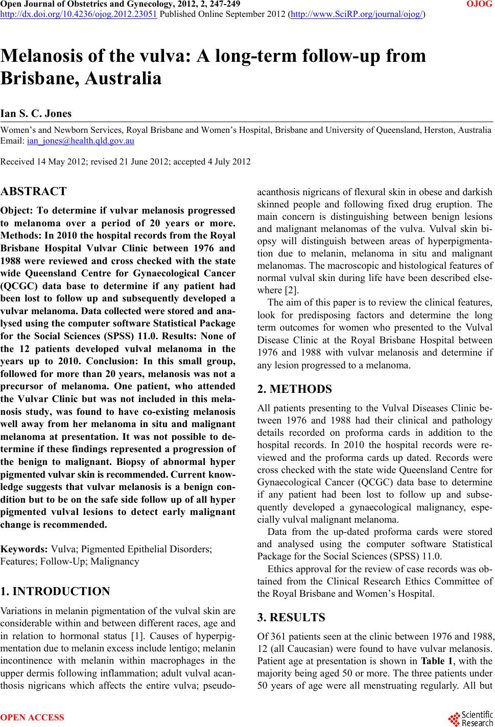 Open Journal of Obstetrics and Gynecology, 2012, 2, 247-249 OJOG http://dx.doi.org/10.4236/ojog.2012.23051 Published Online September 2012 (http://www.SciRP.org/journal/ojog/) Melanosis of the vulva: A long-term follow-up from Brisbane, Australia Ian S. C. Jones Women’s and Newborn Services, Royal Brisbane and Women’s Hospital, Brisbane and University of Queensland, Herston, Australia Email: [email protected] Received 14 May 2012; revised 21 June 2012; accepted 4 July 2012 ABSTRACT Object: To determine if vulvar melanosis progressed to melanoma over a period of 20 years or more. Methods: In 2010 the hospital records from the Royal Brisbane Hospital Vulvar Clinic between 1976 and 1988 were reviewed and cross checked with the state wide Queensland Centre for Gynaecological Cancer (QCGC) data base to determine if any patient had been lost to follow up and subsequently developed a vu lvar melanoma. Data collected were stored and ana - lysed using the computer software Statistical Package for the Social Sciences (SPSS) 11.0. Results: None of the 12 patients developed vulval melanoma in the years up to 2010. Conclusion: In this small group, followed for more than 20 years, melanosis was not a precursor of melanoma. One patient, who attended the Vulvar Clinic but was not included in this mela- nosis study, was found to have co-existing melanosis well away from her melanoma in situ and malignant melanoma at presentation. It was not possible to de- termine if these findings represented a progression of the benign to malignant. Biopsy of abnormal hyper pigmented vulvar skin is recommended. Current know- ledge suggests that vulvar melanosis is a benign con- dition but to be on the safe side follow up of all hyper pigmented vulval lesions to detect early malignant change is recommended. Keywords: Vulva; Pigmented Epithelial Disorders; Features; Follow-Up; Malignancy 1. INTRODUCTION Variations in melanin pigmentation of the vulval skin are considerable within and between different races, age and in relation to hormonal status [1]. Causes of hyperpig- mentation due to melanin excess include lentigo; melanin incontinence with melanin within macrophages in the upper dermis following inflammation; adult vulval acan- thosis nigricans which affects the entire vulva; pseudo- acanthosis nigricans of flexural skin in obese and darkish skinned people and following fixed drug eruption. The main concern is distinguishing between benign lesions and malignant melanomas of the vulva. Vulval skin bi- opsy will distinguish between areas of hyperpigmenta- tion due to melanin, melanoma in situ and malignant melanomas. The macroscopic and histological features of normal vulval skin during life have been described else- where [2]. The aim of this paper is to review the clinical features, look for predisposing factors and determine the long term outcomes for women who presented to the Vulval Disease Clinic at the Royal Brisbane Hospital between 1976 and 1988 with vulvar melanosis and determine if any lesion progressed to a melanoma. 2. METHODS All patients presenting to the Vulval Diseases Clinic be- tween 1976 and 1988 had their clinical and pathology details recorded on proforma cards in addition to the hospital records. In 2010 the hospital records were re- viewed and the proforma cards up dated. Records were cross checked with the state wide Queensland Centre for Gynaecological Cancer (QCGC) data base to determine if any patient had been lost to follow up and subse- quently developed a gynaecological malignancy, espe- cially vulval malignant melanoma. Data from the up-dated proforma cards were stored and analysed using the computer software Statistical Package for the Social Sciences (SPSS) 11.0. Ethics approval for the review of case records was ob- tained from the Clinical Research Ethics Committee of the Royal Brisbane and Women’s Ho spital. 3. RESULTS Of 361 patients seen at the clinic between 1976 and 1988, 12 (all Caucasian) were found to have vulvar melanosis. Patient age at presentation is shown in Tabl e 1, with the majority being aged 50 or more. The three patients under 50 years of age were all menstruating regularly. All but OPEN ACCESS 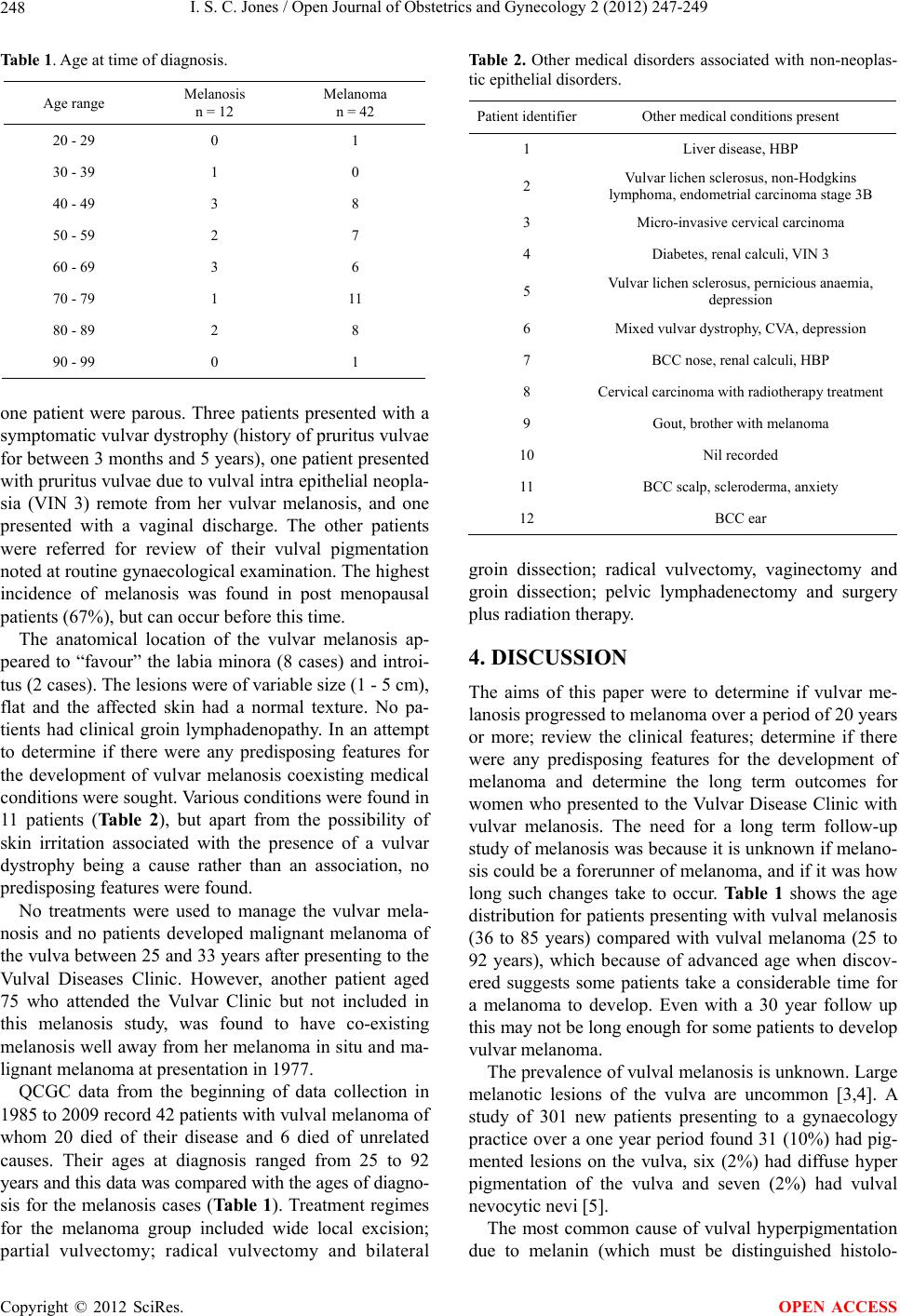 I. S. C. Jones / Open Journal of Obstetrics and Gynecology 2 (2012) 247-249 248 Table 1. Age at time of diagnosis. Age range Melanosis n = 12 Melanoma n = 42 20 - 29 0 1 30 - 39 1 0 40 - 49 3 8 50 - 59 2 7 60 - 69 3 6 70 - 79 1 11 80 - 89 2 8 90 - 99 0 1 one patient were parous. Three patients presented with a symptomatic vulv ar dystrophy (histo ry of pruritus vu lvae for between 3 months and 5 years), one patient presented with pruritus vulvae du e to vulval intra epithelial n eopla- sia (VIN 3) remote from her vulvar melanosis, and one presented with a vaginal discharge. The other patients were referred for review of their vulval pigmentation noted at routine gynaecological examination. The highest incidence of melanosis was found in post menopausal patients (67%), but can o c cur before this time. The anatomical location of the vulvar melanosis ap- peared to “favour” the labia minora (8 cases) and introi- tus (2 cases). The lesions were of variable size (1 - 5 cm), flat and the affected skin had a normal texture. No pa- tients had clinical groin lymphadenopathy. In an attempt to determine if there were any predisposing features for the development of vulvar melanosis coexisting medical conditions were sought. Various conditions were found in 11 patients (Table 2), but apart from the possibility of skin irritation associated with the presence of a vulvar dystrophy being a cause rather than an association, no predisposing features were found. No treatments were used to manage the vulvar mela- nosis and no patients developed malignant melanoma of the vulva between 25 and 33 years after presenting to the Vulval Diseases Clinic. However, another patient aged 75 who attended the Vulvar Clinic but not included in this melanosis study, was found to have co-existing melanosis well away from her melanoma in situ and ma- lignant melanoma at presentation in 1977. QCGC data from the beginning of data collection in 1985 to 2009 record 42 patients with vulval melanoma of whom 20 died of their disease and 6 died of unrelated causes. Their ages at diagnosis ranged from 25 to 92 years and this data was compared with the ages of diagno- sis for the melanosis cases (Tab le 1 ). Treatment regimes for the melanoma group included wide local excision; partial vulvectomy; radical vulvectomy and bilateral Ta b le 2 . Other medical disorders associated with non-neoplas- tic epithelial disorders. Patient identifierOther medical conditions prese nt 1 Liver disease, HBP 2 Vulvar lichen sclerosus, non-Hodgkins lymphoma, endometrial carcin oma stage 3B 3 Micro-invasive cervical carcinoma 4 Diabetes, renal calculi, VIN 3 5 Vulvar lichen sclerosus, pernicious anaemia, depression 6 Mixed vulvar dystrophy, CVA, depression 7 BCC nose, r enal calculi, HBP 8 Cervical carcinoma with radiotherapy treatment 9 Gout, brother with melanoma 10 Nil recorded 11 BCC scalp, scleroderma, anxiety 12 BCC ear groin dissection; radical vulvectomy, vaginectomy and groin dissection; pelvic lymphadenectomy and surgery plus radiation therapy. 4. DISCUSSION The aims of this paper were to determine if vulvar me- lanosis progressed to melanoma over a period of 20 years or more; review the clinical features; determine if there were any predisposing features for the development of melanoma and determine the long term outcomes for women who presented to the Vulvar Disease Clinic with vulvar melanosis. The need for a long term follow-up study of melanosis was because it is unknown if melano- sis could be a forerunner of melanoma, and if it was how long such changes take to occur. Ta b l e 1 shows the age distribution for p atients presenting with vulval melanosis (36 to 85 years) compared with vulval melanoma (25 to 92 years), which because of advanced age when discov- ered suggests some patients take a considerable time for a melanoma to develop. Even with a 30 year follow up this may not be long enough for some patients to develop vulvar melanoma. The prevalence of vulval melan osis is unknown. Large melanotic lesions of the vulva are uncommon [3,4]. A study of 301 new patients presenting to a gynaecology practice over a one year period found 31 (10%) had pig- mented lesions on the vulva, six (2%) had diffuse hyper pigmentation of the vulva and seven (2%) had vulval nevocytic nevi [5]. The most common cause of vulval hyperpigmentation due to melanin (which must be distinguished histolo- Copyright © 2012 SciRes. OPEN ACCESS  I. S. C. Jones / Open Journal of Obstetrics and Gynecology 2 (2012) 247-249 Copyright © 2012 SciRes. 249 gically from haemosiderin), is lentigo [1]. Lentigo, also known as lentigo simplex, is a benign pigmented prolix- feration of epidermal or mucous membrane melanocytes which form smooth, non-infiltrating dark brown lesions on the labia minora and introitus. Lentigo is of unknown aetiology and pathogenesis and lesions do not usually exceed one cm in diameter [1]. However, in the current study melanosis varied in size from one to five cm. Sison-Torre & Ackerman [6] described eight women with extensive pigmentation of the vulva and used the term melanosis of the vulva. Th ey show ed on b iopsy that these lesions were benign and analogous to lentigo. Len- tigines may resemble junctional naev i, but do not exhibit cytological atypia [7]. This raises the question could vulvar melanosis be the big sister to lentigo simplex with the difference being size alone? OPEN ACCESS Vulval pigmentation depends on the presence of cells containing melanin [8]. The detection of such pigmen- tation can be obscured by dermal vascularity, the thick- ness of overlying skin and the position of the melanin containing cells within the skin. The distribution of pig- mentation of dark-skinned races is concentrated in the basal layer, but varies considerably in amount. Micro- scopic patches of melanin pigment were found in almost 80 per cent of 35 Caucasian women at post mortem when there was no evidence of macroscopic hyper pigmenta- tion [8]. The differential diagnosis of vulval hyperpigmentation is lentigo; junction al naevi; following inflammatory con- ditions like lichen planus; adult vulval acanthosis nigri- cans which affect the entire vulva and may be associated with an adenocarcinoma or occasionally lymphoma or epithelial carcinoma; the pseudo-acanthosis nigricans of flexural skin in obese and darkish skinned people (now thought to be related to insulin resistance) and following fixed drug eruption from for example, sulphonamides. The most important risk for these patients is the possibi- lity of misdiagnosing a malignant melanoma. Making a clinical diagnosis based on the anatomical site and skin texture is not foolproof, hence biopsy and histological diagnosis is recommended. Even in the case of the 75 year old woman who presented with co-existing melano- sis, melanoma in situ and malignant melanoma, it was not possible to determine if these findings represented a progression from the benign to malignant. The impor- tance of follow up of all hyper pigmented vulval lesions to detect early malignant change is stressed. Variations in terminology to describe benign hyper- pigmentation due to the presence of melanin include vulvar melanosis, idiopathic lenticular mucocutaneous pigmentation and genital lentiginosis [1]. However vul- var melanosis or its other name melanosis vulvae seem to be terms used by the majority when referring to this con- dition. 5. CONCLUSION The difficulty in finding a suitable term for benign hy- perpigmentation of vulval skin conditions remains. To reliably diagnose a hyper pigmented vulval lesion a histopathology diagnosis is required. Current knowledge suggests that vulvar melanosis is a benign condition but to be on the safe side follow up of all hyper pigmented vulval lesions to detect early malignant change is re- commended. 6. ACKNOWLEDGEMENTS Thanks to the Queensland Centre for Gynaecological Cancer (QCGC) staff for their assistance in providing data and checking their data on the current study patients. REFERENCES [1] Neill, S.M. and Lewis, F.M. (2009) Ridley’s the vulva. 3rd Edition, Wiley-Blackwell, Hoboken. [2] Jones, I.S.C. (1983) A histological assessment of normal vulval skin. Clinical and Experimental Dermatology, 8, 130-138. doi:10.1111/j.1365-2230.1983.tb01818.x [3] Estrada, R. and Kaufman, R. (1993) Benign vulvar mela- nosis. The Journal of Reproductive Medicine, 38, 5-8. [4] Barnhill, R., Albert, L., Shama, S., Goldenhersh, M., Rhodes, A. and Sober, A. (1990) Genital lentiginosis: A clinical and histopathic study. Journal of the American Academy of Dermatology, 22, 453-460. doi:10.1016/0190-9622(90)70064-O [5] Rock, B., Hood, A. and Rock, J. (1990) Prospective study of vulvar nevi. Journal of the American Academy of Dermatology, 22, 104-106. doi:10.1016/0190-9622(90)70017-C [6] Sison-Torre, E.Q. and Ackerman, A.B. (1985) A clinical simulator of mali gnant melanoma: Melanosis of the vulva. The American Journal of Dermatopathology, 7, 51-60. doi:10.1097/00000372-198501001-00013 [7] Janovski, N.A. and Douglas, C.P. (1972) Diseases of the vulva. Harper & Row Publishers, Hagerstown. [8] Jones, I.S.C. (1979) An Assessment of vulval pigmenta- tion. The New Zealand Medical Journal, 89, 348-350. |