

Paper Menu >>
Journal Menu >>
 Open Journal of Obstetrics and Gynecology, 2012, 2, 244-246 OJOG http://dx.doi.org/10.4236/ojog.2012.23050 Published Online September 2012 (http://www.SciRP.org/journal/ojog/) Cesarean scar abscess: A case repor t and a review of the literature* Takako Taguchi1, Seiji Mabuchi1#, Toshio Kimura2, Tadashi Kimura1 1Department of Ob s tetrics and Gynecology, Osaka University Graduate School of Medicine, Suita, Japan 2Department of Gynecology, Ashiya Municipal Hospi t a l , Ashiya, Japan Email: #[email protected]d.osaka-u.ac.jp Received 4 May 2012; revised 8 June 2012; accepted 20 June 2012 ABSTRACT Cesarean section and the resultant Cesarean scar are known to be associated with obstetric complications in subsequent pregnancies. Cesarean scar is also as- sociated with gynecological conditions that can ad- versely affect the patient’s quality of life. We describe a very rare case of Cesarean scar abscess that deve- loped 8 years after a Cesarean delivery, which was managed by emergency hysterectomy. Keywords: Cesarean Section; Cesarean Scar Dehiscence; Abscess 1. INTRODUCTION The increasing rates of Cesarean section and its compli- cations are global issues in developed countries. Cesa- rean section and the resultant Cesarean scar in the lower uterine segment are known to be associated with obstet- ric complications in subsequent pregnancies, such as uterine rupture; Cesarean scar pregnancy (CSP); placenta previa; and placenta accreta, increta, or percreta. Cesar- ean scars are also associated with gynecological condi- tions that can ad versely affect the patient’s quality of life, e.g., abnormal uterine bleeding, chronic pain, or secon- dary infertility [1]. We herein describe a very rare case of Cesarean scar abscess that developed 8 years after a Cesarean delivery, which was m a naged by emergency hysterectomy. 2. CASE REPORT A 44-year-old Japanese woman (gravida 1, para 1) visited our hospital complaining of abdominal pain that had lasted for 2 weeks accompanied by uterine bleeding. Her obstetric history included a lower uterine segment trans- verse Cesarean section 8 years earlier. Her medical his- tory was unremarkable. Physical examinations including pelvic examination revealed a body temperature of 39˚C, marked bloody cervical discharge with an odious smell, cervical motion tenderness, and lower abdominal ten- derness, but no rebound tenderness. Transvaginal ultra- sonography showed a normal-sized uterus with an 8 × 7 cm spherical mass in the lower uterine segment, which was located on the scar cau sed by the previous Cesarean section. The inner part of the mass was irregular (both hyperechogenic and anechogenic). Both ovaries were normal, and there was no intraperitoneal fluid. Labora- tory tests revealed an elevated white blood cell count (12,120/mm3), an elevated C-reactive protein level (14.9 mg/dl), and a negative pregnancy test result. She was admitted, and initial treatment with antibiotics was per- formed on the same day. A subsequent pelvic magnetic resonance imaging (MRI) examination revealed an 11 × 10 × 9 cm exophytic tumor in the lower uterine segment. Both transvaginal ultrasonography and pelvic MRI sug- gested that the tumor was connected to the uterine cavity through a small defect in the lower anterior wall of the uterus, which might have been a Cesarean scar defect. A diagnosis of Cesarean scar abscess was suspected. As her infectious symptoms progressed and she did not want to preserve her fertility, we offered her abdominal hyste- rectomy. Exploratory laparotomy revealed a 12 × 10 × 10 cm elastic mass arising from the lower anterior uterine wall, which was adherent to the right pelvic sidewall, and total abdominal hysterectomy was performed (Figure 1). Grossly, the mass contained bloody purulent discharge and was connected to the uterine cavity by a thin piece of tissue. A pathological examination showed a bundle-like mass of muscle tissue without any findings of degene- rated leiomyoma. The cavity of the tumor and the tissue connecting it to th e uterus were lined with colu mnar cells resembling those found in the endocervical epithelium. Culturing of the abscess contents produced Enterobacter cloacae. The diagnosis of Cesarean scar abscess was confirmed. The patient received intravenous antibiotics for 2 days after the surgery and was discharged. *Conflicts of Interest Statement: The authors declare that no conflicts of interest exist. #Corresponding author. OPEN ACCESS 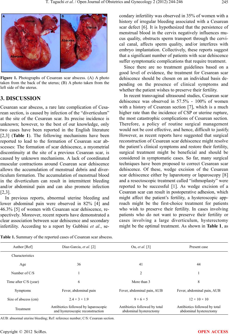 T. Taguchi et al. / Open Journal of Obstetrics and Gynecology 2 (2012) 244-246 245 Figure 1. Photographs of Cesarean scar abscess. (A) A photo taken from the back of the uterus; (B) A photo taken from the left side of the uterus. 3. DISCUSSION Cesarean scar abscess, a rare late complication of Cesa- rean section, is caused by infection of the “diverticulum” at the site of the Cesarean scar. Its precise incidence is unknown; however, to the best of our knowledge, only two cases have been reported in the English literature [2,3] (Table 1). The following mechanisms have been reported to lead to the formation of Cesarean scar ab- scesses: The formation of scar dehiscence, a myometrial discontinuity at the site of a previous Cesarean scar, is caused by unknown mechanisms. A lack of coordinated muscular contractions around Cesarean scar dehiscence allows the accumulation of menstrual debris and diver- ticulum formation. The accumulation of menstrual blood in the diverticulum can result in intermittent bleeding and/or abdominal pain and can also promote infection [2,3]. In previous reports, abnormal uterine bleeding and lower abdominal pain were observed in 82% [4] and 46.3% [5] of women with Cesarean scar dehiscence, re- spectively. Moreover, recent reports have demonstrated a clear association between scar dehiscence and secondary infertility. According to a report by Gubbini et al., se- condary infertility was ob s erved in 35% of women with a history of irregular bleeding associated with a Cesarean scar defect [6]. It is hypothesized that the persistence of menstrual blood in the cervix negatively influences mu- cus quality, obstructs sperm transport through the cervi- cal canal, affects sperm quality, and/or interferes with embryo implantation. Collectively, these reports suggest that a significant number of patients with scar dehiscence suffer symptomatic complications that require treatment. Since there are no treatment guidelines based on a good level of evidence, the treatment for Cesarean scar dehiscence should be chosen on an individual basis de- pending on the presence of clinical symptoms and whether the patient wishes to preserve their fertility. In recent transvaginal ultrasound studies, Cesarean scar dehiscence was observed in 57.5% - 100% of women with a history of Cesarean section [7], which is a much higher rate than the incidence of CSP or uterine rupture, the most catastrophic complications of Cesarean section. Therefore, a policy of routine surgical management would not be cost effective, and hence, difficult to justify. However, as recent reports have suggested that surgical reconstruction of Cesarean scar dehiscence might resolve the patient’s clinical symptoms and restore their fertility, surgical treatment might be beneficial and should be considered in symptomatic cases. So far, many surgical techniques have been proposed to correct Cesarean scar dehiscence. Of these, wedge excision of the Cesarean scar dehiscence either by laparotomy or laparoscopy [8] and a resectoscopic treatment called “isthmoplasty” were reported to be successful [1]. As wedge excision of a Cesarean scar can result in postoperative adhesion, which might affect the patient’s fertility, a hysteroscopic app- roach might be the first-choice treatment for patients who wish to preserve their fertility. In cases involving patients who do not want to preserve their fertility or cases involving a large diverticulum, hysterectomy mig ht b e the optimal treatment. As shown in Table 1, as Table 1. Summary of the reported cases of Cesarean scar abscess. Author [Ref] Diaz-Garcia, et al. [2] Ou, et al. [3] Present case Characteristics Age 36 41 44 Number of C/S 1 2 1 Time after C/S (year) 6 More than 3 8 Symptoms Fever, abdominal pain Fever, abdominal pain, AUB Fever, abdominal pain, AUB Size of abscess (cm) 2.4 × 3 × 1.9 9 × 6 × 5 12 × 10 × 10 Treatment Antibiotics followed b y l a p a r o s copic and hysteroscopic reconstruction Antibiotics followed b y t o t a l abdominal hysterectomy Antibiotics followed b y t o t a l abdominal hysterectomy AUB: abnormal uterine bleeding; Ref: ref erence n umber; C/S: C esarean s ection. Copyright © 2012 SciRes. OPEN ACCESS  T. Taguchi et al. / Open Journal of Obstetrics and Gynecology 2 (2012) 244-246 246 conservative medical treatments using antibiotics failed in all three reported cases, prompt surgical treatment is recommended for Cesarean scar abscess. REFERENCES [1] Florio, P., Filippeschi, M., Moncini, I., Marra, E., Fran- chini, M. and Gubbini, G. (2012) Hysteroscopic treatment of the cesarean-induced isthmocele in restoring infertility. Current Opinion in Obstetrics & Gynecology, 24, 180- 186. [2] Diaz-Garcia, C., Estellés, J.G., Escrivá, A.M., Mora, J.J., Torregrosa, R.R. and Sancho, J.M. (2009) Scar abscess six years after cesarean section: Laparoscopic and hys- teroscopic management. Journal of Minimally Invasive Gyn ecology , 16, 785 - 7 88 . doi:10.1016/j.jmig.2009.07.020 [3] Ou, Y. C., Huang, K.H., Lin, H., Eng, H.L., Lu, H.M. and Changchien, C.C. (2011) Sepsis secondary to cesarean scar diverticulum resembling an infected leiomyoma. Taiwan Journal of Obstetrics and Gynecology, 50, 100- 102. doi:10.1016/j.tjog.2011.01.004 [4] Fabres, C., Aviles, G., De La Jara, C., Escalona, J., Muñoz, J.F., Mackenna, A., Fernández, C., Zegers-Hochschild, F. and Fernández, E. (2003) The cesarean delivery scar pouch: Clinical implications and diagnostic correlation between transvaginal sonography and hysteroscopy. Jour- nal of Ultrasound in Medicine, 22, 695-700. [5] Gubbini, G., Centini, G., Nascetti, D., Marra, E., Moncini, I., Bruni, L., Petraglia, F. and Florio, P. (2011) Surgical hysteroscopic treatment of cesarean-induced isthmocele in restoring fertility: Prospective study. Journal of Mini- mally Invasive Gynecology, 18, 234-237. doi:10.1016/j.jmig.2010.10.011 [6] Gubbini, G., Casadio, P. and Marra, E. (2008) Resecto- scopic correction of the “isthmocele” in women with postmenstrual abnormal uterine bleeding and secondary infertility. Journal of Minimally Invasive Gynecology, 15, 172-175. doi:10.1016/j.jmig.2007.10.004 [7] Menada Valenzano, M., Lijoi, D., Mistrangelo, E., Co- stantini, S. and Ragni, N. (2006) Vaginal ultrasonographic and hysterosonographic evaluation of the low transverse incision after caesarean section: Correlation with gynae- cological symptoms. Gynecologic and Obstetric Investi- gation, 61, 216-222. doi:10.1159/000091497 [8] Donnez, O., Jadoul, P., Squifflet, J. and Donnez, J. (2008) Laparoscopic repair of wide and deep uterine scar dehis- cence after cesarean section. Fertility and Sterility, 89, 974-980. doi:10.1016/j.fertnstert.2007.04.024 Copyright © 2012 SciRes. OPEN ACCESS |