

Paper Menu >>
Journal Menu >>
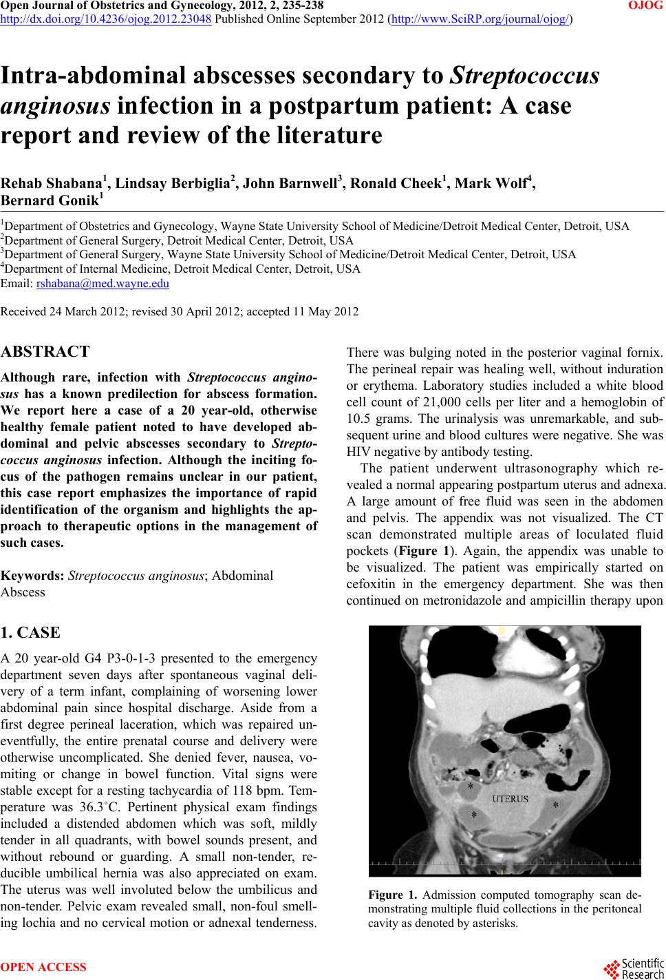 Open Journal of Obstetrics and Gynecology, 2012, 2, 235-238 OJOG http://dx.doi.org/10.4236/ojog.2012.23048 Published Online September 2012 (http://www.SciRP.org/journal/ojog/) Intra-abdominal abscesses secondary to Streptococcus anginosus infection in a postpartum patient: A case report and review of the literature Rehab Shabana1, Lindsay Berbiglia2, John Barnwell3, Ronald Cheek1, Mark Wolf4, Bernard Gonik1 1Department of Ob s tetrics and Gynecology, Wayne State University School of Medicine/Detroit Medical Center, Detroit, USA 2Department of General Surgery, Detroit Medical Center, Detroit, USA 3Department of General Surgery, Wayne State University School of Medicine/Detroit Medical Center, Detroit, USA 4Department of Internal Medicine, Detroit Med ical Cent er, Detroit, USA Email: [email protected] Received 24 March 2012; revised 30 April 2012; accepted 11 May 2012 ABSTRACT Although rare, infection with Streptococcus angino- sus has a known predilection for abscess formation. We report here a case of a 20 year-old, otherwise healthy female patient noted to have developed ab- dominal and pelvic abscesses secondary to Strepto- coccus anginosus infection. Although the inciting fo- cus of the pathogen remains unclear in our patient, this case report emphasizes the importance of rapid identification of the organism and highlights the ap- proach to therapeutic options in the management of such cases. Keywords: Streptoc occu s anginosus; Abdominal Abscess 1. CASE A 20 year-old G4 P3-0-1-3 presented to the emergency department seven days after spontaneous vaginal deli- very of a term infant, complaining of worsening lower abdominal pain since hospital discharge. Aside from a first degree perineal laceration, which was repaired un- eventfully, the entire prenatal course and delivery were otherwise uncomplicated. She denied fever, nausea, vo- miting or change in bowel function. Vital signs were stable except for a resting tachycardia of 118 bpm. Tem- perature was 36.3˚C. Pertinent physical exam findings included a distended abdomen which was soft, mildly tender in all quadrants, with bowel sounds present, and without rebound or guarding. A small non-tender, re- ducible umbilical hernia was also appreciated on exam. The uterus was well involuted below the umbilicus and non-tender. Pelvic exam revealed small, non-foul smell- ing lochia and no cervical motion or adnexal tenderness. There was bulging noted in the posterior vaginal fornix. The perineal repair was healing well, without induration or erythema. Laboratory studies included a white blood cell count of 21,000 cells per liter and a hemoglobin of 10.5 grams. The urinalysis was unremarkable, and sub- sequent urine and blood cultures were negative. She was HIV negative by antibody testing. The patient underwent ultrasonography which re- vealed a normal appearing postpartum uterus and adnexa. A large amount of free fluid was seen in the abdomen and pelvis. The appendix was not visualized. The CT scan demonstrated multiple areas of loculated fluid pockets (Figure 1). Again, the appendix was unable to be visualized. The patient was empirically started on cefoxitin in the emergency department. She was then continued on metronidazole and ampicillin therapy upon Figure 1. Admission computed tomography scan de- monstrating multiple fluid collections in the peritoneal cavity as denoted by asterisks. OPEN ACCESS 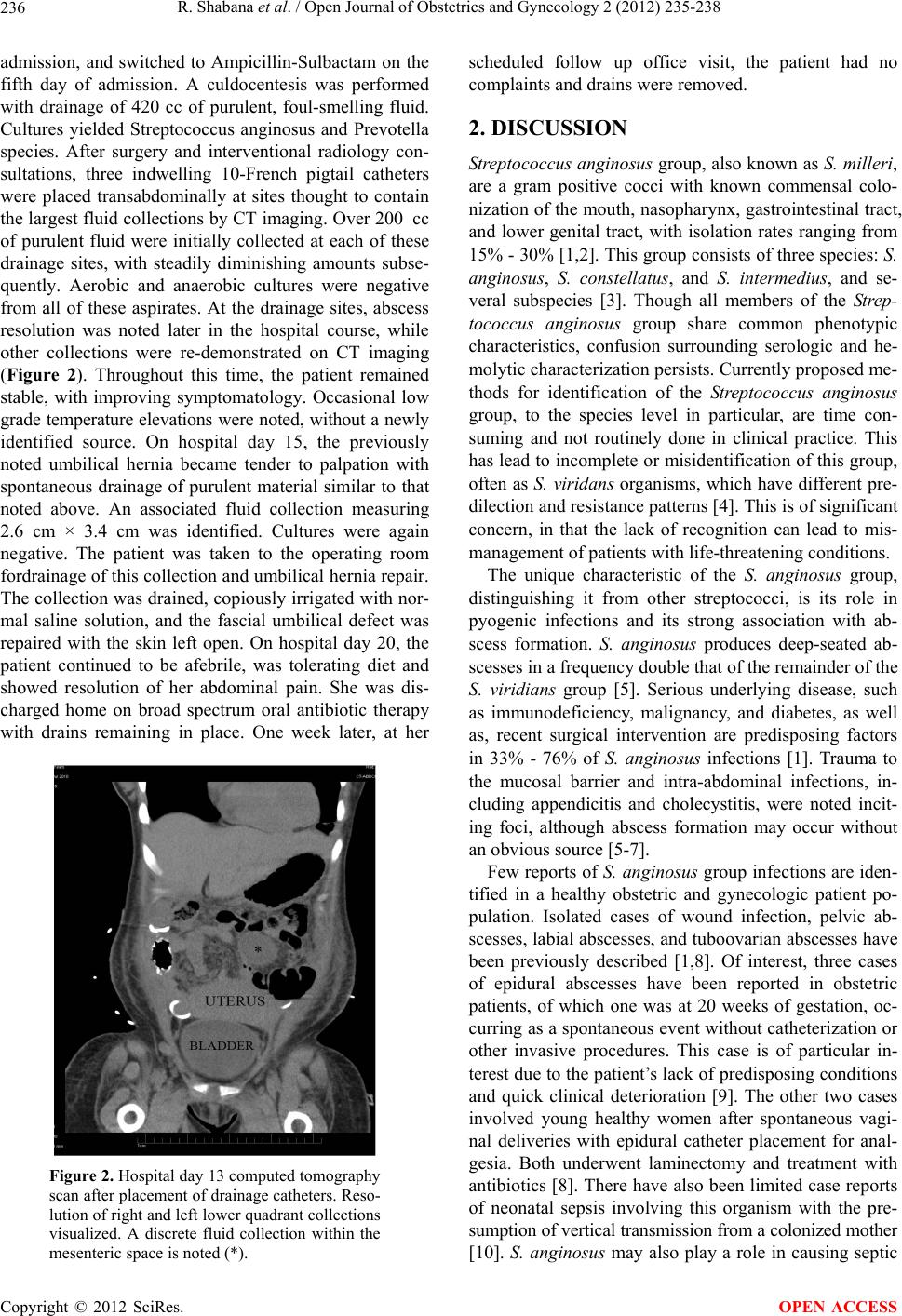 R. Shabana et al. / Open Journal of Obstetrics and Gynecology 2 (2012) 235-238 236 admission, and switched to Ampicillin-Sulbactam on the fifth day of admission. A culdocentesis was performed with drainage of 420 cc of purulent, foul-smelling fluid. Cultures yielded Streptococcus anginosus and Prevotella species. After surgery and interventional radiology con- sultations, three indwelling 10-French pigtail catheters were placed transabdominally at sites thought to contain the largest fluid collections by CT imaging . Over 200 cc of purulent fluid were initially collected at each of these drainage sites, with steadily diminishing amounts subse- quently. Aerobic and anaerobic cultures were negative from all of these aspirates. At the drainage sites, abscess resolution was noted later in the hospital course, while other collections were re-demonstrated on CT imaging (Figure 2). Throughout this time, the patient remained stable, with improving symptomatology. Occasional low grade temperature elevations were noted, without a newly identified source. On hospital day 15, the previously noted umbilical hernia became tender to palpation with spontaneous drainage of purulent material similar to that noted above. An associated fluid collection measuring 2.6 cm × 3.4 cm was identified. Cultures were again negative. The patient was taken to the operating room fordrainage of this collection an d umbilical h ernia repair. The collection was drained, copiously irrigated with nor- mal saline solution, and the fascial umbilical defect was repaired with the skin left open. On hospital day 20, the patient continued to be afebrile, was tolerating diet and showed resolution of her abdominal pain. She was dis- charged home on broad spectrum oral antibiotic therapy with drains remaining in place. One week later, at her Figure 2. Hospital day 13 computed tomography scan after placement of drainage catheters. Reso- lution of right and left lower quadrant collections visualized. A discrete fluid collection within the mesenteric space is noted (*). scheduled follow up office visit, the patient had no complaints and drains were removed. 2. DISCUSSION Streptococcus anginosus group, also known as S. milleri, are a gram positive cocci with known commensal colo- nization of the mouth, nasopharynx, gastrointestinal tract, and lower genital tract, with isolation rates ranging from 15% - 30% [1,2 ]. This group consists of three species: S. anginosus, S. constellatus, and S. intermedius, and se- veral subspecies [3]. Though all members of the Strep- tococcus anginosus group share common phenotypic characteristics, confusion surrounding serologic and he- molytic characterization persists. Currently proposed me- thods for identification of the Streptococcus anginosus group, to the species level in particular, are time con- suming and not routinely done in clinical practice. This has lead to incomplete or misidentification of this gro up, often as S. viridans organisms, which have different pre- dilection and resistance patterns [4]. This is of significant concern, in that the lack of recognition can lead to mis- management of patients with life-threatening conditions. The unique characteristic of the S. anginosus group, distinguishing it from other streptococci, is its role in pyogenic infections and its strong association with ab- scess formation. S. anginosus produces deep-seated ab- scesses in a frequency double that of the remainder of the S. viridians group [5]. Serious underlying disease, such as immunodeficiency, malignancy, and diabetes, as well as, recent surgical intervention are predisposing factors in 33% - 76% of S. anginosus infections [1]. Trauma to the mucosal barrier and intra-abdominal infections, in- cluding appendicitis and cholecystitis, were noted incit- ing foci, although abscess formation may occur without an obvious source [5-7]. Few reports of S. anginosus group infections are iden- tified in a healthy obstetric and gynecologic patient po- pulation. Isolated cases of wound infection, pelvic ab- scesses, labial abscesses, and tuboovarian abscesses have been previously described [1,8]. Of interest, three cases of epidural abscesses have been reported in obstetric patients, of which one was at 20 weeks of gestation, oc- curring as a spontane ous event with out catheterization or other invasive procedures. This case is of particular in- terest due to the patient’s lack of predisposing conditions and quick clinical deterioration [9]. The other two cases involved young healthy women after spontaneous vagi- nal deliveries with epidural catheter placement for anal- gesia. Both underwent laminectomy and treatment with antibiotics [8]. Th ere have also been limited case reports of neonatal sepsis involving this organism with the pre- sumption of vertical transmission from a colonized mother [10]. S. anginosus may also play a role in causing septic Copyright © 2012 SciRes. OPEN ACCESS  R. Shabana et al. / Open Journal of Obstetrics and Gynecology 2 (2012) 235-238 237 abortions via ascending infection [11]. The case presented is of particular interest for several reasons including the lack of obvious inciting cause, the disparity between the patient’s eventually recognized disease state and her presenting symptomatology, and the choice of management. Treatment with systemic antibi- otics and serial interventional radiology-directed drain- age versus early surgical exploration remains controver- sial. It has been noted in previous studies, that the ma- jority of infectious S. anginosus cases originate from skin/soft tissue or intraabdominal sources. Of 186 cases of S. anginosus noted in a retrospective study, 64 had skin/soft tissue manifestations and 41 had intraabdominal manifestations. These in cluded liver abscess, cho langitis, peritonitis, pancreatic abscess, subphrenic abscess, and most commonly appendicitis, the source in 23 of the 41 cases [1]. Another study detailing 33 cases of S. angino- sus and bacteremia found the majority were due to in- traabdominal sources [7]. The inciting factors in these cases included cholecystitis/cholangitis (18%) and ap- pendicitis (12%) [7]. Given our patient’s extensive in- traabdominal disease process, appendicitis was considered a possible inciting focus. The patient underwent several imaging procedures, including ultrasonography and se- rial CT scanning, none of which allowed visualization of the appendix. Though not visualized, there was also no noted area of increased inflammation surrounding the area of the appendix to suggest appendicitis. Of note, S. anginosus is a commensal organism of the appendix and was isolated from a quarter of normal-appearing appen- dices, not in the setting of appendicitis [12]. Lastly, re- garding a source of the infection, it should be recalled that the patient had a small perineal laceration repaired in an uncomplicated manner at the time of delivery. As pre- viously noted, S. anginosus species are known to colo- nize the lower female genital tract. Therefore, trauma to this mucosal barrier may have been the site for entrance of the pathogen. Our patient did not have any signs of in- flammation, induration or purulence at the laceration site upon subsequent presen tation to the hospital. Of interest, certain S. anginosus biotypes have a specific affinity for the vaginal mucosa. They are identified by the fact they produce acid from raffinose and melibiose in the labora- tory. Unfortunately our clinical specimen was not tested with these substrates to help better define the source of the organism. A final point of interest in this case is the manage ment of multiple intra-abdominal abscesses with percutaneous drainage instead of surgical intervention. On presentatio n the patient had significant abdominal tenderness, but lacked other gastrointestinal symptoms. She was tolerat- ing a regular diet and complained of only mildly de- creased appetite. She was afebrile and hemodynamically stable. Also, at the time of presentation, the patient had been symptomatic for seven days, making surgical inter- vention a potentially more complicated approach. Given the noted clinical findings and the apparent amenability of the fluid collections to drainage, the decision was made to proceed wit h percutaneous drai na ge. On review, there are several studies detailing the use of radiology guided percutaneous drainage catheters for intraabdominal abscesses and success rates comparable to that of open intervention, with overall success rates between 33% and 100% [13]. Three studies were re- viewed detailing the management of intraabdominal ab- scesses, leading to similar conclusions that percutaneous drainage had a higher success rate determined by abscess and sepsis resolution, and lower rates of complications, when compared to operative intervention [14-16]. It should be noted that second attempts at percutaneous drainage were required in a significant number of pa- tients described in the above studies and that the duration of drain management ranged from 6 to 60 days. Our pa- tient underwent a total of five drainage procedures and three drainage catheter placement procedures, which resulted in resolution of the fluid collections. Common adverse events described with catheter management in- cluded drains falling out or becoming clogged or dam- aged, cellulitis at the drain site, and less commonly fis- tula formation, bleeding, need for re-hospitalization, and inability to place the drain. Recognized limitations in the evaluation of this case include the initiation of antibiotics prior to obtaining cultures and the polymicrobial nature of the infection. Intravenous antibiotic therapy was initiated prior to the culdocentesis procedure from which the S. anginosus cultures were obtained. This may have limited culture results, suppressing growth of other involved pathogens. Prevotella species was also isolated from the culdocente- sis specimen. Its ability to cause abscesses and bactere- mia is well documented, particularly in the mouth fol- lowing invasive proc edures. Since both S. anginosus and Prevotella both can colonize the oral cavity, a more care- ful assessment of the patient’s teeth and mouth would have been helpful despite there being no presenting sym- ptomatology related to these structures. With regard to antibiotic selection , the initial empiric use of broad spec- trum antibiotics is justifiable given the presenting cir- cumstances. Later, culture-based identification of the causative pathogens would have allowed for therapeutic modifications in the event of a suboptimal response, or if microbial resistance was identified. Fortunately, S. angi- nosus is sensitive to a wide variety of Gram-positive fo- cused antimicrobial agents. In summary, this case report details an unusual viru- lent infection presenting in an otherwise young healthy postpartum patient caused by S. anginosus. Because of the pathologic potential for this organism, it highlights Copyright © 2012 SciRes. OPEN ACCESS  R. Shabana et al. / Open Journal of Obstetrics and Gynecology 2 (2012) 235-238 Copyright © 2012 SciRes. OPEN ACCESS 238 [7] Salavert, M., Gomez, L., Rodriguez-Carballeira, M., Xer- cavins, M., Freixas, N. and Garau, J. (1996) Seven-year review of bacteremia caused by Streptococcus milleri and other viridans streptococci. European Journal of Clinical Microbiology & Infectious Diseases, 15, 365-371. doi:10.1007/BF01690091 the importance of careful laboratory identification and timely reporting. Given its predilectio n for puru lence and abscess formation, once identified as a cause for infec- tion, careful observation for disease progression is re- quired. Transabdominal percutaneous drainage of acces- sible abscess pockets, in the otherwise stable parturient, seems to be a reasonable consideration as a first line approach. There are presently insufficient data available in the obstetric literature detailing colonization rates and the incidence of disease. This limits insight as to the need for preemptive identification or prophylactic interven- tio ns, as we do with other known puerper al Streptococcus pathogens S. agal act i ae and S. pyogenes. [8] Treszezamsky, A.D. and Feldman, D., Sarabanchong, V.O. (2011) Concurrent postpartum uterine and abdominal wall dehiscence and Stretococcus anginosus infection. Obstetrics & Gynecology, 118, 449-451. doi:10.1097/AOG.0b013e31821619e9 [9] Lampen, R. and Bearman, G. (2005) Epidural abscess caused by Streptococcus milleri in a pregnant woman. BMC Infectious Diseases, 5, 100. doi:10.1186/1471-2334-5-100 [10] Cox, R.A., Chen, K., Coykendall, A.L., Wesbecher, P. and Herson, V.C. (1987) Fatal infection in neonates of 26 weeks’ gestation due to Streptococcus milleri: Report of two cases. Journal of Clinical Pathology, 40, 190-193. doi:10.1136/jcp.40.2.190 REFERENCES [1] Singh, K.P., Morris, A., Lang, S.D., MacCulloch, D.M. and Bremner, D.A. (1988) Clinically significant Strepto- coccus anginosus (Streptococcus milleri) infec tions: A re- view of 186 cases. New Zealand Medical Journal, 101, 813-816. [11] MacGowan, A.P. and Terry, P.B. (1987) Streptococcus milleri and second trimester abortion. Journal of Clinical Pathology, 40, 292-293. doi:10.1136/jcp.40.3.292 [2] Ruoff, K.L. (1988) Streptococcus anginosus (Streptococ- cus milleri): The unrecognized pathogen. Clinical Micro- biology Reviews, 1, 102-128. [12] Poole, P.M. and Wilson, G. (1977) Streptococcus milleri in the appendix. Journal of Clinical Pathology, 30, 937- 942. doi:10.1136/jcp.30.10.937 [3] Claridge, J.E., et al. (2001) Streptococcus intermedius, Streptococcus constellatus, and Streptococcus anginosus (Streptococcus milleri group) are of different clinical im- portance and are not equally associated with abscess. Clinical Infectious Diseases, 32, 1511-1515. doi:10.1086/320163 [13] Gohl, J., Gmeinwieser, J. and Gusinde, J. (1999) Intraab- dominal abscesses. Intervention versus surgical treatment. Zentralblatt fur Chirurgie, 124, 187-194. [14] Cinat, M.E., Wilson, S.E. and Din, A.M. (2002) Deter- minants for successful percutaneous image-guided d rain- age of intra-abdominal abscess. Archives of Surgery, 137, 845-849. doi:10.1001/archsurg.137.7.845 [4] Facklam, R. (2002) What happened to the streptococci: Overview of taxonomic and nomenclature changes. Cli- nical Microbiology Reviews, 15, 613-630. doi:10.1128/CMR.15.4.613-630.2002 [15] Johnson, W.C., Gerzof, S.G., Robbins, A.H. and Nabseth, D.C. (1981) Treatment of abdominal abscesses: Com- parative evaluation of operative drainage versus percuta- neous catheter drainage guided by computed tomography or ultrasound. Annals of Surgery, 194, 510-520. doi:10.1097/00000658-198110000-00014 [5] Admon, D., Ephros, M.A., Gavish, D. and Raz, R. (1987) Infection with Streptococcus milleri. Journal of Infection, 14, 55-60. doi:10.1016/S0163-4453(87)90834-6 [6] Jacobs, J.A., Pietersen, H.G., Stobberingh, E.E. and Soeters, P.B. (1994) Bacteremia involving the “Strepto- coccus milleri” group: Analysis of 19 cases. Clinical In- fectious Diseases, 19, 704-713. doi:10.1093/clinids/19.4.704 [16] Van Sonnenberg, E., et al. (1984) Percutaneous drainage of 250 abdominal abscesses and fluid collections. Part I: Results, failures, and complications. Radiology, 151 , 337- 341. |