

Paper Menu >>
Journal Menu >>
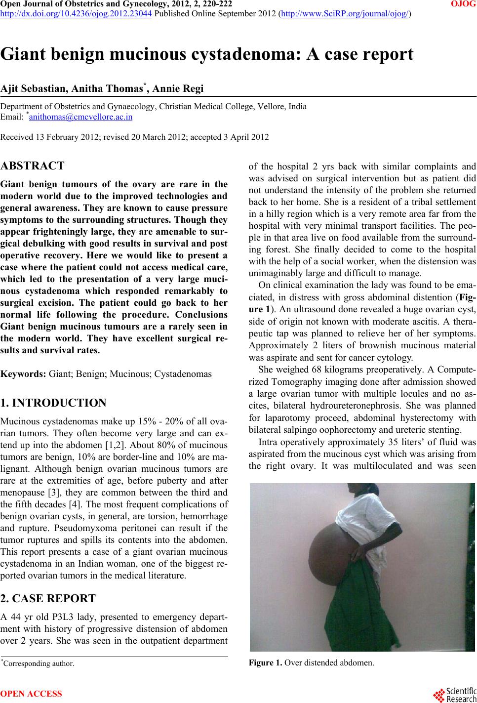 Open Journal of Obstetrics and Gynecology, 2012, 2, 220-222 OJOG http://dx.doi.org/10.4236/ojog.2012.23044 Published Online September 2012 (http://www.SciRP.org/journal/ojog/) Giant benign mucinous cystadenoma: A case report Ajit Sebastian, Anitha Thomas*, Annie Regi Department of Obstetrics and Gynaecology, Christian Medical College, Vellore, India Email: *[email protected] Received 13 February 2012; revised 20 March 2012; accepted 3 April 2012 ABSTRACT Giant benign tumours of the ovary are rare in the modern world due to the improved technologies and general awareness. They are known to cause pressure sy mpto ms to the surrounding structures. Though they appear frighteningly large, they are amenable to sur- gical debulking with good results in survival and post operative recovery. Here we would like to present a case where the patient could not access medical care, which led to the presentation of a very large muci- nous cystadenoma which responded remarkably to surgical excision. The patient could go back to her normal life following the procedure. Conclusions Giant benign mucinous tumours are a rarely seen in the modern world. They have excellent surgical re- sults and survival rates. Keywords: Giant; Benign; Mucinous; Cystadenomas 1. INTRODUCTION Mucinous cystadenomas make up 15% - 20% of all ova- rian tumors. They often become very large and can ex- tend up into the abdomen [1,2]. About 80% of mucinous tumors are benign, 10% are border-line and 10% are ma- lignant. Although benign ovarian mucinous tumors are rare at the extremities of age, before puberty and after menopause [3], they are common between the third and the fifth decades [4]. The most frequent complications of benign ovarian cysts, in general, are torsion, hemorrhage and rupture. Pseudomyxoma peritonei can result if the tumor ruptures and spills its contents into the abdomen. This report presents a case of a giant ovarian mucinous cystadenoma in an Indian woman, one of the biggest re- ported ovarian tumors in the medical literature. 2. CASE REPORT A 44 yr old P3L3 lady, presented to emergency depart- ment with history of progressive distension of abdomen over 2 years. She was seen in the outpatient department of the hospital 2 yrs back with similar complaints and was advised on surgical intervention but as patient did not understand the intensity of the problem she returned back to her home. She is a resident of a tribal settlement in a hilly region which is a very remote area far from the hospital with very minimal transport facilities. The peo- ple in that area live on food available from the surround- ing forest. She finally decided to come to the hospital with the help of a social worker, when the distension was unimaginably large and difficult to manage. On clinical examinatio n the lad y was found to be ema- ciated, in distress with gross abdominal distention (Fig- ure 1). An ultrasound done revealed a huge ovarian cyst, side of origin not known with moderate ascitis. A thera- peutic tap was planned to relieve her of her symptoms. Approximately 2 liters of brownish mucinous material was aspirate and sent for cancer cytology. She weighed 68 kilograms preoperatively. A Compute- rized Tomography imaging done after admission showed a large ovarian tumor with multiple locules and no as- cites, bilateral hydroureteronephrosis. She was planned for laparotomy proceed, abdominal hysterectomy with bilateral salpingo oophorectomy and ureteric stenting . Intra operatively approximately 35 liters’ of fluid was aspirated from the mucinous cyst which was arising from the right ovary. It was multiloculated and was seen Figure 1. Over distended abdomen. *Corresponding a uthor. OPEN ACCESS 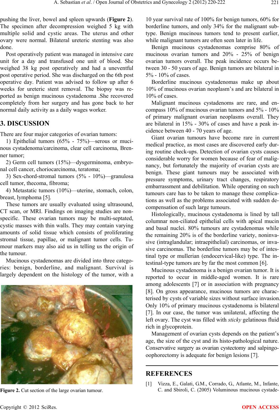 A. Sebastian et al. / Open Journal of Obstetrics and Gynecology 2 (2012) 220-222 221 pushing the liver, bowel and spleen upwards (Figure 2). The specimen after decompression weighed 5 kg with multiple solid and cystic areas. The uterus and other ovary were normal. Bilateral ureteric stenting was also done. Post operatively patient was managed in intensive care unit for a day and transfused one unit of blood. She weighed 38 kg post operatively and had a uneventful post operative period. She was discharged on the 6th post operative day. Patient was advised to follow up after 6 weeks for ureteric stent removal. The biopsy was re- ported as benign mucinous cystadenoma .She recovered completely from her surgery and has gone back to her normal daily activity as a daily wages worker. 3. DISCUSSION There are four major categories of ovarian tumors: 1) Epithelial tumors (65% - 75%)—serous or muci- nous cystadenoma/carcinoma, clear cell carcinoma, Br en- ner tumor; 2) Germ cell tumors (15%)—dysgerminoma, embryo- nal cell cancer, choriocarcinoma, teratoma; 3) Sex-chord-stromal tumors (5% - 10%)—granulosa cell tumor, thecoma, fibroma; 4) Metastatic tumors (10%)—uterine, stomach, colon, breast, lymphoma [5]. These tumors are usually evaluated using ultrasound, CT scan, or MRI. Findings on imaging studies are non- specific. These ovarian tumors may be multi-septated, cystic masses with thin walls. They may contain varying amounts of solid tissue which consists of proliferating stromal tissue, papillae, or malignant tumor cells. Tu- mour markers may also aid us in telling us the origin of the tumour. Mucinous cystadenomas are divided into three catego- ries: benign, borderline, and malignant. Survival is largely dependent on the histology of the tumor, with a Figure 2. Cut section of the large ovarian tumour. 10 year survival rate of 100% for benign tumors, 60% for borderline tumors, and only 34% for the malignant sub- type. Benign mucinous tumors tend to present earlier, while malignant tumors are often seen later in life. Benign mucinous cystadenomas comprise 80% of mucinous ovarian tumors and 20% - 25% of benign ovarian tumors overall. The peak incidence occurs be- tween 30 - 50 years of age. Benign tumors are bilateral in 5% - 10% of cases. Borderline mucinous cystadenomas make up about 10% of mucinous ovarian neoplasm’s and are bilateral in 10% of cases. Malignant mucinous cystadenoms are rare, and en- compass 10% of mucinous ovarian tumors and 5% - 10% of primary malignant ovarian neoplasms overall. They are bilateral in 15% - 30% of cases and have a peak in- cidence between 40 - 70 years of age. Giant ovarian tumours have become rare in current medical practice, as most cases are discovered early dur- ing routine check-ups. Detection of ovarian cysts causes considerable worry for women because of fear of malig- nancy, but fortunately the majority of ovarian cysts are benign. These giant tumours may be associated with pressure symptoms, urinary tract changes, respiratory embarrassment and debilitation. While operating on su ch tumours care has to be taken to manage these complica- tions as well as the problems associated with sudden de- compensation of such large tumours. Histologically, mucinous cystadenoma is lined by tall columnar non-ciliated epithelial cells with apical mucin and basal nuclei. 80% tumours are cystadenomas while the remaining 20% is of the borderline variety, noninva- sive (intraglandular; intraepithelial) carcinomas, or inva- sive carcinomas. The borderline tumors may be of intes- tinal type or mullerian (endocervical-like) type. The in- testinal-type tumors are by far the most common [6]. Mucinous cystadenoma is a benign ovarian tumor. It is reported to occur in middle-aged women. It is rare among adolescents [7] or in association with pregnancy [8]. On gross appearance, mucinous tumors are charac- terised by cysts of variable sizes without surface invasion. Only 10% of primary mucinous cystadenoma is bilateral [7]. In our case, the tumor was unilateral, affecting the left ovary. The cyst was filled with sticky gelatinous fluid rich in glycoprotein. Management of ovarian cysts depends on the patient’s age, the size of the cyst and its histo-pathological nature. Conservative surgery as ovarian cystectomy and salpingo- oophorectomy is adequate for benign lesions [7]. REFERENCES [1] Vi zza, E., Galati, G.M., Corrado, G., Atlante, M., Infante, C. and Sbiroli, C. (2005) Voluminous mucinous cystade- Copyright © 2012 SciRes. OPEN ACCESS  A. Sebastian et al. / Open Journal of Obstetrics and Gynecology 2 (2012) 220-222 Copyright © 2012 SciRes. OPEN ACCESS 222 noma of the ovary in a 13-year-old girl. Journal of Pedia- tric and Adolescent Gynecology, 18, 419-422. doi:10.1016/j.jpag.2005.09.009 [2] Mittal, S., Gupta, N., Sharma, A.K. and Dadhwal, V. (2008) Laparoscopic management of a large recurrent be- nign mucinous cystadenoma of the ovary. Archives of Gynecology and Obstetrics, 277, 379-380. doi:10.1007/s00404-007-0556-5 [3] Crum, C.P., Lester, S.C. and Cotran, R.S. (2007) Patho- logy of female genital system and breast. In: Kuma, V., et al., Eds., Robbins’ basic pathology, 8th Edition, Saun- ders/Elsevier, Philadelphia. [4] Ioffe, O.B., Simsir, A. and Silverberg, S.G. (2000) Pa- thology. In: Berek, J.S. and Hacker, N.F., Eds., Practical gynaecologic oncology, Lippincott Williams & Wilkins Company, Philadelphia, 213-214. [5] Scully, R.E. (1987) Classification of human ovarian tu- mors. Environmental Health Perspectives, 73, 15-25. doi:10.1289/ehp.877315 [6] Hart, W.R. (2005) Mucinous tumors of the ovary: A re- view. International Journal of Gynecological Pathology, 24, 4-25. [7] Alobaid, A.S. (2008) Mucinous cystadenoma of the ovary in a 12-year-old girl. Saudi Medical Journal, 29, 126-128. [8] Yenicesu, G.I., Cetin, M., Arici, S. (2009) A huge ovarian mucinous cystadenoma complicating pregnancy: A case report. Cumhuriyet Medical Journal, 31, 174-177. |