

Paper Menu >>
Journal Menu >>
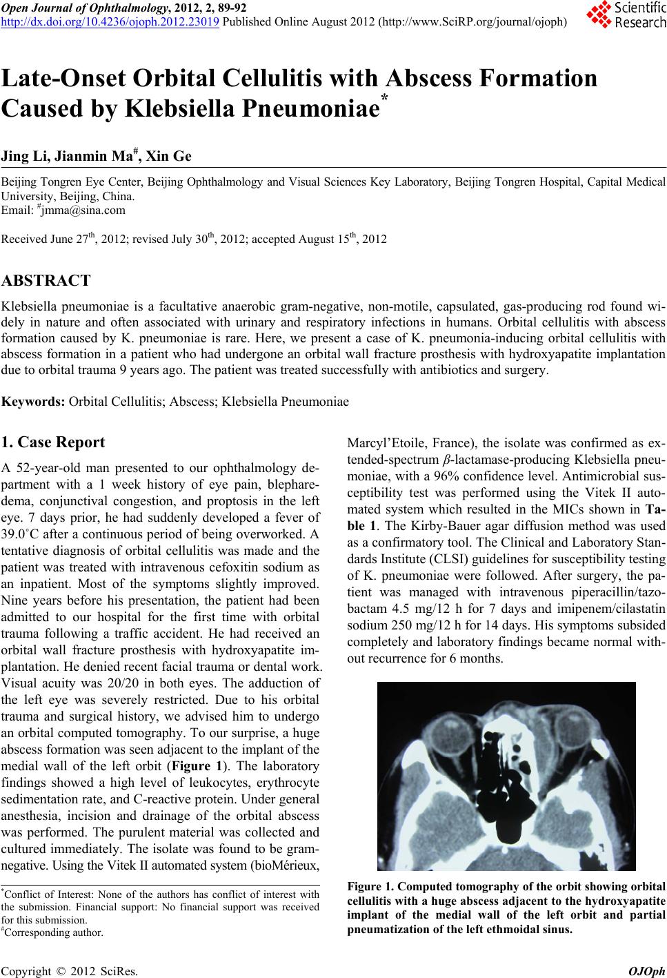 Open Journal of Ophthalmology, 2012, 2, 89-92 http://dx.doi.org/10.4236/ojoph.2012.23019 Published Online August 2012 (http://www.SciRP.org/journal/ojoph) 89 Late-Onset Orbital Cellulitis with Abscess Formation Caused by Klebsiella Pneumoniae* Jing Li, Jianmin Ma#, Xin Ge Beijing Tongren Eye Center, Beijing Ophthalmology and Visual Sciences Key Laboratory, Beijing Tongren Hospital, Capital Medical University, Beijing, China. Email: #[email protected] Received June 27th, 2012; revised July 30th, 2012; accepted August 15th, 2012 ABSTRACT Klebsiella pneumoniae is a facultative anaerobic gram-negative, non-motile, capsulated, gas-producing rod found wi- dely in nature and often associated with urinary and respiratory infections in humans. Orbital cellulitis with abscess formation caused by K. pneumoniae is rare. Here, we present a case of K. pneumonia-inducing orbital cellulitis with abscess formation in a patient who had undergone an orbital wall fracture prosthesis with hydroxyapatite implantation due to orbital trauma 9 years ago. The patient was treated successfully with antibiotics and surgery. Keywords: Orbital Cellulitis; Abscess; Klebsiella Pneumoniae 1. Case Report A 52-year-old man presented to our ophthalmology de- partment with a 1 week history of eye pain, blephare- dema, conjunctival congestion, and proptosis in the left eye. 7 days prior, he had suddenly developed a fever of 39.0˚C after a continuous period of being overworked. A tentative diagnosis of orbital cellulitis was made and the patient was treated with intravenous cefoxitin sodium as an inpatient. Most of the symptoms slightly improved. Nine years before his presentation, the patient had been admitted to our hospital for the first time with orbital trauma following a traffic accident. He had received an orbital wall fracture prosthesis with hydroxyapatite im- plantation. He denied recent facial trauma or dental work. Visual acuity was 20/20 in both eyes. The adduction of the left eye was severely restricted. Due to his orbital trauma and surgical history, we advised him to undergo an orbital computed tomography. To our surprise, a huge abscess formation was seen adjacent to the implant of the medial wall of the left orbit (Figure 1). The laboratory findings showed a high level of leukocytes, erythrocyte sedimentation rate, and C-reactive protein. Under general anesthesia, incision and drainage of the orbital abscess was performed. The purulent material was collected and cultured immediately. The isolate was found to be gram- negative. Using the Vitek II automated system (bioMérieux, Marcyl’Etoile, France), the isolate was confirmed as ex- tended-spectrum β-lactamase-producing Klebsiella pneu- moniae, with a 96% confidence level. Antimicrobial sus- ceptibility test was performed using the Vitek II auto- mated system which resulted in the MICs shown in Ta- ble 1. The Kirby-Bauer agar diffusion method was used as a confirmatory tool. The Clinical and Laboratory Stan- dards Institute (CLSI) guidelines for susceptibility testing of K. pneumoniae were followed. After surgery, the pa- tient was managed with intravenous piperacillin/tazo- bactam 4.5 mg/12 h for 7 days and imipenem/cilastatin sodium 250 mg/12 h for 14 days. His symptoms subsided completely and laboratory findings became normal with- out recurrence for 6 months. Figure 1. Computed tomogr aphy of the orbit show ing orbital cellulitis with a huge abscess adjacent to the hydroxyapatite implant of the medial wall of the left orbit and partial pneumatization of the left ethmoidal sinus. *Conflict of Interest: None of the authors has conflict of interest with the submission. Financial support: No financial support was received for this submission. #Corresponding author. Copyright © 2012 SciRes. OJOph 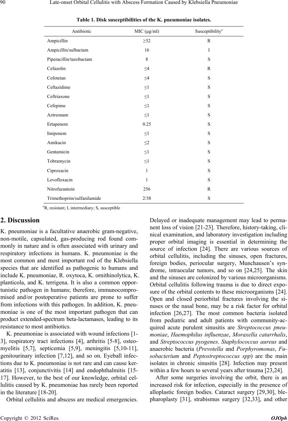 Late-onset Orbital Cellulitis with Abscess Formation Caused by Klebsiella Pneumoniae 90 Table 1. Disk susceptibilities of the K. pneumoniae isolates. Antibiotic MIC (μg/ml) Susceptibilityα Ampicillin ≥32 R Ampicillin/sulbactam 16 I Piperacillin/tazobactam 8 S Cefazolin ≤4 R Cefotetan ≤4 S Ceftazidime ≤1 S Ceftriaxone ≤1 S Cefepime ≤1 S Aztreonam ≤1 S Ertapenem 0.25 S Imipenem ≤1 S Amikacin ≤2 S Gentamicin ≤1 S Tobramycin ≤1 S Ciproxacin 1 S Levofloxacin 1 S Nitrofurantoin 256 R Trimethoprim/sulfanilamide 2/38 S αR, resistant; I, intermediary; S, susceptible 2. Discussion K. pneumoniae is a facultative anaerobic gram-negative, non-motile, capsulated, gas-producing rod found com- monly in nature and is often associated with urinary and respiratory infections in humans. K. pneumoniae is the most common and most important rod of the Klebsiella species that are identified as pathogenic to humans and include K. pneumoniae, R. oxytoca, K. ornithinolytica, K. planticola, and K. terrigena. It is also a common oppor- tunistic pathogen in humans; therefore, immunocompro- mised and/or postoperative patients are prone to suffer from infections with this pathogen. In addition, K. pneu- moniae is one of the most important pathogen that can product extended-spectrum beta-lactamases, leading to its resistance to most antibiotics. K. pneumoniae is associated with wound infections [1- 3], respiratory tract infections [4], arthritis [5-8], osteo- myelitis [5,7], septicemia [5,9], meningitis [5,10-11], genitourinary infection [7,12], and so on. Eyeball infec- tions due to K. pneumoniae is not rare and can cause ker- atitis [13], conjunctivitis [14] and endophthalmitis [15- 17]. However, to the best of our knowledge, orbital cel- lulitis caused by K. pneumoniae has rarely been reported in the literature [18-20]. Orbital cellulitis and abscess are medical emergencies. Delayed or inadequate management may lead to perma- nent loss of vision [21-23]. Therefore, history-taking, cli- nical examination, and laboratory investigation including proper orbital imaging is essential in determining the source of infection [24]. There are various sources of orbital cellulitis, including the sinuses, open fractures, foreign bodies, periocular surgery, Munchausen’s syn- drome, intraocular tumors, and so on [24,25]. The skin and the sinuses are colonized by various microorganisms. Orbital cellulitis following trauma is due to direct expo- sure of the orbital contents to these microorganisms [24]. Open and closed periorbital fractures involving the si- nuses or the nasal bone, may be a risk factor for orbital infection [26,27]. The most common bacteria isolated from pediatric and adult patients with community-ac- quired acute purulent sinusitis are Streptococcus pneu- moniae, Haemophilus influenzae, Moraxella catarrhalis, and Streptococcus pyogenes. Staphylococcus aureus and anaerobic bacteria (Prevotella and Porphyromonas, Fu- sobacterium and Peptostreptococcus spp) are the main isolates in chronic sinusitis [28]. Infection may present within a few hours to several years after trauma [23,24]. After some surgeries involving the orbit, there is an increased risk for infection, especially in the presence of alloplastic foreign bodies. Cataract surgery [29,30], ble- pharoplasty [31], strabismus surgery [32,33], and other Copyright © 2012 SciRes. OJOph  Late-onset Orbital Cellulitis with Abscess Formation Caused by Klebsiella Pneumoniae 91 procedures may all expose the orbit to infection [34,35]. Therefore, patients undergoing any interventional proce- dures should be closely monitored for signs of infection and should be treated aggressively if evidence of an in- fection is found [24]. Because K. pneumoniae is almost extended-spectrum β-lactamase-producing bacillus, it is resistant to most anti- biotics. Susceptibility testing is necessary to help physi- cians choose the appropriate antibiotics in clinic. In our case, the results of the susceptibility testing showed that piperacillin/tazobactam, cefazolin, cefotetan, ceftazidime, ceftriaxone, cefepime, aztreonam, ertapenem, imipenem, amikacin, gentamicin, tobramycin, ciproxacin, levoflox- acin are all effective. However, the extended-spectrum β-lactamase-producing bacteria, which show susceptible to penicillins, cephalosporins and aztreonam often be- come resistant to them. Therefore, β-lactamase inhibitors and carbapenems are recommended in clinical practice. Our patient was treated with intravenous piperacillin/ tazobactam for 7 days and oral imipenem for 14 days after surgery without recurrence for 6 months. 3. Conclusion To our knowledge, this is the first case of late-onset or- bital cellulitis and abscess formation induced by K. pneu- moniae after an orbital wall fracture prosthesis with hy- droxyapatite implantation reported in the literature. Or- bital cellulitis and abscess formation due to K. pneumo- niae should be considered, whenever patients suffer the orbital trauma and undergo surgery. REFERENCES [1] E. Rezaei, H. Safari, M. Naderinasab, et al., “Common Pathogens in Burn Wound and Changes in Their Drug Sensitivity,” Burns, Vol. 37, No. 5, 2011, pp. 804-806. doi:10.1016/j.burns.2011.01.019 [2] M. I. Ahmed, “Prevalence of Nosocomial Wound Infec- tion among Postoperative Patients and Antibiotics Pat- terns at Teaching Hospital in Sudan,” North American Journal of Medical Sciences, Vol. 4, No. 1, 2012, pp. 29- 34. doi:10.4103/1947-2714.92900 [3] A. K. Seth, M. R. Geringer, A. N. Gurjala, et al., “Under- standing the Host Inflammatory Response to Wound In- fection: An in Vivo Study of Klebsiella Pneumoniae in a Rabbit Ear Wound Model,” Wound Repair and Regen- eration, Vol. 20, No. 2, 2012, pp. 214-225. doi:10.1111/j.1524-475X.2012.00764.x [4] S. Ghafourian, Z. B. Sekawi, N. Sadeghifard, et al., “The prevalence of ESBLs Producing Klebsiella Pneumoniae Isolates in Some Major Hospitals, Iran,” Open Microbi- ology Journal, Vol. 5, 2011, pp. 91-95. doi:10.2174/1874285801105010091 [5] Z. Ghorashi, N. Nezami, H. Hoseinpour-Feizi, et al., “Arthritis, Osteomyelitis, Septicemia and Meningitis Cau- sed by Klebsiella in a Low-Birth-Weight Newborn: A Ca- se Report,” Journal of Medical Case Reports, Vol. 5, 2011, p. 241. doi:10.1186/1752-1947-5-241 [6] S. Schelenz, K. Bramham and D. Goldsmith, “Septic Ar- thritis Due to Extended Spectrum Beta Lactamase Pro- ducing Klebsiella Pneumoniae,” Joint Bone Spine, Vol. 74, No. 3, 2007, pp. 275-278. doi:10.1016/j.jbspin.2006.08.007 [7] L. C. Chew, “Septic Monoarthritis and Osteomyelitis in an Elderly Man Following Klebsiella Pneumoniae Geni- tourinary Infection: Case Report,” Annals of the Academy of Medicine Singapore, Vol. 35, No. 2, 2006, pp. 100- 103. [8] J. E. Kohler, M. P. Hutchens, P. M. Sadow, et al., “Kleb- siella Pneumoniae Necrotizing Fasciitis and Septic Ar- thritis: An Appearance in the Western Hemisphere,” Sur- gical Infections, Vol. 8, No. 2, 2007, pp. 227-232. doi:10.1089/sur.2006.007 [9] N. Y. Lee, W. H. Huang, K. C. Tsui, et al., “Carbapenem Therapy for Bacteremia Due to Extended-Spectrum (Be- ta)-Lactamase-Producing Escherichia Coli or Klebsiella Pneumoniae,” Diagnostic Microbiology and Infectious Disease, Vol. 70, No. 1, 2011, pp. 150-153. doi:10.1016/j.diagmicrobio.2010.12.008 [10] A. Stucki, M. Cottagnoud, F. Acosta, et al., “Efficacy of Doripenem against Escherichia Coli and Klebsiella Pneu- moniae in Experimental Meningitis,” Journal of Antim- icrobial Chemotherapy, Vol. 67, No. 3, 2012, pp. 661- 665. doi:10.1093/jac/dkr482 [11] W. N. Chang, C. H. Lu, C. R. Huang, et al., “Clinical Characteristics of Post-Neurosurgical Klebsiella Pneu- moniae Meningitis in Adults and a Clinical Comparison to the Spontaneous Form in a Taiwanese Population,” Journal of Clinical Neuroscience, Vol. 17, No. 3, 2010, pp. 334-338. doi:10.1016/j.jocn.2009.06.019 [12] D. A. Rosen, J. S. Pinkner, J. N. Walker, et al., “Molecu- lar Variations in Klebsiella Pneumoniae and Escherichia coli FimH Affect Function and Pathogenesis in the Uri- nary Tract,” Infection and Immunity, Vol. 76, No. 7, 2008, pp. 3346-3356. doi:10.1128/IAI.00340-08 [13] S. Zarei-Ghanavati, M. R. Sedaghat and A. Ghavami- Shahri, “Acute Klebsiella Pneumoniae Interface Keratitis after Deep Anterior Lamellar Keratoplasty,” Japanese Journal of Ophthalmology, Vol. 55, No. 1, 2011, pp. 74- 76. doi:10.1007/s10384-010-0836-7 [14] T. Aung and T. K. Chan, “Nosocomial Klebsiella Pneu- moniae Conjunctivitis Resulting in Infectious Keratitis and Bilateral Corneal Perforation,” Cornea, Vol. 17, No. 5, 1998, pp. 558-561. doi:10.1097/00003226-199809000-00015 [15] A. H. Kashani and D. Eliott, “Bilateral Klebsiella Pneu- moniae (K1 Serotype) Endogenous Endophthalmitis as the Presenting Sign of Disseminated Infection,” Ophthal- mic Surgery, Lasers & Imaging, Vol. 42, 2011, pp. e12- e14. [16] L. P. Ang, H. M. Lee, K. G. Au Eong, et al., “Endoge- nous Klebsiella Endophthalmitis,” Eye, Vol. 14, No. 6, 2000, pp. 855-860. doi:10.1038/eye.2000.236 [17] Y. J. Chen, H. K. Kuo, P. C. Wu, et al., “A 10-Year Com- parison of Endogenous Endophthalmitis Outcomes: An Copyright © 2012 SciRes. OJOph  Late-onset Orbital Cellulitis with Abscess Formation Caused by Klebsiella Pneumoniae Copyright © 2012 SciRes. OJOph 92 East Asian Experience with Klebsiella Pneumoniae Infec- tion,” Retina, Vol. 24, No. 3, 2004, pp. 383-390. doi:10.1097/00006982-200406000-00008 [18] S. J. Yang, S. Y. Park, Y. J. Lee, et al., “Klebsiella Pneu- moniae Orbital Cellulitis with Extensive Vascular Occlu- sions in a Patient with Type 2 Diabetes,” Korean Journal of Internal Medicine, Vol. 25, No. 1, 2010, pp. 114-117. doi:10.3904/kjim.2010.25.1.114 [19] I. A. Chaudhry, F. A. Shamsi, E. Elzaridi, et al., “Inpa- tient Preseptal Cellulitis: Experience from a Tertiary Eye Care Centre,” British Journal of Ophthalmology, Vol. 92, No. 10, 2008, pp. 1337-1341. doi:10.1136/bjo.2007.128975 [20] C. T. Lin and Y.Y. Tsai, “Klebsiella Pneumoniae Orbital Cellulitis,” Chinese Medical Journal (Taipei), Vol. 64, No. 9, 2001, pp. 551-554. [21] A. Hauser and S. Fogarasi, “Periorbital and Orbital Cellu- litis,” Pediatrics in Review, Vol. 31, No. 6, 2010, pp. 242- 249. [22] M. Coskun, Ö. IIhan, U. Keskin, et al., “Central Retinal Artery Occlusion Secondary to Orbital Cellulitis and Ab- scess Following Dacryocystitis,” European Journal of Ophthalmology, Vol. 21, No. 5, 2011, pp. 649-652. doi:10.5301/EJO.2011.6493 [23] K. Starska, M. Lukomski and D. Starska-Dawidowskda, “Preseptal Orbital Abscess as a Post-Traumatic Late Complication—Review of the Literature and Case Re- port,” Otolaryngologia Polska, the Polish Otolaryngology, Vol. 61, No. 3, 2007, pp. 331-334. [24] S. Rumelt and P. A. Rubin, “Potential Sources for Orbital Cellulitis,” International Ophthalmology Clinics, Vol. 36, No. 3, 1996, pp. 207-221. doi:10.1097/00004397-199603630-00019 [25] P. Laloyaux, D. Vanpee and J. B. Gillet, “Orbital Cellu- litis with Abscess Formation Caused by Frontal Sinusi- tis,” Journal of Emergency Medicine, Vol. 18, No. 2, 2000, pp. 253-254. doi:10.1016/S0736-4679(99)00204-8 [26] G. J. Ben Simon, S. Bush, D. Selva, et al., “Orbital Cellu- litis: A Rare Complication after Orbital Blowout Frac- ture,” Ophthalmology, Vol. 112, No. 11, 2005, pp. 2030- 2034. doi:10.1016/j.ophtha.2005.06.012 [27] S. P. Bord and J. Linden, “Trauma to the Globe and Or- bit,” Emergency Medicine Clinics of North America, Vol. 26, No. 1, 2008, pp. 97-123. doi:10.1016/j.emc.2007.11.006 [28] I. Brook, “Microbiology of Sinusitis,” Proceedings of the American Thoracic Society, Vol. 8, No. 1, 2011, pp. 90- 100. doi:10.1513/pats.201006-038RN [29] V. Kumar and D. Saunders, “Orbital Cellulitis after Phaco- emulsification and Intraocular Lens Implantation,” Jour- nal of Cataract and Refractive Surgery, Vol. 30, No. 4, 2004, pp. 918-920. doi:10.1016/j.jcrs.2003.11.026 [30] S. Y. Liang, G. Moloney, B. A. O’Donnell, et al., “Or- bital Cellulitis as a Postoperative Complication of Sub- Tenon Anaesthesia in Cataract Surgery,” Clinical and Ex- perimental Ophthalmology, Vol. 34, No. 9, 2006, pp. 897-899. doi:10.1111/j.1442-9071.2006.01361.x [31] V. Juthani, C. I. Zoumalan, R. D. Lisman, et al., “Success- ful Management of Methicillin-Resistant Staphylococcus aureus Orbital Cellulitis after Blepharoplasty,” Plastic and Reconstructive Surgery, Vol. 126, No. 6, 2010, pp. 305e-307e. doi:10.1097/PRS.0b013e3181f63f67 [32] E. Hoyama, V. Limawararut, I, Leibovitch, et al., “Blind- ing Orbital Cellulitis: A Complication of Strabismus Sur- gery,” Ophthalmic Plastic and Reconstructive Surgery, Vol. 22, No. 6, 2006, pp. 472-473. doi:10.1097/01.iop.0000245486.61228.8d [33] A. Basheikh and R. Superstein, “A Child with Bilateral Orbital Cellulitis One Day after Strabismus Surgery,” Journal of AAPOS, Vol. 13, No. 5, 2009, pp. 488-490. doi:10.1016/j.jaapos.2009.05.016 [34] M. P. Hatton and M. L. Durand, “Orbital Cellulitis with Abscess Formation Following Surgical Treatment of Ca- naliculitis,” Ophthalmic Plastic and Reconstructive Sur- gery, Vol. 24, No. 4, 2008, pp. 314-316. doi:10.1097/IOP.0b013e31817e9be7 [35] M. M. Marcet, J. J. Woog, A. R. Bellows, et al., “Orbital Complications after Aqueous Drainage Device Proce- dures,” Ophthalmic Plastic and Reconstructive Surgery, Vol. 21, No. 1, 2005, pp. 67-69. doi:10.1097/01.IOP.0000150351.91768.16 |