

Paper Menu >>
Journal Menu >>
 Open Journal of Ophthalmology, 2012, 2, 64-70 http://dx.doi.org/10.4236/ojoph.2012.23014 Published Online August 2012 (http://www.SciRP.org/journal/ojoph) Investigation of Four Genes Responsible for Autosomal Recessive Congenital Cataract and Highly Expressed in the Brain in Four Unrelated Tunisian Families Manèl Chograni1, Myriam Chaabouni1,2, Faouzi Maazoul2, Habiba Chaabouni Bouhamed1,2* 1Laboratoire de Génétique Humaine, Faculté de Médecine de Tunis, University Tunis Elmanar, Tunis, Tunisia; 2Congenital and Hereditary Disorders Department, Charles Nicolle Hospital, Tunis, Tunisia. Email: *[email protected] Received March 14th, 2012; revised April 28th, 2012; accepted May 11th, 2012 ABSTRACT Purpose: To identify the causative gene for phenotypes associating autosomal recessive congenital cataract, mental re- tardation and congenital cataract, mental retardation and microcephaly in four unrelated Tunisian families. Methods: Four genes (EPHA2, GALK1, GCNT2, and CRYBB1) were selected based on their expression in human brain and their known or putative function. Linkage analysis were performed for the four genes in multiple affected and unaffected families’ members and results were explored by the GeneMapper ID v3.2 software. Results: No linkage was identified for the four studied genes in the four families. Affected members of each family did not share common haplotypes in corresponding candidate regions containing selected gene. Conclusion: Although the four studied genes were reported responsible for autosomal recessive congenital cataract and highly expressed in the human brain, we report no linkage for EPHA 2, GALK1, GCNT2, and CRYBB1 genes in four families with congenital cataract, mental retardation and con- genital cataract, mental retardation and microcephaly. Keywords: Congenital Cataract; Mental Retardation; Microcephaly; Autosomal Recessive; Association; Linkage Study 1. Introduction Congenital cataracts are one of the major causes of vision loss in children worldwide and are responsible for ap- proximately one third of blindness in infants [1]. Con- genital cataracts can occur in an isolated fashion or as one component of a syndrome affecting multiple tissues. Nonsyndromic congenital cataracts have an estimated frequency of 1 to 6 per 10,000 live births. They vary markedly in severity and morphology, affecting the nu- clear, cortical, polar, or subcapsular parts of the lens or, in severe cases, the entire lens, with a variety of types of opacity. Approximately one third of congenital cataract cases are familial [2]. Few autosomal recessive cataract loci have been mapped. To date, 13 loci residing on chromosomes 1p34.4-p32.2, 1q21.1, 3p22-24.2, 6p23-24, 9q13-22, 16q21-22, 19q13, 19q13.4, 20p12.1, 21q22.3, 22q11, 22q12.1 and 17q, have been mapped, with six of these also causing auto- somal dominant cataracts [3-12]. EPHA2 (Ephrin-receptor type-A2) belongs to the tyro- sine kinase family of proteins and is an epithelial cell ki- nase that has been associated with autosomal dominant cataracts and recently it was implicated in ARCC in hu- man [11]. EPHA2 is expressed in a variety of different regions during development. Expression has been ob- served in the hindbrain, specifically in rhombomere 4, during early embryogenesis [13]. Galactokinase (GALK1) is involved in the first step of metabolism of galactose, the conversion of galactose to galactose-1-phosphate at the expense of ATP. In the ab- sence of GALK1 the accumulating galactose is converted to galactitol by aldose reductase. Stambolian and col- leagues first identified mutations in families with cata- racts [14]. And recently GALK1 found to be mutated in Pakistani families with ARCC [12]. Glucosaminyl (N-acetyl) Transferase 2 Gene (GCNT2) had been reported for ARCC in Arab families from Israel [4]. GCNT2 is highly expressed in fetal brain and kidney and adult brain but expressed ubiquitously in various adult tissues [15]. Crystallin genes, which encode major structural pro- teins in the lens, are considered as obvious candidate ge- nes of congenital cataracts owing to both their high levels of lenticular expression and their confirmed functions in maintaining lens transparency. Increasing evidence sug- *Corresponding author. Copyright © 2012 SciRes. OJOph 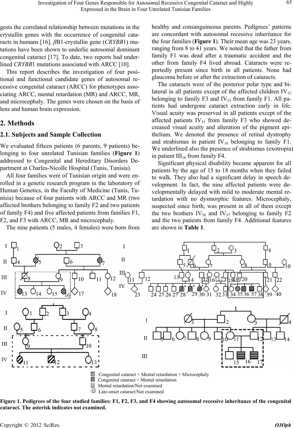 Investigation of Four Genes Responsible for Autosomal Recessive Congenital Cataract and Highly Expressed in the Brain in Four Unrelated Tunisian Families 65 gests the correlated relationship between mutations in the crystallin genes with the occurrence of congenital cata- racts in humans [16]. βB1-crystallin gene (CRYBB1) mu- tations have been shown to underlie autosomal dominant congenital cataract [17]. To date, two reports had under- lined CRYBB1 mutations associated with ARCC [10]. This report describes the investigation of four posi- tional and functional candidate genes of autosomal re- cessive congenital cataract (ARCC) for phenotypes asso- ciating ARCC, mental retardation (MR) and ARCC, MR, and microcephaly. The genes were chosen on the basis of lens and human brain expression. 2. Methods 2.1. Subjects and Sample Collection We evaluated fifteen patients (6 parents, 9 patients) be- longing to four unrelated Tunisian families (Figure 1) addressed to Congenital and Hereditary Disorders De- partment at Charles-Nicolle Hospital (Tunis, Tunisia). All four families were of Tunisian origin and were en- rolled in a genetic research program in the laboratory of Human Genetics, in the Faculty of Medicine (Tunis, Tu- nisia) because of four patients with ARCC and MR (two affected brothers belonging to family F2 and two patients of family F4) and five affected patients from families F1, F2, and F3 with ARCC, MR and microcephaly. The nine patients (5 males, 4 females) were born from healthy and consanguineous parents. Pedigrees’ patterns are concordant with autosomal recessive inheritance for the four families (Figure 1). Their mean age was 23 years, ranging from 8 to 41 years. We noted that the father from family F1 was dead after a traumatic accident and the other from family F4 lived abroad. Cataracts were re- portedly present since birth in all patients. None had glaucoma before or after the extraction of cataracts. The cataracts were of the posterior polar type and bi- lateral in all patients except of the affected children IV12 belonging to family F3 and IV13 from family F1. All pa- tients had undergone cataract extraction early in life. Visual acuity was preserved in all patients except of the affected patients IV11 from family F3 who showed de- creased visual acuity and alteration of the pigment epi- thelium. We denoted the presence of retinal dystrophy and strabismus in patient IV16 belonging to family F1. We underlined also the presence of strabismus (exotropia) in patient III16 from family F4. Significant physical disability became apparent for all patients by the age of 15 to 18 months when they failed to walk. They also had a significant delay in speech de- velopment. In fact, the nine affected patients were de- velopmentally delayed with mild to moderate mental re- tardation with no dysmorphic features. Microcephaly, suspected since birth, was present in all of them except the two brothers IV36 and IV37 belonging to family F2 and the two patients from family F4. Additional features are shown in Table 1. Congenital cataract + Mental retardation + Microcephaly Congenital cataract + Mental retardation Mental retardation/Not examined Late-onset cataract/Not examined Figure 1. Pedigrees of the four studied families: F1, F2, F3, and F4 showing autosomal recessive inheritance of the congenital cataract. The asterisk indicates not examined. Copyright © 2012 SciRes. OJOph 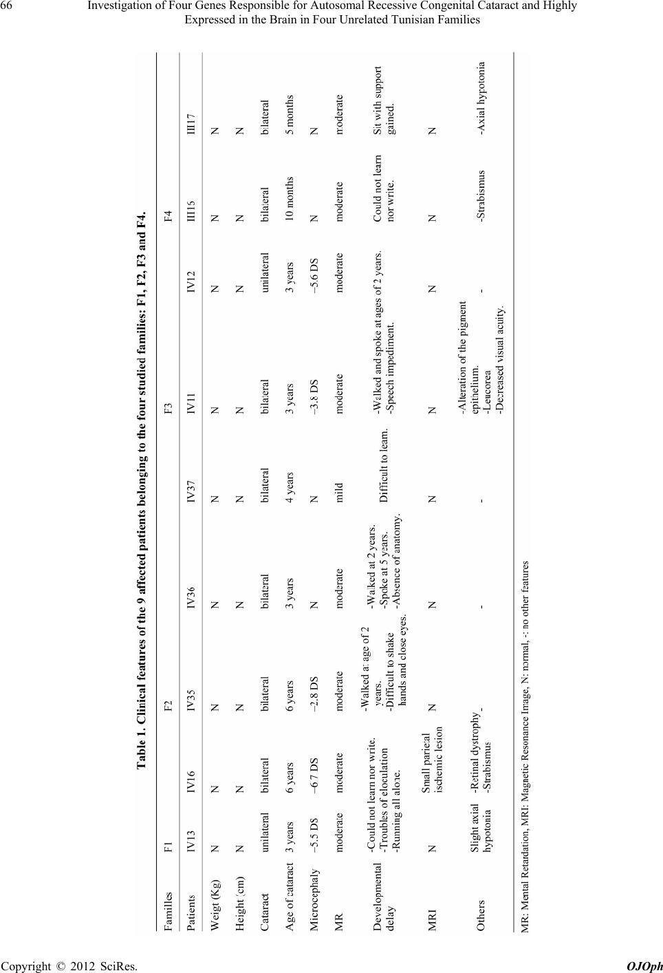 Investigation of Four Genes Responsible for Autosomal Recessive Congenital Cataract and Highly Expressed in the Brain in Four Unrelated Tunisian Families 66 Copyright © 2012 SciRes. OJOph  Investigation of Four Genes Responsible for Autosomal Recessive Congenital Cataract and Highly Expressed in the Brain in Four Unrelated Tunisian Families Copyright © 2012 SciRes. OJOph 67 Magnetic resonance imaging (MRI) of the brain was normal in all screened patients except for the presence of a small ischemic parietal lesion in patient IV16 from fam- ily F1. Biological investigations (karyotyping with R-banding) revealed normal karyotypes: 46, XX for females, 46, XY for males (600 bands resolution) and normal metabolic screening including Fehling reaction and thin layer chro- matography of reducing sugars, plasmatic amino acid and urine organic acid chromatography for all patients. Genomic DNA of affected and unaffected members (9 siblings, 6 parents) was extracted from peripheral blood leukocytes by the standard proteinase-K extraction con- sisting on: lysis of red blood cells by RBC (Red Blood Cells) Lysis Buffer (155 mM NH4Cl, 10 mM KHCO3, 0.5 EDTA, pH 7.5) and white blood cells by a WBC (White Blood Cells) Lysis Buffer (1 mM Na-EDTA, 5mM Tris HCl, pH 7.5), treatment of the lysate with a mixture of detergent composed of SDS (Sodium Dodecyl Sulfate or sacrosyl and proteinase K) in order to liberate the DNA and digest the associated proteins, precipitation of the DNA in the form of filaments by absolute ethanol and finally diluation of the DNA in T10E1 Buffer (Tris 10 mM, EDTA 0.1-1 mM), and stored in 10 ml Vacuum tube sterile containing 100 µl of 0.1 M EDTA.K3. Patients and parents for minors gave informed consent. In this study, the researches carried out on human are in compliance with the Helsinki Declaration and ethics com- mittee Charles Nicolle hospital, Tunis has given approval for this study. 2.2. Molecular and Genotyping Analysis On the basis of the pedigree of the four studied families (Figure 1), we suspected autosomal recessive inheritance for phenotypes associating congenital cataract, mental re- tardation and congenital cataract, mental retardation, and microcephaly. All individuals were genotyped for eight microsatellite loci within a 10-cM region on 1p36.21-p35.2 previously reported as linked to EPHA2 (D1S2697-D1S1592-D1 S2644-D1S2864-D1S2787-D1S507-D1S434-D1S2667) [11,13], at six microsatellite loci on 17q22-q25.3 linked to GALK1 (D17S944-D17S1825-D17S1301-D17S1839- D17S785-D17S1847) [12], at five microsatellite loci on 6p25-p23 reported as linked to GCNT2 (D6S1574-D6 S309-D6S470-D6S1034-D6S289) [4], and at six micro- satellite loci on 22q11.2-q12.1 previously reported as linked to CRYBB1 (D22S539-D22S686-D22S345-D22 S419-D22S1167-D22S1144) [10]. Suitable microsatellite primers for polymerase chain reaction (PCR) amplification of each candidate region containing the candidate gene were designed using NCBI (http://www.ncbi.nlm.nih.gov/unists). PCR was performed by using 100 ng of DNA template, 20 pmol each of forward (FAM labelled) and reverse primers (Biomatik, Canada), 1.5 units of Taq DNA poly- merase (Promega, Madison, WI) and 1.25 mM dNTPs (Promega, Madison, WI) in a total volume of 25 μl. PCR consisted on 30 cycles and was carried out in an auto- mated thermal cycle GeneAmp PCR System 9700 (Ap- plied Biosystems, California) under the following condi- tions: initial denaturation at 96˚C for 5 min and denatura- tion at 96˚C for 30 s, annealing at 52˚C - 60˚C for 30 s, and elongation at 72˚C for 30 s followed by one cycle of final extension at 72˚C for 7 min. Genotyping was per- formed on a genetic analyser (PRISM 3130; ABI) with accompanying software (GeneScan; ABI, Foster City, CA). 2.3. Linkage Analysis Two-point LOD scores were calculated using the MLINK program of the LINKAGE package (ver. 4.1P; http:www.hgmp.mrc.ac.uk; provided in the public do- main by the Human Genome Mapping Project Resources Centre, Cambridge, UK), and multipoint and haplotype analyses were performed with GeneMapper ID v3.2 soft- ware. 3. Results Segregation analysis using the polymorphic markers on chromosome 1 for EPHA2, on chromosome 17 for GALK1, on chromosome 6 for GCNT2 and on chromosome 22 for CRYBB1 within minimum 10-cM region allowed us to exclude implication in studied phenotypes (congenital cataract, mental retardation and congenital cataract, men- tal retardation, and microcephaly) of all regions analyzed seeing that the affected members and their parents did not share a common haplotype. There is no linkage of anyone of the four genes (EPHA2, GALK1, GCNT2, and CRYBB1) to the associa- tion between ARCC, MR and ARCC, MR, and micro- cephaly in the four studied families (F1, F2, F3, and F4). 4. Discussion Congenital cataracts are common major abnormalities of the eye, which frequently cause blindness in infants. It may occur as an isolated anomaly, as part of generalized ocular development defects, or as a component of a mul- tisystem disorder [18]. In fact, association of cataract with congenital anomalies, mental retardation and micro- cephaly is reported in several cases with chromosomal anomalies and syndromes from genic origins [19-21]. Until today no candidate gene has been reported re- sponsible for phenotypes associating ARCC, MR and ARCC, MR, and microcephaly, so we tried to investigate 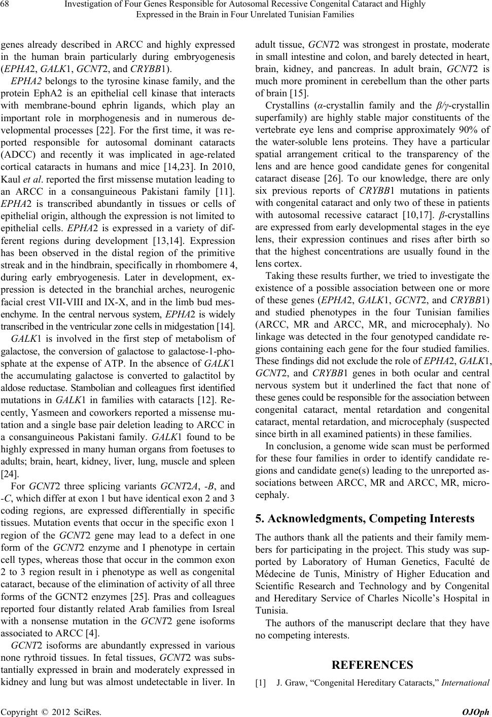 Investigation of Four Genes Responsible for Autosomal Recessive Congenital Cataract and Highly Expressed in the Brain in Four Unrelated Tunisian Families 68 genes already described in ARCC and highly expressed in the human brain particularly during embryogenesis (EPHA2, GALK1, GCNT2, and CRYBB1). EPHA2 belongs to the tyrosine kinase family, and the protein EphA2 is an epithelial cell kinase that interacts with membrane-bound ephrin ligands, which play an important role in morphogenesis and in numerous de- velopmental processes [22]. For the first time, it was re- ported responsible for autosomal dominant cataracts (ADCC) and recently it was implicated in age-related cortical cataracts in humans and mice [14,23]. In 2010, Kaul et al. reported the first missense mutation leading to an ARCC in a consanguineous Pakistani family [11]. EPHA2 is transcribed abundantly in tissues or cells of epithelial origin, although the expression is not limited to epithelial cells. EPHA2 is expressed in a variety of dif- ferent regions during development [13,14]. Expression has been observed in the distal region of the primitive streak and in the hindbrain, specifically in rhombomere 4, during early embryogenesis. Later in development, ex- pression is detected in the branchial arches, neurogenic facial crest VII-VIII and IX-X, and in the limb bud mes- enchyme. In the central nervous system, EPHA2 is widely transcribed in the ventricular zone cells in midgestation [14]. GALK1 is involved in the first step of metabolism of galactose, the conversion of galactose to galactose-1-pho- sphate at the expense of ATP. In the absence of GALK1 the accumulating galactose is converted to galactitol by aldose reductase. Stambolian and colleagues first identified mutations in GALK1 in families with cataracts [12]. Re- cently, Yasmeen and coworkers reported a missense mu- tation and a single base pair deletion leading to ARCC in a consanguineous Pakistani family. GALK1 found to be highly expressed in many human organs from foetuses to adults; brain, heart, kidney, liver, lung, muscle and spleen [24]. For GCNT2 three splicing variants GCNT2A, -B, and -C, which differ at exon 1 but have identical exon 2 and 3 coding regions, are expressed differentially in specific tissues. Mutation events that occur in the specific exon 1 region of the GCNT2 gene may lead to a defect in one form of the GCNT2 enzyme and I phenotype in certain cell types, whereas those that occur in the common exon 2 to 3 region result in i phenotype as well as congenital cataract, because of the elimination of activity of all three forms of the GCNT2 enzymes [25]. Pras and colleagues reported four distantly related Arab families from Isreal with a nonsense mutation in the GCNT2 gene isoforms associated to ARCC [4]. GCNT2 isoforms are abundantly expressed in various none rythroid tissues. In fetal tissues, GCNT2 was subs- tantially expressed in brain and moderately expressed in kidney and lung but was almost undetectable in liver. In adult tissue, GCNT2 was strongest in prostate, moderate in small intestine and colon, and barely detected in heart, brain, kidney, and pancreas. In adult brain, GCNT2 is much more prominent in cerebellum than the other parts of brain [15]. Crystallins (α-crystallin family and the β/γ-crystallin superfamily) are highly stable major constituents of the vertebrate eye lens and comprise approximately 90% of the water-soluble lens proteins. They have a particular spatial arrangement critical to the transparency of the lens and are hence good candidate genes for congenital cataract disease [26]. To our knowledge, there are only six previous reports of CRYBB1 mutations in patients with congenital cataract and only two of these in patients with autosomal recessive cataract [10,17]. β-crystallins are expressed from early developmental stages in the eye lens, their expression continues and rises after birth so that the highest concentrations are usually found in the lens cortex. Taking these results further, we tried to investigate the existence of a possible association between one or more of these genes (EPHA2, GALK1, GCNT2, and CRYBB1) and studied phenotypes in the four Tunisian families (ARCC, MR and ARCC, MR, and microcephaly). No linkage was detected in the four genotyped candidate re- gions containing each gene for the four studied families. These findings did not exclude the role of EPHA2, GALK1, GCNT2, and CRYBB1 genes in both ocular and central nervous system but it underlined the fact that none of these genes could be responsible for the association between congenital cataract, mental retardation and congenital cataract, mental retardation, and microcephaly (suspected since birth in all examined patients) in these families. In conclusion, a genome wide scan must be performed for these four families in order to identify candidate re- gions and candidate gene(s) leading to the unreported as- sociations between ARCC, MR and ARCC, MR, micro- cephaly. 5. Acknowledgments, Competing In ter est s The authors thank all the patients and their family mem- bers for participating in the project. This study was sup- ported by Laboratory of Human Genetics, Faculté de Médecine de Tunis, Ministry of Higher Education and Scientific Research and Technology and by Congenital and Hereditary Service of Charles Nicolle’s Hospital in Tunisia. The authors of the manuscript declare that they have no competing interests. REFERENCES [1] J. Graw, “Congenital Hereditary Cataracts,” International Copyright © 2012 SciRes. OJOph  Investigation of Four Genes Responsible for Autosomal Recessive Congenital Cataract and Highly Expressed in the Brain in Four Unrelated Tunisian Families 69 Journal of Developmental Biology, Vol. 48, No. 8-9. 2004, pp. 1031-1044. doi:10.1387/ijdb.041854jg [2] J. F. Hejtmancik and N. Smaoui, “Molecular Genetics of Cataract,” Developments in Ophthalmology, Vol. 37, 2004, pp. 67-82. doi:10.1159/000072039 [3] A. Foster and G. J. Johnson, “Magnitude and Causes of Blindness in the Developing World,” International Oph- thalmology, Vol. 14, No. 3, 1990, pp. 135-140. doi:10.1007/BF00158310 [4] S. P. Ponnam, K. Ramesha, S. Tejwani, B. Ramamurthy and C. Kannabiran, “Mutation of the Gap Junction Pro- tein Alpha 8 (GJA8) Gene Causes Autosomal Recessive Cataract,” Journal of Medical Genetics, Vol. 44, No. 7, 2007, p. e85. doi:10.1136/jmg.2007.050138 [5] E. Pras, J. Raz, V. Yahalom, M. Frydman, H. J. Garzozi, E. Pras and J. F. Hejtmancik, “A Nonsense Mutation in the Glucosaminyl (Nacetyl) Transferase 2 Gene (GCNT2) Association with Autosomal Recessive Congenital Cata- racts,” Investigative Ophthalmology & Visual Science, Vol. 45, No. 6, 2004, pp. 1940-1945. [6] N. Smaoui, O. Beltaief, S. BenHamed, R. M’Rad, F. Ma- azoul, A. Ouertani, H. Chaabouni and J. F. Hejtmancik, “A Homozygous Splice Mutation in the HSF4 Gene Is Associated with an Autosomal Recessive Congenital Ca- taract,” Investigative Ophthalmology & Visual Science, Vol. 45, No. 8, 2004, pp. 2716-2721. doi:10.1167/iovs.03-1370 [7] E. Pras, E. Levy-Nissenbaum, T. Bakhan, H. Lahat, E. Assia, N. Geffen-Carmi, M. Frydman, B. Goldman and E. Pras, “A Missense Mutation in the LIM2 Gene Is Associ- ated with Autosomal Recessive Presenile Cataract in an Inbred Iraqi Jewish Family,” American Journal of Human Genetics, Vol. 70, No. 5, 2002, pp. 1363-1367. doi:10.1086/340318 [8] E. Pras, M. Frydman, E. Levy-Nissenbaum, T. Bakhan, J. Raz, E. I. Assia, B. Goldman and E. Pras, “A Nonsense Mutation (W9X) in CRYAA Causes Autosomal Reces- sive Cataract in an Inbred Jewish Persian Family,” Inves- tigative Ophthalmology & Visual Science, Vol. 41, No. 11, 2000, pp. 3511-3515. [9] R. D. Ramachandran, V. Perumalsamy and J. F. Hejtman- cik, “Autosomal Recessive Juvenile Onset Cataract Asso- ciated with Mutation in BFSP1,” Human Genetics, Vol. 121, No. 3-4, 2007, pp. 475-482. doi:10.1007/s00439-006-0319-6 [10] S. A. Riazuddin, A. Yasmeen, W. Yao, Y. V. Sergeev, Q. Zhang, F. Zulfiqar, A. Riaz, S. Riazuddin and J. F. Hejt- mancik, “Mutations in BetaB3-Crystallin Associated with Autosomal Recessive Cataract in two Pakistani Families,” Investigative Ophthalmology & Visual Science, Vol. 46, No. 6, 2005, pp. 2100-2106. doi:10.1167/iovs.04-1481 [11] D. Cohen, U. Bar-Yosef, J. Levy, L. Gradstein, N. Belfair, R. Ofir, S. Joshua, T. Lifshitz, R. Carmi and O. S. Birk, “Homozygous CRYBB1 Deletion Mutation Underlies Autosomal Recessive Congenital Cataract,” Investigative Ophthalmology & Visual Science, Vol. 48, No. 5, 2007, pp. 2208-2213. doi:10.1167/iovs.06-1019 [12] H. Kaul, S. A. Riazuddin, M. Shahid, S. Kousar, N. H. Butt, A. U. Zafar, S. N. Khan, T. Husnain, J. Akram, J. F. Hejtmancik and S. Riazuddin, “Autosomal Recessive Con- genital Cataract Linked to EPHA2 in a Consanguineous Pakistani Family,” Molecular Vision, Vol. 16, 2010, pp. 511-517. [13] A. Yasmeen, S. A. Riazuddin, H. Kaul, S. Mohsin, M. Khan, Z. A. Qazi, I. A. Nasir, A. U. Zafar, S. N. Khan, T. Husnain, J. Akram, J. F. Hejtmancik and S. Riazuddin, “Autosomal Recessive Congenital Cataract in Consan- guineous Pakistani Families Is Associated with Mutations in GALK1,” Molecular Vision, Vol. 16, 2010, pp. 682- 688. [14] A. Shiels, T. M. Bennett, H. L. Knopf, G. Maraini, A. Li, X. Jiao and J. F. Hejtmancik, “The EPHA2 Gene Is Asso- ciated with Cataracts Linked to Chromosome 1p,” Mo- lecular Vision, Vol. 14, 2008, pp. 2042-2055. [15] J. C. Ruiz and E. J. Robertson, “The Expression of the Receptor-Protein Tyrosine Kinase Gene, Eck, Is Highly Restricted during Early Mouse Development,” Mecha- nisms of Development, Vol. 46, No. 2, 1994, pp. 87-100. doi:10.1016/0925-4773(94)90078-7 [16] T. Mori, A. Wanaka, A. Taguchi, K. Matsumoto and M. Tohyama, “Differential Expressions of the Eph Family of Receptor Tyrosine Kinase Genes (Sek, Elk, Eck) in the Developing Nervous System of the Mouse,” Molecular Brain Research, Vol. 29, No. 2, 1995, pp. 325-335. doi:10.1016/0169-328X(94)00263-E [17] D. Stambolian, Y. Ai, D. Sidjanin, K. Nesburn, G. Sathe, M. Rosenberg and D. J. Bergsma, “Cloning of the Galac- tokinase cDNA and Identification of Mutations in Two Families with Cataracts,” Nature Genetics, Vol. 10, No. 3, 1995, pp. 307-312. doi:10.1038/ng0795-307 [18] K. Sasaki, K. Kurata-Miura, M. Ujita, K. Angata, S. Na- kagawa, S. Sekine, T. Nishi and M. Fukuda, “Expression Cloning of cDNA Encoding a Human Beta-1,3-N-Acetyl Glucosaminyl Transferase That Is Essential for Poly-N- Acetyllactosamine Synthesis,” Proceedings of the Na- tional Academy of Sciences, Vol. 94, No. 26, 1997, pp. 14294-14299. doi:10.1073/pnas.94.26.14294 [19] L. Fu and J. J. Liang, “Alteration of Protein-Protein In- teractions of Congenital Cataract Crystalline Mutants,” Investigative Ophthalmology & Visual Science, Vol. 44, No. 33, 2003, pp. 1155-1159. doi:10.1167/iovs.02-095 0 [20] D. S. Mackay, O. B. Boskovska, H. L. Knopf, K. J. Lam- pi and A. Shiels, “A Nonsense Mutation in CRYBB1 As- sociated with Autosomal Dominant Cataract Linked to Human Chromosome 22q,” American Journal of Human Genetics, Vol. 71, No. 5, 2002, pp. 1216-1221. doi:10.1086/344212 [21] E. Meyer, F. Rahman, J. Owens, S. Pasha, N. V. Morgan, R. C. Trembath, E. M. Stone, A. T. Moore and E. R. Ma- her, “Initiation Codon Mutation in BetaB1-Crystallin (CRYBB1) Associated with Autosomal Recessive Nu- clear Pulverulent Cataract,” Molecular Vision, Vol. 15, 2009, pp. 1014-1019. [22] H. Eiberg, A. M. Lund, M. Warburg and T. Rosenberg, “Assignment of Congenital Cataract Volkmann Type (CCV) to Chromosome 1p36,” Human Genetics, Vol. 96, Copyright © 2012 SciRes. OJOph  Investigation of Four Genes Responsible for Autosomal Recessive Congenital Cataract and Highly Expressed in the Brain in Four Unrelated Tunisian Families Copyright © 2012 SciRes. OJOph 70 No. 1, 1995, pp. 33-38. doi:10.1007/BF00214183 [23] G. Jun, H. Guo, B. E. Klein, R. Klein, J. J. Wang, P. Mitchell, H. Miao, K. E. Lee, T. Joshi, M. Buck, P. Chugha, D. Bardenstein, A. P. Klein, J. E. Bailey-Wilson, X. Gong, T. D. Spector, T. Andrew, C. J. Hammond, R. C. Elston, S. K. Iyengar and B. Wang, “EPHA2 Is Associ- ated with Age-Related Cortical Cataract in Mice and Hu- mans,” PLoS Genet, Vol. 5, No. 7, 2009, e1000584. doi:10.1371/journal.pgen.1000584 [24] A. C. Ionides, V. Berry, D. S. Mackay, A. T. Moore, S. S. Bhattacharya and A. Shiels, “A Locus for Autosomal Do- minant Posterior Polar Cataract on Chromosome 1p,” Human Molecular Genetics, Vol. 6, No. 1, 1997, pp. 47- 51. doi:10.1093/hmg/6.1.47 [25] W. He and S. Li, “Congenital Cataract: Gene Mapping,” Human Genetics, Vol. 106, No. 1, 2000, pp. 1-13. doi:10.1007/s004390051002 [26] W. He, C. M. Tuck-Muller, J. E. Martínez, S. Li, E. R. Rowley and W. Wertelecki, “Molecular Characterization of a Ring Chromosome 16 from a Patient with Bilateral Cataracts,” American Journal of Human Genetics, Vol. 107, No. 1, 2002, pp. 12-17. doi:10.1002/ajmg.10091 |