

Paper Menu >>
Journal Menu >>
 Open Journal of Ophthalmology, 2012, 2, 97-102 http://dx.doi.org/10.4236/ojoph.2012.23021 Published Online August 2012 (http://www.SciRP.org/journal/ojoph) 97 Visual Allesthesia in a Patient with Glioblastoma Multiforme Antonios Reptsis1, Iordanis Demirtzoglou1*, Athanasios Nikolakopoulos1, Diamant is Almaliotis2, Angeliki Cheva, Vasileios Karampatakis2 1Eye Clinic, Papanikolaou General Hospital, Thessaloniki, Greece; 2Laboratory of Experimental Ophthalmology, Aristotle University of Thessaloniki, Thessaloniki, Greece. Email: *[email protected] Received March 10th, 2012; revised April 24th, 2012; accepted May 8th, 2012 ABSTRACT Purpose: To report a rare case of visual allesthesia in a patient with glioblastoma multiforme. Material-Methods: A 46-year-old male presented in emergency ophthalmologic department complaining for difficulties in performing tasks related to color discrimination in his occupation (PC technician). The patient underwent a thorough ophthalmological examination and then he was referred to the neurological department for further evaluation. Results: The patient pre- sented an atypical pattern of color perception disturbance. His best corrected visual acuity decreased progressively dur- ing hospitalization. He also experienced visual allesthesia paroxysmally (illusory left homonymous transpositions of subjects viewed in the right homonymous visual field). The visual field evaluation revealed homonymous left he- mianopsia. Magnetic resonance imaging revealed glioblastoma multiforme confirmed by biopsy. Conclusion: A thor- ough ophthalmological and neuro-imaging control is suggested in patients with sudden color perception disturbance. Patients with temporal or occipital cortex damage may experience visual allesthesia. Keywords: Glioblastoma Multiforme; Visual Allesthesia; Visual Field Defect 1. Introduction Glioblastoma mu ltiforme (GBM) is the most common and most malignant of the primary brain tumors. Several studies [1,2] assess GBM prognosis associated with dif- ferent mutations. GBM occurs most often in subcortical white matter of the temporal (31%), parietal (24%), fron- tal (23%), and occipital (16%) lobes [3]. Combined fron- totemporal location is particularly typical. Brainstem, cerebellum, and spinal cord are less common sites for GBM. The etiology of GBM remains unknown. Familial gliomas account for approximately 5% of malignant glio- mas and less than 1% of gliomas are associated with ge- netic disorders such as tuberous sclerosis, neurofibroma- tosis type 1 and type 2, Turcot syndrome, Li-Fraumeni syndrome [4]. Patients with no treatment die usually within 3 months. Patients with combination of surgical resection, radio- therapy and chemotherapy present a median survival of approximately 12 months, with fewer than 25% of pa- tients surviving up to 2 years and fewer than 10% of pa- tients surviving up to 5 years. While GBM occurs in all age groups, its peak incidence is at 45 - 70 years with a mean age of 53 years [2]. Neurologic symptoms and signs reflect the location, size and rate of tumor growth, including non specific headaches, nausea and vomiting, personality changes, cognitive impairment, hemiparesis, sensory loss, visual loss (visual field defects, as cortically based hemianopsia, may present in occipital lobe tumor location), aphasia and seizures. We report a case of acquired dyschromatopsy as the initiative symptom and visual allesthesia in a patient with GBM. 2. Case Report A 46-year-old male was referred by a private practice physician to the emergency department of our clinic. During a schedule color graphics installation on PC, he replaced 4 schedules considering them defective unless a technician colleague informed him that schedules were not defective. Color perception disturbance was the ini- tial symptom in the presented patient with GBM and the patient revealed the experience of visual allesthesia a few days after hospitalization. Patient did not use medications except for arterial hy- pertension and no history of familial tumors. Best cor- rected visual acuity was 10/10 (–1.00 sph, –1.50 cyl/10 degrees) for r ight eye and 10/10 (–1.25 sph, –2.50 cyl/2 0 *Corresponding a uthor. Copyright © 2012 SciRes. OJOph 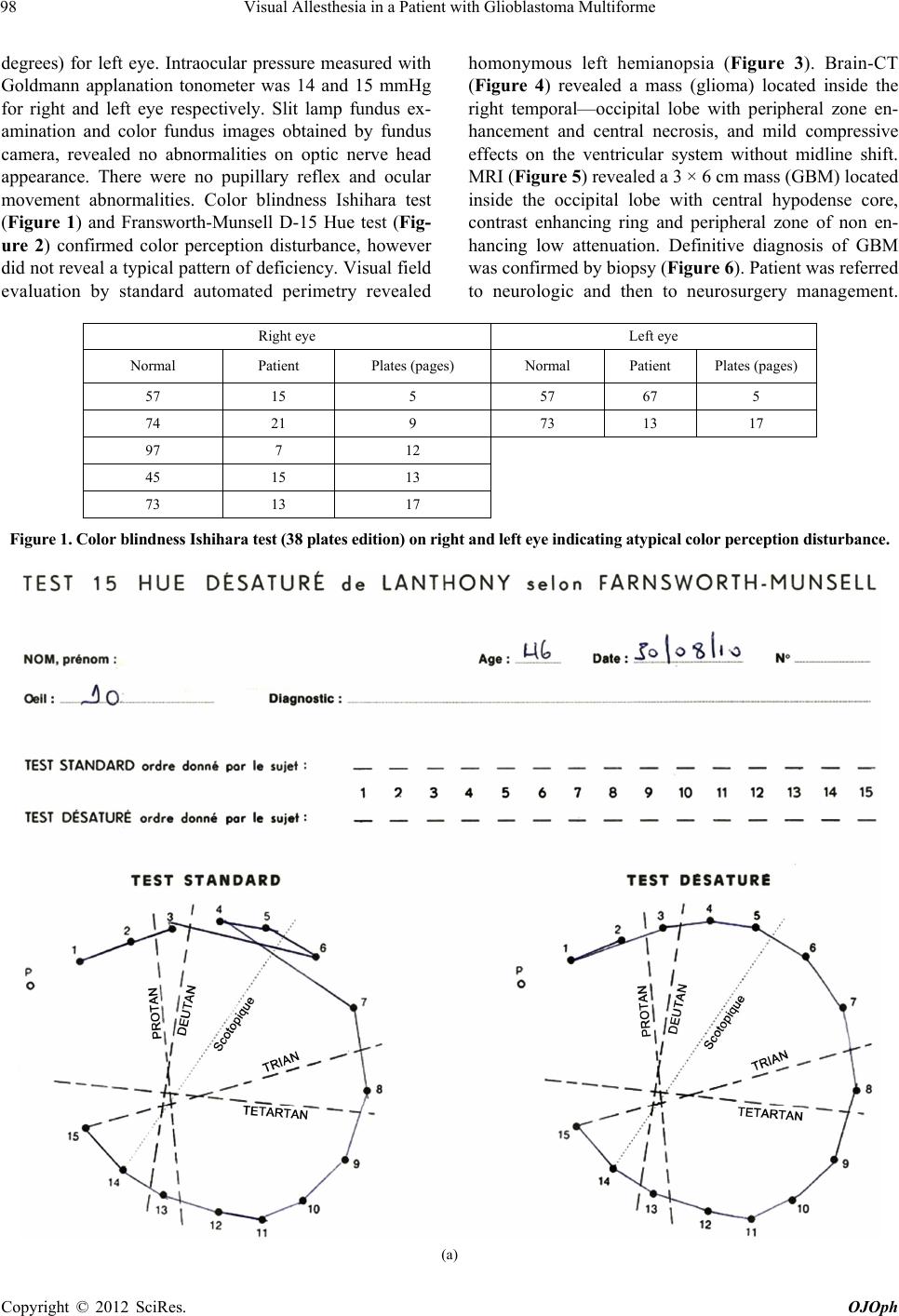 Visual Allesthesia in a Patient with Glioblastoma Multiforme 98 degrees) for left eye. Intraocular pressure measured with Goldmann applanation tonometer was 14 and 15 mmHg for right and left eye respectively. Slit lamp fundus ex- amination and color fundus images obtained by fundus camera, revealed no abnormalities on optic nerve head appearance. There were no pupillary reflex and ocular movement abnormalities. Color blindness Ishihara test (Figure 1) and Fransworth-Munsell D-15 Hue test (Fig- ure 2) confirmed color perception disturbance, however did not reveal a typical pattern of deficiency. Visual field evaluation by standard automated perimetry revealed homonymous left hemianopsia (Figure 3). Brain-CT (Figure 4) revealed a mass (glioma) located inside the right temporal—occipital lobe with peripheral zone en- hancement and central necrosis, and mild compressive effects on the ventricular system without midline shift. MRI (Figure 5) revealed a 3 × 6 cm mass (GBM) located inside the occipital lobe with central hypodense core, contrast enhancing ring and peripheral zone of non en- hancing low attenuation. Definitive diagnosis of GBM was confirmed by biopsy (Figure 6). Patient was referred to neurologic and then to neurosurgery management. Right eye Left eye Normal Patient Plates (pages) Normal Patient Plates (pages) 57 15 5 57 67 5 74 21 9 73 13 17 97 7 12 45 15 13 73 13 17 Figure 1. Color blindness Ishihara test (38 plates edition) on right and left eye indicating atypical color perception disturbance. (a) Copyright © 2012 SciRes. OJOph 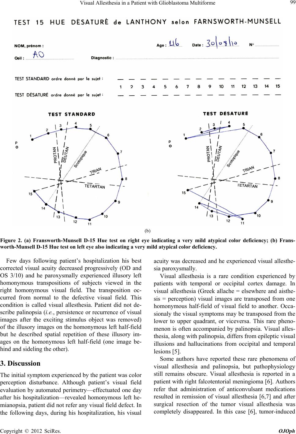 Visual Allesthesia in a Patient with Glioblastoma Multiforme 99 (b) Figure 2. (a) Fransworth-Munsell D-15 Hue test on right eye indicating a very mild atypical color deficiency; (b) Frans- worth-Munsell D-15 Hue test on left eye also indicating a very mild atypical color deficiency. Few days following patient’s hospitalization his best corrected visual acuity decreased progressively (OD and OS 3/10) and he paroxysmally experienced illusory left homonymous transpositions of subjects viewed in the right homonymous visual field. The transposition oc- curred from normal to the defective visual field. This condition is called visual allesthesia. Patient did not de- scribe palinopsia (i.e., persistence or recurrence of visual images after the exciting stimulus object was removed) of the illusory images on the homonymous left half-field but he described spatial repetition of these illusory im- ages on the homonymous left half-field (one image be- hind and sideling the other). 3. Discussion The initial symptom experienced by the patien t was color perception disturbance. Although patient’s visual field evaluation by automated perimetry—effectuated one day after his hospitalization—revealed homonymous left he- mianopsia, patient did not refer any visual field defect. In the following days, during his hospitalization, his visual acuity was decreased and he experienced visual allesthe- sia paroxysmally. Visual allesthesia is a rare condition experienced by patients with temporal or occipital cortex damage. In visual allesthesia (Greek allache = elsewhere and aisthe- sis = perception) visual images are transposed from one homonymous half-field of visual field to another. Occa- sionaly the visual symptoms may be transposed from the lower to upper quadrant, or viceversa. This rare pheno- menon is often accompanied by palinopsia. Visual alles- thesia, along with palinopsia, differs from epileptic v isual illusions and hallucinations from occipital and temporal lesions [5]. Some authors have reported these rare phenomena of visual allesthesia and palinopsia, but pathophysiology still remains obscure. Visual allesthesia is reported in a patient with right falcotentorial meningioma [6]. Authors refer that administration of anticonvulsant medications resulted in remission of visual allesthesia [6,7] and after surgical resection of the tumor visual allesthesia was completely disappeared. In this case [6], tumor-induced Copyright © 2012 SciRes. OJOph 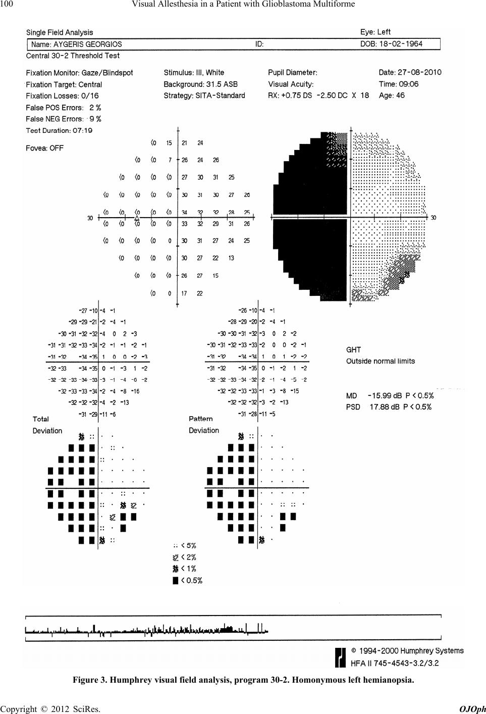 Visual Allesthesia in a Patient with Glioblastoma Multiforme 100 Figure 3. Humphrey visual field analysis, program 30-2. Homonymous left hemianopsia. Copyright © 2012 SciRes. OJOph 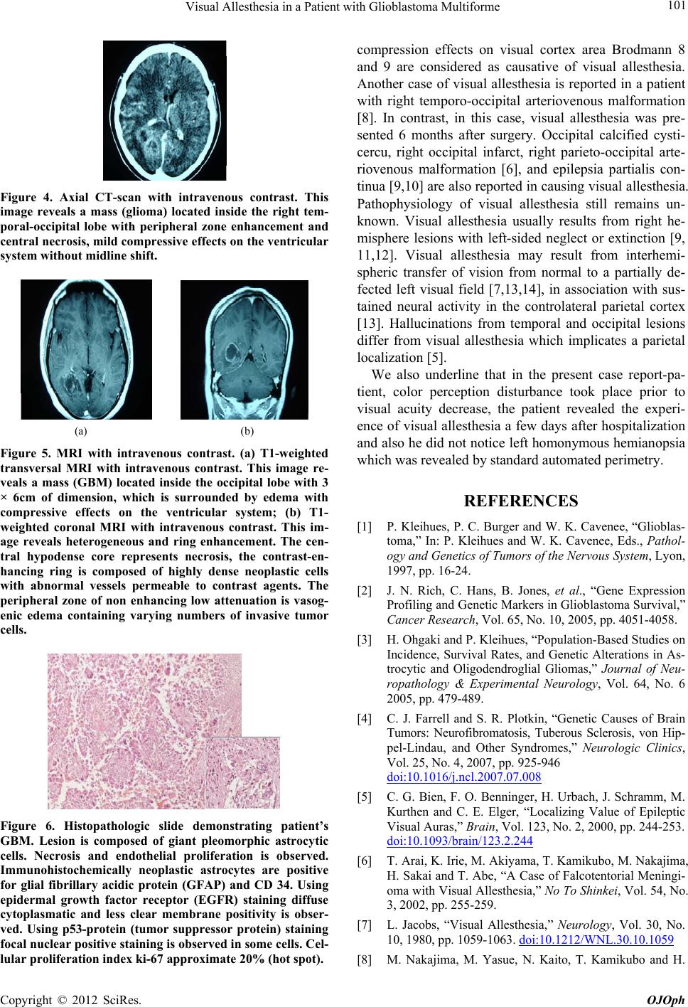 Visual Allesthesia in a Patient with Glioblastoma Multiforme 101 Figure 4. Axial CT-scan with intravenous contrast. This image reveals a mass (glioma) located inside the right tem- poral-occipital lobe with peripheral zone enhancement and central necrosis, mild compressive effects on the ventricular system without midline shift. (a) (b) Figure 5. MRI with intravenous contrast. (a) T1-weighted transversal MRI with intravenous contrast. This image re- veals a mass (GBM) located inside the occipital lobe with 3 × 6cm of dimension, which is surrounded by edema with compressive effects on the ventricular system; (b) T1- weighted coronal MRI with intravenous contrast. This im- age reveals heterogeneous and ring enhancement. The cen- tral hypodense core represents necrosis, the contrast-en- hancing ring is composed of highly dense neoplastic cells with abnormal vessels permeable to contrast agents. The peripheral zone of non enhancing low attenuation is vasog- enic edema containing varying numbers of invasive tumor cells. Figure 6. Histopathologic slide demonstrating patient’s GBM. Lesion is composed of giant pleomorphic astrocytic cells. Necrosis and endothelial proliferation is observed. Immunohistochemically neoplastic astrocytes are positive for glial fibrillary acidic protein (GFAP) and CD 34. Using epidermal growth factor receptor (EGFR) staining diffuse cytoplasmatic and less clear membrane positivity is obser- ved. Using p53-protein (tumor suppressor protein) staining focal nuclear positive staining is observed in some cells. Cel- lular proliferation index ki-67 approximate 20% (hot spot). compression effects on visual cortex area Brodmann 8 and 9 are considered as causative of visual allesthesia. Another case of visual allesthesia is reported in a patient with right temporo-occipital arteriovenous malformation [8]. In contrast, in this case, visual allesthesia was pre- sented 6 months after surgery. Occipital calcified cysti- cercu, right occipital infarct, right parieto-occipital arte- riovenous malformation [6], and epilepsia partialis con- tinua [9,10] are also reported in causing visual allesthesia. Pathophysiology of visual allesthesia still remains un- known. Visual allesthesia usually results from right he- misphere lesions with left-sided neglect or extinction [9, 11,12]. Visual allesthesia may result from interhemi- spheric transfer of vision from normal to a partially de- fected left visual field [7,13,14], in association with sus- tained neural activity in the controlateral parietal cortex [13]. Hallucinations from temporal and occipital lesions differ from visual allesthesia which implicates a parietal localization [5]. We also underline that in the present case report-pa- tient, color perception disturbance took place prior to visual acuity decrease, the patient revealed the experi- ence of visual allesthesia a few days after hospitalization and also he did not notice left homonymous hemianopsia which was revealed by standard automated perimetry. REFERENCES [1] P. Kleihues, P. C. Burger and W. K. Cavenee, “Glioblas- toma,” In: P. Kleihues and W. K. Cavenee, Eds., Pathol- ogy and Genetics of Tumors of the Nervous System, Lyon, 1997, pp. 16-24. [2] J. N. Rich, C. Hans, B. Jones, et al., “Gene Expression Profiling and Genetic Markers in Glioblastoma Survival,” Cancer Research, Vol. 65, No. 10, 2005, pp. 4051-4058. [3] H. Ohgaki and P. Kleihues, “Population-Based Studies on Incidence, Survival Rates, and Genetic Alt erations in As- trocytic and Oligodendroglial Gliomas,” Journal of Neu- ropathology & Experimental Neurology, Vol. 64, No. 6 2005, pp. 479-489. [4] C. J. Farrell and S. R. Plotkin, “Genetic Causes of Brain Tumors: Neurofibromatosis, Tuberous Sclerosis, von Hip- pel-Lindau, and Other Syndromes,” Neurologic Clinics, Vol. 25, No. 4, 2007, pp. 925-946 doi:10.1016/j.ncl.2007.07.008 [5] C. G. Bien, F. O. Benninger, H. Urbach, J. Schramm, M. Kurthen and C. E. Elger, “Localizing Value of Epileptic Visual Auras,” Brain, Vol. 123, No. 2, 2000, pp. 244-253. doi:10.1093/brain/123.2.244 [6] T. Arai, K. Irie, M. Akiyama, T. Kamikubo, M. Nakajima, H. Sakai and T. Abe, “A Case of Falcotentorial Meningi- oma with Visual Allesthesia,” No To Shinkei, Vol. 54, No. 3, 2002, pp. 255-259. [7] L. Jacobs, “Visual Allesthesia,” Neurology, Vol. 30, No. 10, 1980, pp. 1059-1063. doi:10.1212/WNL.30.10.1059 [8] M. Nakajima, M. Yasue, N. Kaito, T. Kamikubo and H. Copyright © 2012 SciRes. OJOph 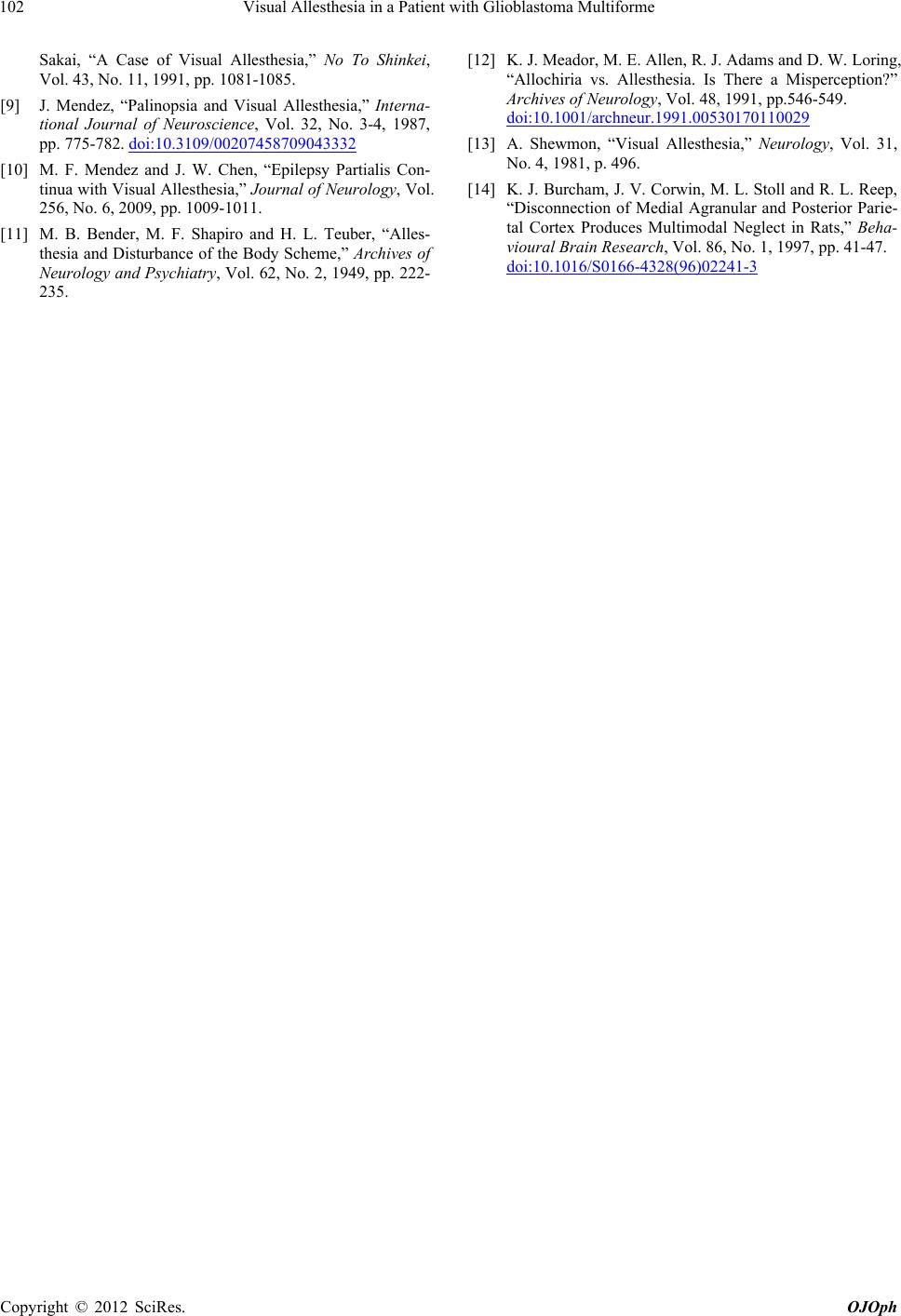 Visual Allesthesia in a Patient with Glioblastoma Multiforme 102 Sakai, “A Case of Visual Allesthesia,” No To Shinkei, Vol. 43, No. 11, 1991, pp. 1081-1085. [9] J. Mendez, “Palinopsia and Visual Allesthesia,” Interna- tional Journal of Neuroscience, Vol. 32, No. 3-4, 1987, pp. 775-782. doi:10.3109/00207458709043332 [10] M. F. Mendez and J. W. Chen, “Epilepsy Partialis Con- tinua with Visual Allesthesia,” Journal of Neurology, Vol. 256, No. 6, 2009, pp. 1009-1011. [11] M. B. Bender, M. F. Shapiro and H. L. Teuber, “Alles- thesia and Disturbance of the Body Scheme,” Archives of Neurology and Psychiatry, Vol. 62, No. 2, 1949, pp. 222- 235. [12] K. J. Meador, M. E. Alle n, R. J. Adams and D. W. Loring, “Allochiria vs. Allesthesia. Is There a Misperception?” Archives of Neurology, Vol. 48, 1991, pp.546-549. doi:10.1001/archneur.1991.00530170110029 [13] A. Shewmon, “Visual Allesthesia,” Neurology, Vol. 31, No. 4, 1981, p. 496. [14] K. J. Burcham, J. V. Corwin, M. L. Stoll and R. L. Reep, “Disconnection of Medial Agranular and Posterior Parie- tal Cortex Produces Multimodal Neglect in Rats,” Beha- vioural Brain Research, Vol. 86, No. 1, 1997, pp. 41-47. doi:10.1016/S0166-4328(96)02241-3 Copyright © 2012 SciRes. OJOph |