

Paper Menu >>
Journal Menu >>
 Vol.2, No.6, 563-570 (2010) Natural Science http://dx.doi.org/10.4236/ns.2010.26071 Copyright © 2010 SciRes. OPEN ACCESS Binding of naturally occurring hydroxycinnamic acids to bovine serum albumin Lucie Trnková1,2, Iva Boušová1*, Vladimír Kubíček1, Jaroslav Dršata1,2 1Charles University in Prague, Faculty of Pharmacy, Hradec Králové, Czech Republic; *Corresponding Author: [email protected] 2University of Hradec Králové, Faculty of Education, Hradec Králové, Czech Republic Received 22 February 2010; revised 12 April 2010; accepted 13 May 2010. ABSTRACT Hydroxycinnamic acids (HCAs) possess numer- ous biological effects including antioxidant, an- tiallergic, antimicrobial, and immunomodulatory activities and due to these properties are widely used in folk medicine. Nevertheless, they can interact with protein molecules and cause some structural and functional changes. The possib- ility of HCAs binding to bovine serum albumin (BSA) under physiological conditions was inve- stigated by the UV-VIS absorption spectroscopy and fluorescence quenching method. Apart from rosmarinic acid, all tested HCAs quenched tryptophan fluorescence of BSA in the studied range of concentrations (0-20 µM) mainly by static quenching mechanism (formation of non- fluorescent HCA-BSA complexes). The binding constants, number of binding sites and free en- ergy changes were determined. The binding affinities of HCAs were ranked in the order: chlorogenic acid > sinapic acid ≥ caffeic acid > ferulic acid > o-coumaric acid > p-coumaric acid ≥ m-coumaric acid, which was confirmed by spectral overlaps of BSA emission spectrum with absorption spectrum of HCA. All free en- ergy changes possessed negative sign indicat- ing the spontaneity of HCA-BSA interaction. Keywords: Bovine Serum Albumin; Hydroxycinnamic Acid; Fluorescence Quenching; Protein-Ligand Binding 1. INTRODUCTION Recently, considerable attention has been focused on the study of the interaction between small molecules (drugs) and biological macromolecules (e.g. proteins), especially discussing the thermodynamic quality, binding force qu- ality, and mechanism of interactions [1-3]. These studies play crucial role in promoting research on proteins be- cause they can provide useful information for study of pharmacological and biological effects of drugs as well as conformational changes of proteins caused by drugs. Serum albumin is one of the most abundant proteins in circulatory system of a wide variety of organisms and one of the most extensively studied proteins at all [4,5]. Bovine serum albumin (BSA) consists of 583 amino acids in a single polypeptide chain cross-linked with 17 disulfide bonds. It is composed of three homologous domains (I-III), each of which comprises of two subdo- mains (A and B). BSA has two tryptophan residues, which significantly contribute to the intrinsic fluores- cence of this protein: Trp-134 is located near the surface in domain IB and Trp-212 is buried in a hydrophobic (non-polar) pocket in the internal part of domain IIA [6]. HSA differs from BSA by 24% of primary structure and the most important difference from spectroscopic point of view seems to be presence of only one tryptophan residue (Trp-214) in its molecule [4,5]. Serum albumin possesses a wide range of physiological functions in- volving the binding, transport and deposition of many endogenous and exogenous ligands present in blood cir- culation [4,7]. Perhaps, its most outstanding property is the ability to bind a variety of ligands. It is well known that many drugs bind to serum albumin and their effec- tiveness depends on the binding ability [5,8]. On the other hand, drugs can cause various changes in protein conformation influencing its physiological function and such impaired proteins may be consequently pathologi- cally accumulated in body tissues. Plant polyphenols represent a heterogeneous group of natural compounds with one or more hydroxyl groups attached to the benzene ring. These substances possess several important physiological roles in plants, such as defense against herbivores and pathogens, pigmentation, and attraction of pollinating insects [9]. The most widely distributed polyphenolic compounds in plant tissues are hydroxycinnamic acids. Some of the most common 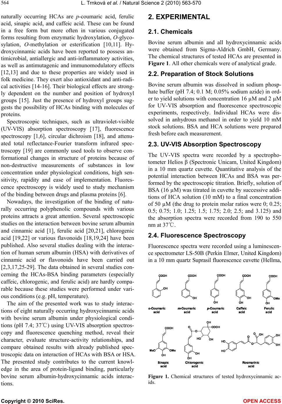 L. Trnková et al. / Natural Science 2 (2010) 563-570 Copyright © 2010 SciRes. OPEN ACCESS 564 naturally occurring HCAs are p-coumaric acid, ferulic acid, sinapic acid, and caffeic acid. These can be found in a free form but more often in various conjugated forms resulting from enzymatic hydroxylation, O-glyco- sylation, O-methylation or esterification [10,11]. Hy- droxycinnamic acids have been reported to possess an- timicrobial, antiallergic and anti-inflammatory activities, as well as antimutagenic and immunomodulatory effects [12,13] and due to these properties are widely used in folk medicine. They exert also antioxidant and anti-radi- cal activities [14-16]. Their biological effects are strong- ly dependent on the number and position of hydroxyl groups [15]. Just the presence of hydroxyl groups sug- gests the possibility of HCAs binding with molecules of proteins. Spectroscopic techniques, such as ultraviolet-visible (UV-VIS) absorption spectroscopy [17], fluorescence spectroscopy [1,6], circular dichroism [18], and attenu- ated total reflectance-Fourier transform infrared spec- troscopy [19] are commonly used tools to observe con- formational changes in structure of proteins because of non-destructive measurements of substances in low concentration under physiological conditions, high sen- sitivity, rapidity and ease of implementation. Fluores- cence spectroscopy is widely used to study mechanism of the binding between drugs and plasma proteins [6]. Nowadays, the investigation of the binding of natu- rally occurring polyphenolic compounds with various proteins attracts a great attention. Several spectroscopic studies on the interaction between bovine serum albumin and cinnamic acid [1], ferulic acid [20,21], chlorogenic acid [19,22] or various flavonoids [18,19,24] have been published. Also several studies dealing with the interac- tion of human serum albumin (HSA) with derivatives of cinnamic acid or flavonoids have been carried out [2,3,17,25-29]. The data obtained in several studies con- cerning the HCAs-BSA binding parameters (especially caffeic, chlorogenic, and ferulic acid) are hardly compa- rable because these studies were performed under vari- ous conditions (e.g. pH, temperature). The aim of the presented work was to study interac- tions of eight naturally occurring hydroxycinnamic acids with bovine serum albumin under physiological condi- tions (pH 7.4; 37℃) using UV-VIS absorption spectros- copy and fluorescence quenching method, reveal their character, evaluate structure-activity relationships, and compare obtained results with already published spec- troscopic data on interaction of HCAs with BSA or HSA. The presented study contributes to the current knowl- edge in the area of protein-ligand binding, particularly bovine serum albumin-hydroxycinnamic acids interac- tions. 2. EXPERIMENTAL 2.1. Chemicals Bovine serum albumin and all hydroxycinnamic acids were obtained from Sigma-Aldrich GmbH, Germany. The chemical structures of tested HCAs are presented in Figure 1. All other chemicals were of analytical grade. 2.2. Preparation of Stock Solutions Bovine serum albumin was dissolved in sodium phosp- hate buffer (pH 7.4; 0.1 M; 0.05% sodium azide) in ord- er to yield solutions with concentration 16 µM and 2 µM for UV-VIS absorption and fluorescence spectroscopic experiments, respectively. Individual HCAs were dis- solved in anhydrous methanol in order to yield 10 mM stock solutions. BSA and HCA solutions were prepared fresh before each measurement. 2.3. UV-VIS Absorption Spectroscopy The UV-VIS spectra were recorded by a spectropho- tometer Helios β (Spectronic Unicam, United Kingdom) in a 10 mm quartz cuvette. Quantitative analysis of the potential interaction between HCAs and BSA was per- formed by the spectroscopic titration. Briefly, solution of BSA (16 µM) was titrated in cuvette by successive addi- tions of HCA solution (10 mM) to a final concentration of 50 µM (the drug to protein molar ratios were 0; 0.25; 0.5; 0.75; 1.0; 1.25; 1.5; 1.75; 2.0; 2.5; and 3.125) and the absorption spectra were recorded from 190 to 550 nm at 37℃. 2.4. Fluorescence Spectroscopy Fluorescence spectra were recorded using a luminescen- ce spectrometer LS-50B (Perkin Elmer, United Kingdom) in a 10 mm quartz Suprasil fluorescence cuvette (Hellma, Figure 1. Chemical structures of tested hydroxycinnamic ac- ids. 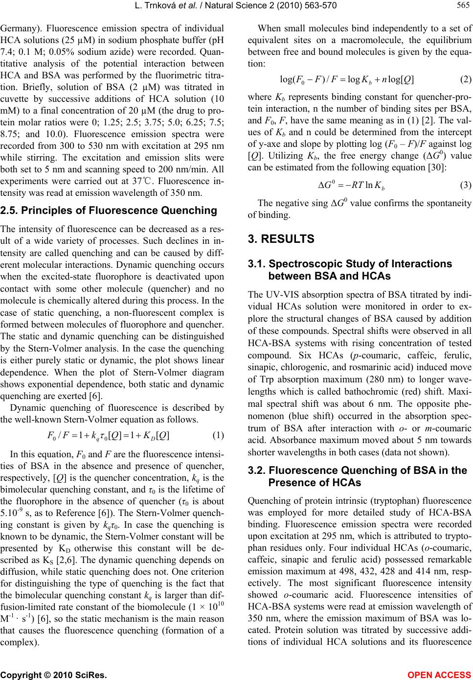 L. Trnková et al. / Natural Science 2 (2010) 563-570 Copyright © 2010 SciRes. OPEN ACCESS 565 565 Germany). Fluorescence emission spectra of individual HCA solutions (25 µM) in sodium phosphate buffer (pH 7.4; 0.1 M; 0.05% sodium azide) were recorded. Quan- titative analysis of the potential interaction between HCA and BSA was performed by the fluorimetric titra- tion. Briefly, solution of BSA (2 µM) was titrated in cuvette by successive additions of HCA solution (10 mM) to a final concentration of 20 µM (the drug to pro- tein molar ratios were 0; 1.25; 2.5; 3.75; 5.0; 6.25; 7.5; 8.75; and 10.0). Fluorescence emission spectra were recorded from 300 to 530 nm with excitation at 295 nm while stirring. The excitation and emission slits were both set to 5 nm and scanning speed to 200 nm/min. All experiments were carried out at 37℃. Fluorescence in- tensity was read at emission wavelength of 350 nm. 2.5. Principles of Fluorescence Quenching The intensity of fluorescence can be decreased as a res- ult of a wide variety of processes. Such declines in in- tensity are called quenching and can be caused by diff- erent molecular interactions. Dynamic quenching occurs when the excited-state fluorophore is deactivated upon contact with some other molecule (quencher) and no molecule is chemically altered during this process. In the case of static quenching, a non-fluorescent complex is formed between molecules of fluorophore and quencher. The static and dynamic quenching can be distinguished by the Stern-Volmer analysis. In the case the quenching is either purely static or dynamic, the plot shows linear dependence. When the plot of Stern-Volmer diagram shows exponential dependence, both static and dynamic quenching are exerted [6]. Dynamic quenching of fluorescence is described by the well-known Stern-Volmer equation as follows. ][1][1/ 00 QKQkFF Dq (1) In this equation, F0 and F are the fluorescence intensi- ties of BSA in the absence and presence of quencher, respectively, [Q] is the quencher concentration, kq is the bimolecular quenching constant, and τ0 is the lifetime of the fluorophore in the absence of quencher (τ0 is about 5.10-9 s, as to Reference [6]). The Stern-Volmer quench- ing constant is given by kqτ0. In case the quenching is known to be dynamic, the Stern-Volmer constant will be presented by KD otherwise this constant will be de- scribed as KS [2,6]. The dynamic quenching depends on diffusion, while static quenching does not. One criterion for distinguishing the type of quenching is the fact that the bimolecular quenching constant kq is larger than dif- fusion-limited rate constant of the biomolecule (1 × 10¹⁰ M-1 · s-1) [6], so the static mechanism is the main reason that causes the fluorescence quenching (formation of a complex). When small molecules bind independently to a set of equivalent sites on a macromolecule, the equilibrium between free and bound molecules is given by the equa- tion: ]log[log/)log( 0QnKFFF b (2) where Kb represents binding constant for quencher-pro- tein interaction, n the number of binding sites per BSA, and F0, F, have the same meaning as in (1) [2]. The val- ues of Kb and n could be determined from the intercept of y-axe and slope by plotting log (F0 – F)/F against log [Q]. Utilizing Kb, the free energy change (ΔG0) value can be estimated from the following equation [30]: b KRTGln 0 (3) The negative sing ΔG0 value confirms the spontaneity of binding. 3. RESULTS 3.1. Spectroscopic Study of Interactions between BSA and HCAs The UV-VIS absorption spectra of BSA titrated by indi- vidual HCAs solution were monitored in order to ex- plore the structural changes of BSA caused by addition of these compounds. Spectral shifts were observed in all HCA-BSA systems with rising concentration of tested compound. Six HCAs (p-coumaric, caffeic, ferulic, sinapic, chlorogenic, and rosmarinic acid) induced move of Trp absorption maximum (280 nm) to longer wave- lengths which is called bathochromic (red) shift. Maxi- mal spectral shift was about 6 nm. The opposite phe- nomenon (blue shift) occurred in the absorption spec- trum of BSA after interaction with o- or m-coumaric acid. Absorbance maximum moved about 5 nm towards shorter wavelengths in both cases (data not shown). 3.2. Fluorescence Quenching of BSA in the Presence of HCAs Quenching of protein intrinsic (tryptophan) fluorescence was employed for more detailed study of HCA-BSA binding. Fluorescence emission spectra were recorded upon excitation at 295 nm, which is attributed to trypto- phan residues only. Four individual HCAs (o-coumaric, caffeic, sinapic and ferulic acid) possessed remarkable emission maximum at 498, 432, 428 and 414 nm, resp- ectively. The most significant fluorescence intensity showed o-coumaric acid. Fluorescence intensities of HCA-BSA systems were read at emission wavelength of 350 nm, where the emission maximum of BSA was lo- cated. Protein solution was titrated by successive addi- tions of individual HCA solutions and its fluorescence 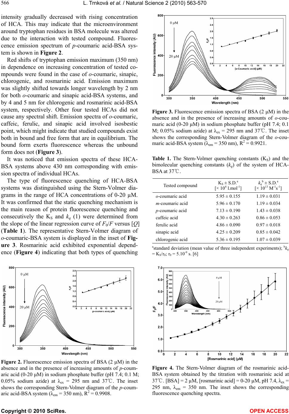 L. Trnková et al. / Natural Science 2 (2010) 563-570 Copyright © 2010 SciRes. OPEN ACCESS 566 intensity gradually decreased with rising concentration of HCA. This may indicate that the microenvironment around tryptophan residues in BSA molecule was altered due to the interaction with tested compound. Fluores- cence emission spectrum of p-coumaric acid-BSA sys- tem is shown in Figure 2. Red shifts of tryptophan emission maximum (350 nm) in dependence on increasing concentration of tested co- mpounds were found in the case of o-coumaric, sinapic, chlorogenic, and rosmarinic acid. Emission maximum was slightly shifted towards longer wavelength by 2 nm for both o-coumaric and sinapic acid-BSA systems, and by 4 and 5 nm for chlorogenic and rosmarinic acid-BSA system, respectively. Other four tested HCAs did not cause any spectral shift. Emission spectra of o-coumaric, caffeic, ferulic, and sinapic acid involved isosbestic point, which might indicate that studied compounds exist both in bound and free form that are in equilibrium. The bound form exerts fluorescence whereas the unbound form does not (Figure 3). It was noticed that emission spectra of these HCA- BSA systems above 430 nm corresponding with emis- sion spectra of individual HCAs. The type of fluorescence quenching of HCA-BSA systems was distinguished using the Stern-Volmer dia- grams in the range of HCA concentrations of 0-20 µM. It was confirmed that the static quenching mechanism is the main reason of protein fluorescence quenching and consecutively the KS and kq (1) were determined from the slope of the linear regression curve of F0/F versus [Q] (Table 1). The representative Stern-Volmer diagram of o-coumaric-BSA system is displayed in the inset of Fig- ure 3. Rosmarinic acid exhibited exponential depend- ence (Figure 4) indicating that both types of quenching Figure 2. Fluorescence emission spectra of BSA (2 μM) in the absence and in the presence of increasing amounts of p-coum- aric acid (0-20 μM) in sodium phosphate buffer (pH 7.4; 0.1 M; 0.05% sodium azide) at λex = 295 nm and 37℃. The inset shows the corresponding Stern-Volmer diagram of the p-coum- aric acid-BSA system (λem = 350 nm), R2 = 0.9908. Figure 3. Fluorescence emission spectra of BSA (2 μM) in the absence and in the presence of increasing amounts of o-cou- maric acid (0-20 μM) in sodium phosphate buffer (pH 7.4; 0.1 M; 0.05% sodium azide) at λex = 295 nm and 37℃. The inset shows the corresponding Stern-Volmer diagram of the o-cou- maric acid-BSA system (λem = 350 nm), R2 = 0.9921. Table 1. The Stern-Volmer quenching constants (KS) and the bimolecular quenching constants (kq) of the system of HCA- BSA at 37℃. Tested compound KS ± S.D.a [× 104 l.mol-1] kqb ± S.D.a [× 1013 M-1s-1] o-coumaric acid 5.95 ± 0.155 1.19 ± 0.031 m-coumaric acid 5.96 ± 0.170 1.19 ± 0.034 p-coumaric acid 7.13 ± 0.190 1.43 ± 0.038 caffeic acid 4.30 ± 0.263 0.86 ± 0.053 ferulic acid 4.86 ± 0.090 0.97 ± 0.018 sinapic acid 4.25 ± 0.209 0.85 ± 0.042 chlorogenic acid 5.36 ± 0.195 1.07 ± 0.039 astandard deviation (mean value of three independent experiments); bkq = KS/τ0; τ0 = 5.10-9 s. [6] Figure 4. The Stern-Volmer diagram of the rosmarinic acid- BSA system obtained by the titration with rosmarinic acid at 37℃. [BSA] = 2 µM, [rosmarinic acid] = 0-20 µM, pH 7.4, λex = 295 nm, λem = 350 nm. The inset shows the corresponding fluorescence quenching spectra. 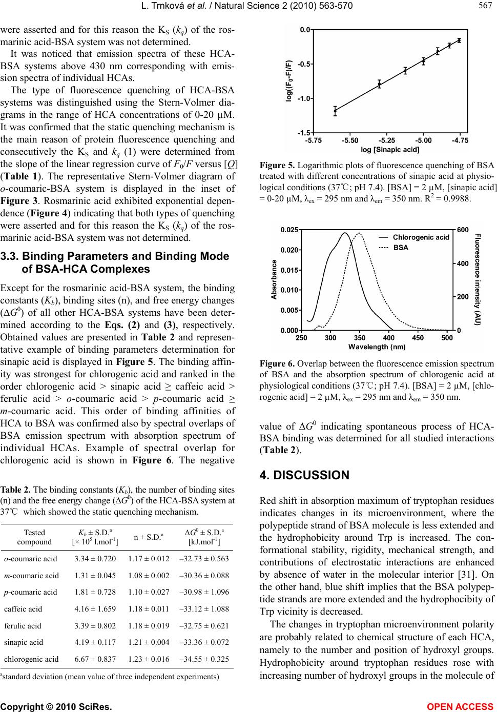 L. Trnková et al. / Natural Science 2 (2010) 563-570 Copyright © 2010 SciRes. OPEN ACCESS 567 567 were asserted and for this reason the KS (kq) of the ros- marinic acid-BSA system was not determined. It was noticed that emission spectra of these HCA- BSA systems above 430 nm corresponding with emis- sion spectra of individual HCAs. The type of fluorescence quenching of HCA-BSA systems was distinguished using the Stern-Volmer dia- grams in the range of HCA concentrations of 0-20 µM. It was confirmed that the static quenching mechanism is the main reason of protein fluorescence quenching and consecutively the KS and kq (1) were determined from the slope of the linear regression curve of F0/F versus [Q] (Table 1). The representative Stern-Volmer diagram of o-coumaric-BSA system is displayed in the inset of Figure 3. Rosmarinic acid exhibited exponential depen- dence (Figure 4) indicating that both types of quenching were asserted and for this reason the KS (kq) of the ros- marinic acid-BSA system was not determined. 3.3. Binding Parameters and Binding Mode of BSA-HCA Complexes Except for the rosmarinic acid-BSA system, the binding constants (Kb), binding sites (n), and free energy changes (∆G0) of all other HCA-BSA systems have been deter- mined according to the Eqs. (2) and (3), respectively. Obtained values are presented in Table 2 and represen- tative example of binding parameters determination for sinapic acid is displayed in Figure 5. The binding affin- ity was strongest for chlorogenic acid and ranked in the order chlorogenic acid > sinapic acid ≥ caffeic acid > ferulic acid > o-coumaric acid > p-coumaric acid ≥ m-coumaric acid. This order of binding affinities of HCA to BSA was confirmed also by spectral overlaps of BSA emission spectrum with absorption spectrum of individual HCAs. Example of spectral overlap for chlorogenic acid is shown in Figure 6. The negative Table 2. The binding constants (Kb), the number of binding sites (n) and the free energy change (∆G0) of the HCA-BSA system at 37℃ which showed the static quenching mechanism. Tested compound Kb ± S.D.a [× 105 l.mol-1] n ± S.D.a ΔG0 ± S.D.a [kJ.mol-1] o-coumaric acid 3.34 ± 0.720 1.17 ± 0.012 –32.73 ± 0.563 m-coumaric acid 1.31 ± 0.045 1.08 ± 0.002 –30.36 ± 0.088 p-coumaric acid 1.81 ± 0.728 1.10 ± 0.027 –30.98 ± 1.096 caffeic acid 4.16 ± 1.659 1.18 ± 0.011 –33.12 ± 1.088 ferulic acid 3.39 ± 0.802 1.18 ± 0.019 –32.75 ± 0.621 sinapic acid 4.19 ± 0.117 1.21 ± 0.004 –33.36 ± 0.072 chlorogenic acid 6.67 ± 0.837 1.23 ± 0.016 –34.55 ± 0.325 astandard deviation (mean value of three independent experiments) Figure 5. Logarithmic plots of fluorescence quenching of BSA treated with different concentrations of sinapic acid at physio- logical conditions (37℃; pH 7.4). [BSA] = 2 µM, [sinapic acid] = 0-20 µM, λex = 295 nm and λem = 350 nm. R2 = 0.9988. Figure 6. Overlap between the fluorescence emission spectrum of BSA and the absorption spectrum of chlorogenic acid at physiological conditions (37℃; pH 7.4). [BSA] = 2 µM, [chlo- rogenic acid] = 2 µM, λex = 295 nm and λem = 350 nm. value of ΔG0 indicating spontaneous process of HCA- BSA binding was determined for all studied interactions (Table 2). 4. DISCUSSION Red shift in absorption maximum of tryptophan residues indicates changes in its microenvironment, where the polypeptide strand of BSA molecule is less extended and the hydrophobicity around Trp is increased. The con- formational stability, rigidity, mechanical strength, and contributions of electrostatic interactions are enhanced by absence of water in the molecular interior [31]. On the other hand, blue shift implies that the BSA polypep- tide strands are more extended and the hydrophocibity of Trp vicinity is decreased. The changes in tryptophan microenvironment polarity are probably related to chemical structure of each HCA, namely to the number and position of hydroxyl groups. Hydrophobicity around tryptophan residues rose with increasing number of hydroxyl groups in the molecule of 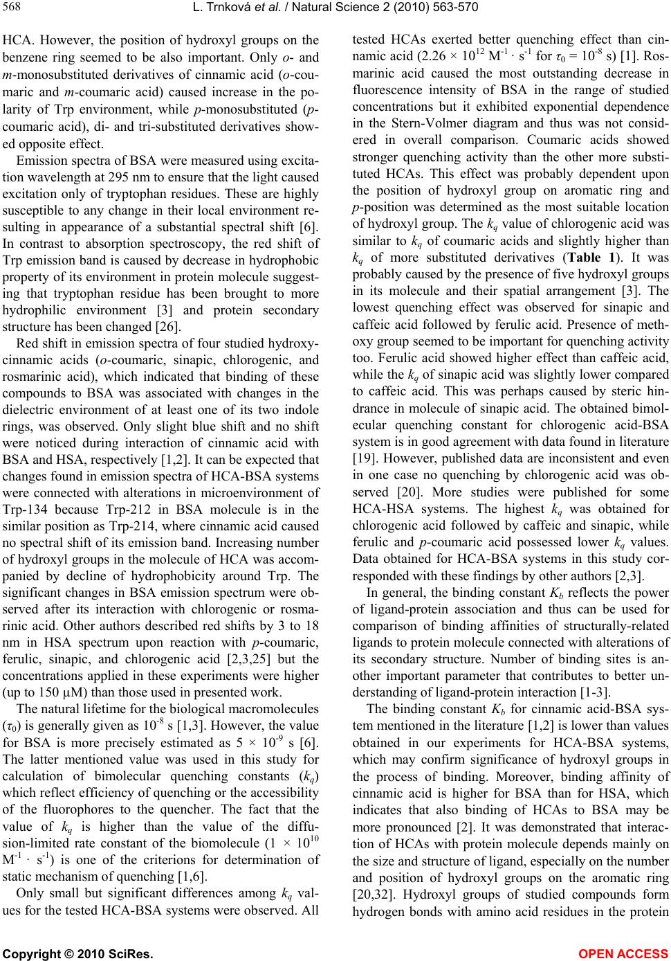 L. Trnková et al. / Natural Science 2 (2010) 563-570 Copyright © 2010 SciRes. OPEN ACCESS 568 HCA. However, the position of hydroxyl groups on the benzene ring seemed to be also important. Only o- and m-monosubstituted derivatives of cinnamic acid (o-cou- maric and m-coumaric acid) caused increase in the po- larity of Trp environment, while p-monosubstituted (p- coumaric acid), di- and tri-substituted derivatives show- ed opposite effect. Emission spectra of BSA were measured using excita- tion wavelength at 295 nm to ensure that the light caused excitation only of tryptophan residues. These are highly susceptible to any change in their local environment re- sulting in appearance of a substantial spectral shift [6]. In contrast to absorption spectroscopy, the red shift of Trp emission band is caused by decrease in hydrophobic property of its environment in protein molecule suggest- ing that tryptophan residue has been brought to more hydrophilic environment [3] and protein secondary structure has been changed [26]. Red shift in emission spectra of four studied hydroxy- cinnamic acids (o-coumaric, sinapic, chlorogenic, and rosmarinic acid), which indicated that binding of these compounds to BSA was associated with changes in the dielectric environment of at least one of its two indole rings, was observed. Only slight blue shift and no shift were noticed during interaction of cinnamic acid with BSA and HSA, respectively [1,2]. It can be expected that changes found in emission spectra of HCA-BSA systems were connected with alterations in microenvironment of Trp-134 because Trp-212 in BSA molecule is in the similar position as Trp-214, where cinnamic acid caused no spectral shift of its emission band. Increasing number of hydroxyl groups in the molecule of HCA was accom- panied by decline of hydrophobicity around Trp. The significant changes in BSA emission spectrum were ob- served after its interaction with chlorogenic or rosma- rinic acid. Other authors described red shifts by 3 to 18 nm in HSA spectrum upon reaction with p-coumaric, ferulic, sinapic, and chlorogenic acid [2,3,25] but the concentrations applied in these experiments were higher (up to 150 µM) than those used in presented work. The natural lifetime for the biological macromolecules (τ0) is generally given as 10-8 s [1,3]. However, the value for BSA is more precisely estimated as 5 × 10⁻⁹ s [6]. The latter mentioned value was used in this study for calculation of bimolecular quenching constants (kq) which reflect efficiency of quenching or the accessibility of the fluorophores to the quencher. The fact that the value of kq is higher than the value of the diffu- sion-limited rate constant of the biomolecule (1 × 10¹⁰ M-1 · s-1) is one of the criterions for determination of static mechanism of quenching [1,6]. Only small but significant differences among kq val- ues for the tested HCA-BSA systems were observed. All tested HCAs exerted better quenching effect than cin- namic acid (2.26 × 10¹² M-1 · s-1 for τ0 = 10-8 s) [1]. Ros- marinic acid caused the most outstanding decrease in fluorescence intensity of BSA in the range of studied concentrations but it exhibited exponential dependence in the Stern-Volmer diagram and thus was not consid- ered in overall comparison. Coumaric acids showed stronger quenching activity than the other more substi- tuted HCAs. This effect was probably dependent upon the position of hydroxyl group on aromatic ring and p-position was determined as the most suitable location of hydroxyl group. The kq value of chlorogenic acid was similar to kq of coumaric acids and slightly higher than kq of more substituted derivatives (Table 1). It was probably caused by the presence of five hydroxyl groups in its molecule and their spatial arrangement [3]. The lowest quenching effect was observed for sinapic and caffeic acid followed by ferulic acid. Presence of meth- oxy group seemed to be important for quenching activity too. Ferulic acid showed higher effect than caffeic acid, while the kq of sinapic acid was slightly lower compared to caffeic acid. This was perhaps caused by steric hin- drance in molecule of sinapic acid. The obtained bimol- ecular quenching constant for chlorogenic acid-BSA system is in good agreement with data found in literature [19]. However, published data are inconsistent and even in one case no quenching by chlorogenic acid was ob- served [20]. More studies were published for some HCA-HSA systems. The highest kq was obtained for chlorogenic acid followed by caffeic and sinapic, while ferulic and p-coumaric acid possessed lower kq values. Data obtained for HCA-BSA systems in this study cor- responded with these findings by other authors [2,3]. In general, the binding constant Kb reflects the power of ligand-protein association and thus can be used for comparison of binding affinities of structurally-related ligands to protein molecule connected with alterations of its secondary structure. Number of binding sites is an- other important parameter that contributes to better un- derstanding of ligand-protein interaction [1-3]. The binding constant Kb for cinnamic acid-BSA sys- tem mentioned in the literature [1,2] is lower than values obtained in our experiments for HCA-BSA systems, which may confirm significance of hydroxyl groups in the process of binding. Moreover, binding affinity of cinnamic acid is higher for BSA than for HSA, which indicates that also binding of HCAs to BSA may be more pronounced [2]. It was demonstrated that interac- tion of HCAs with protein molecule depends mainly on the size and structure of ligand, especially on the number and position of hydroxyl groups on the aromatic ring [20,32]. Hydroxyl groups of studied compounds form hydrogen bonds with amino acid residues in the protein 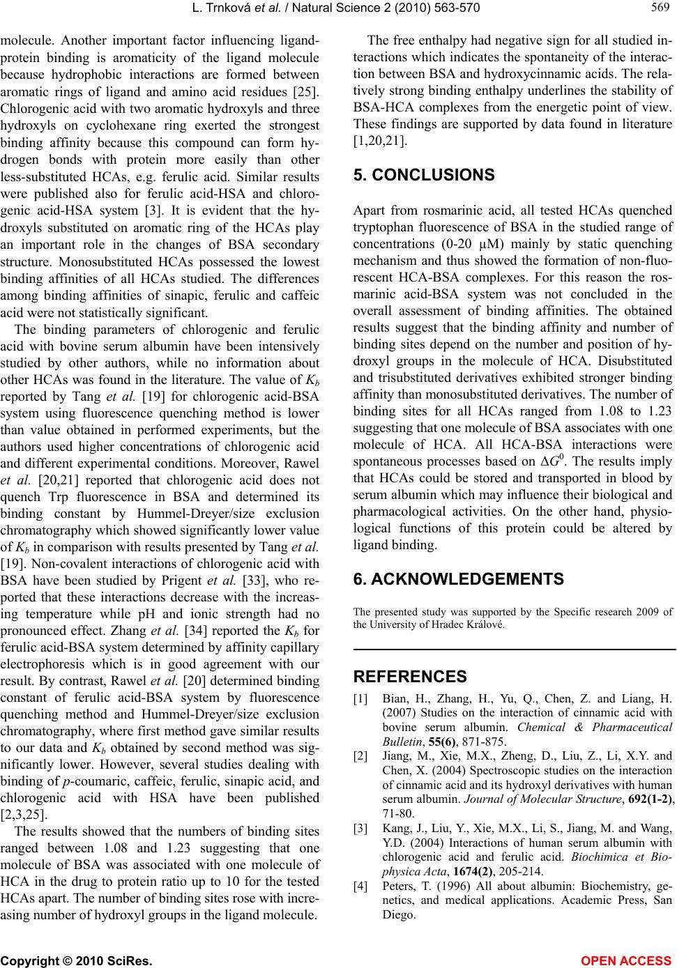 L. Trnková et al. / Natural Science 2 (2010) 563-570 Copyright © 2010 SciRes. OPEN ACCESS 569 569 molecule. Another important factor influencing ligand- protein binding is aromaticity of the ligand molecule because hydrophobic interactions are formed between aromatic rings of ligand and amino acid residues [25]. Chlorogenic acid with two aromatic hydroxyls and three hydroxyls on cyclohexane ring exerted the strongest binding affinity because this compound can form hy- drogen bonds with protein more easily than other less-substituted HCAs, e.g. ferulic acid. Similar results were published also for ferulic acid-HSA and chloro- genic acid-HSA system [3]. It is evident that the hy- droxyls substituted on aromatic ring of the HCAs play an important role in the changes of BSA secondary structure. Monosubstituted HCAs possessed the lowest binding affinities of all HCAs studied. The differences among binding affinities of sinapic, ferulic and caffeic acid were not statistically significant. The binding parameters of chlorogenic and ferulic acid with bovine serum albumin have been intensively studied by other authors, while no information about other HCAs was found in the literature. The value of Kb reported by Tang et al. [19] for chlorogenic acid-BSA system using fluorescence quenching method is lower than value obtained in performed experiments, but the authors used higher concentrations of chlorogenic acid and different experimental conditions. Moreover, Rawel et al. [20,21] reported that chlorogenic acid does not quench Trp fluorescence in BSA and determined its binding constant by Hummel-Dreyer/size exclusion chromatography which showed significantly lower value of Kb in comparison with results presented by Tang et al. [19]. Non-covalent interactions of chlorogenic acid with BSA have been studied by Prigent et al. [33], who re- ported that these interactions decrease with the increas- ing temperature while pH and ionic strength had no pronounced effect. Zhang et al. [34] reported the Kb for ferulic acid-BSA system determined by affinity capillary electrophoresis which is in good agreement with our result. By contrast, Rawel et al. [20] determined binding constant of ferulic acid-BSA system by fluorescence quenching method and Hummel-Dreyer/size exclusion chromatography, where first method gave similar results to our data and Kb obtained by second method was sig- nificantly lower. However, several studies dealing with binding of p-coumaric, caffeic, ferulic, sinapic acid, and chlorogenic acid with HSA have been published [2,3,25]. The results showed that the numbers of binding sites ranged between 1.08 and 1.23 suggesting that one molecule of BSA was associated with one molecule of HCA in the drug to protein ratio up to 10 for the tested HCAs apart. The number of binding sites rose with incre- asing number of hydroxyl groups in the ligand molecule. The free enthalpy had negative sign for all studied in- teractions which indicates the spontaneity of the interac- tion between BSA and hydroxycinnamic acids. The rela- tively strong binding enthalpy underlines the stability of BSA-HCA complexes from the energetic point of view. These findings are supported by data found in literature [1,20,21]. 5. CONCLUSIONS Apart from rosmarinic acid, all tested HCAs quenched tryptophan fluorescence of BSA in the studied range of concentrations (0-20 µM) mainly by static quenching mechanism and thus showed the formation of non-fluo- rescent HCA-BSA complexes. For this reason the ros- marinic acid-BSA system was not concluded in the overall assessment of binding affinities. The obtained results suggest that the binding affinity and number of binding sites depend on the number and position of hy- droxyl groups in the molecule of HCA. Disubstituted and trisubstituted derivatives exhibited stronger binding affinity than monosubstituted derivatives. The number of binding sites for all HCAs ranged from 1.08 to 1.23 suggesting that one molecule of BSA associates with one molecule of HCA. All HCA-BSA interactions were spontaneous processes based on ΔG0. The results imply that HCAs could be stored and transported in blood by serum albumin which may influence their biological and pharmacological activities. On the other hand, physio- logical functions of this protein could be altered by ligand binding. 6. ACKNOWLEDGEMENTS The presented study was supported by the Specific research 2009 of the University of Hradec Králové. REFERENCES [1] Bian, H., Zhang, H., Yu, Q., Chen, Z. and Liang, H. (2007) Studies on the interaction of cinnamic acid with bovine serum albumin. Chemical & Pharmaceutical Bulletin, 55(6), 871-875. [2] Jiang, M., Xie, M.X., Zheng, D., Liu, Z., Li, X.Y. and Chen, X. (2004) Spectroscopic studies on the interaction of cinnamic acid and its hydroxyl derivatives with human serum albumin. Journal of Molecular Structure, 692(1-2), 71-80. [3] Kang, J., Liu, Y., Xie, M.X., Li, S., Jiang, M. and Wang, Y.D. (2004) Interactions of human serum albumin with chlorogenic acid and ferulic acid. Biochimica et Bio- physica Acta, 1674(2), 205-214. [4] Peters, T. (1996) All about albumin: Biochemistry, ge- netics, and medical applications. Academic Press, San Diego. 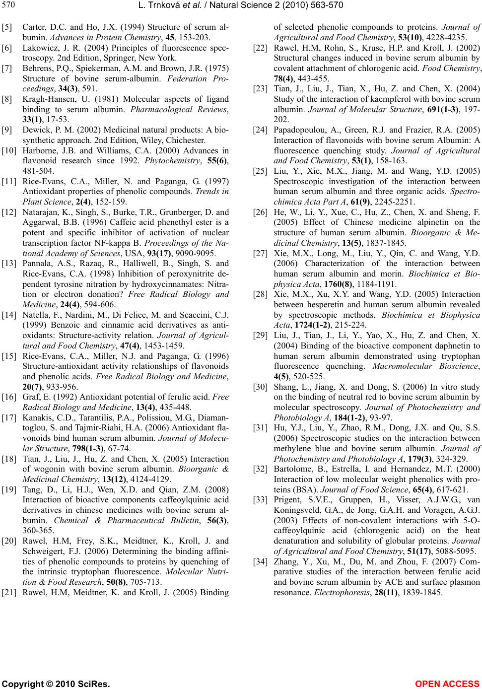 L. Trnková et al. / Natural Science 2 (2010) 563-570 Copyright © 2010 SciRes. OPEN ACCESS 570 [5] Carter, D.C. and Ho, J.X. (1994) Structure of serum al- bumin. Advances in Protein Chemistry, 45, 153-203. [6] Lakowicz, J. R. (2004) Principles of fluorescence spec- troscopy. 2nd Edition, Springer, New York. [7] Behrens, P.Q., Spiekerman, A.M. and Brown, J.R. (1975) Structure of bovine serum-albumin. Federation Pro- ceedings, 34(3), 591. [8] Kragh-Hansen, U. (1981) Molecular aspects of ligand binding to serum albumin. Pharmacological Reviews, 33(1), 17-53. [9] Dewick, P. M. (2002) Medicinal natural products: A bio- synthetic approach. 2nd Edition, Wiley, Chichester. [10] Harborne, J.B. and Williams, C.A. (2000) Advances in flavonoid research since 1992. Phytochemistry, 55(6), 481-504. [11] Rice-Evans, C.A., Miller, N. and Paganga, G. (1997) Antioxidant properties of phenolic compounds. Trends in Plant Science, 2(4), 152-159. [12] Natarajan, K., Singh, S., Burke, T.R., Grunberger, D. and Aggarwal, B.B. (1996) Caffeic acid phenethyl ester is a potent and specific inhibitor of activation of nuclear transcription factor NF-kappa B. Proceedings of the Na- tional Academy of Sciences, USA, 93(17), 9090-9095. [13] Pannala, A.S., Razaq, R., Halliwell, B., Singh, S. and Rice-Evans, C.A. (1998) Inhibition of peroxynitrite de- pendent tyrosine nitration by hydroxycinnamates: Nitra- tion or electron donation? Free Radical Biology and Medicine, 24(4), 594-606. [14] Natella, F., Nardini, M., Di Felice, M. and Scaccini, C.J. (1999) Benzoic and cinnamic acid derivatives as anti- oxidants: Structure-activity relation. Journal of Agricul- tural and Food Chemistry, 47(4), 1453-1459. [15] Rice-Evans, C.A., Miller, N.J. and Paganga, G. (1996) Structure-antioxidant activity relationships of flavonoids and phenolic acids. Free Radical Biology and Medicine, 20(7), 933-956. [16] Graf, E. (1992) Antioxidant potential of ferulic acid. Free Radical Biology and Medicine, 13(4) , 435-448. [17] Kanakis, C.D., Tarantilis, P.A., Polissiou, M.G., Diaman- toglou, S. and Tajmir-Riahi, H.A. (2006) Antioxidant fla- vonoids bind human serum albumin. Journal of Molecu- lar Structure, 798(1-3), 67-74. [18] Tian, J., Liu, J., Hu, Z. and Chen, X. (2005) Interaction of wogonin with bovine serum albumin. Bioorganic & Medicinal Chemistry, 13(12), 4124-4129. [19] Tang, D., Li, H.J., Wen, X.D. and Qian, Z.M. (2008) Interaction of bioactive components caffeoylquinic acid derivatives in chinese medicines with bovine serum al- bumin. Chemical & Pharmaceutical Bulletin, 56(3), 360-365. [20] Rawel, H.M, Frey, S.K., Meidtner, K., Kroll, J. and Schweigert, F.J. (2006) Determining the binding affini- ties of phenolic compounds to proteins by quenching of the intrinsic tryptophan fluorescence. Molecular Nutri- tion & Food Research, 50(8), 705-713. [21] Rawel, H.M, Meidtner, K. and Kroll, J. (2005) Binding of selected phenolic compounds to proteins. Journal of Agricultural and Food Chemistry, 53(10), 4228-4235. [22] Rawel, H.M, Rohn, S., Kruse, H.P. and Kroll, J. (2002) Structural changes induced in bovine serum albumin by covalent attachment of chlorogenic acid. Food Chemistry, 78(4), 443-455. [23] Tian, J., Liu, J., Tian, X., Hu, Z. and Chen, X. (2004) Study of the interaction of kaempferol with bovine serum albumin. Journal of Molecular Structure, 691(1-3), 197- 202. [24] Papadopoulou, A., Green, R.J. and Frazier, R.A. (2005) Interaction of flavonoids with bovine serum Albumin: A fluorescence quenching study. Journal of Agricultural and Food Chemistry, 53(1), 158-163. [25] Liu, Y., Xie, M.X., Jiang, M. and Wang, Y.D. (2005) Spectroscopic investigation of the interaction between human serum albumin and three organic acids. Spectro- chimica Acta Part A, 61(9), 2245-2251. [26] He, W., Li, Y., Xue, C., Hu, Z., Chen, X. and Sheng, F. (2005) Effect of Chinese medicine alpinetin on the structure of human serum albumin. Bioorganic & Me- dicinal Chemistry, 13(5), 1837-1845. [27] Xie, M.X., Long, M., Liu, Y., Qin, C. and Wang, Y.D. (2006) Characterization of the interaction between human serum albumin and morin. Biochimica et Bio- physica Acta, 1760(8), 1184-1191. [28] Xie, M.X., Xu, X.Y. and Wang, Y.D. (2005) Interaction between hesperetin and human serum albumin revealed by spectroscopic methods. Biochimica et Biophysica Acta, 1724(1-2), 215-224. [29] Liu, J., Tian, J., Li, Y., Yao, X., Hu, Z. and Chen, X. (2004) Binding of the bioactive component daphnetin to human serum albumin demonstrated using tryptophan fluorescence quenching. Macromolecular Bioscience, 4(5), 520-525. [30] Shang, L., Jiang, X. and Dong, S. (2006) In vitro study on the binding of neutral red to bovine serum albumin by molecular spectroscopy. Journal of Photochemistry and Photobiology A, 184(1-2), 93-97. [31] Hu, Y.J., Liu, Y., Zhao, R.M., Dong, J.X. and Qu, S.S. (2006) Spectroscopic studies on the interaction between methylene blue and bovine serum albumin. Journal of Photochemistry and Photobiology A, 179(3), 324-329. [32] Bartolome, B., Estrella, I. and Hernandez, M.T. (2000) Interaction of low molecular weight phenolics with pro- teins (BSA). Journal of Food Science, 65(4), 617-621. [33] Prigent, S.V.E., Gruppen, H., Visser, A.J.W.G., van Koningsveld, G.A., de Jong, G.A.H. and Voragen, A.G.J. (2003) Effects of non-covalent interactions with 5-O- caffeoylquinic acid (chlorogenic acid) on the heat denaturation and solubility of globular proteins. Journal of Agricultural and Food Chemistry, 51(17), 5088-5095. [34] Zhang, Y., Xu, M., Du, M. and Zhou, F. (2007) Com- parative studies of the interaction between ferulic acid and bovine serum albumin by ACE and surface plasmon resonance. Electrophoresis, 28(11), 1839-1845. |