

Paper Menu >>
Journal Menu >>
 Open Journal of Synthesis Theory and Applications, 2012, 1, 9-12 http://dx.doi.org/10.4236/ojsta.2012.12002 Published Online July 2012 (http://www.SciRP.org/journal/ojsta) Improved Solid-Phase Peptide Synthesis of Wild-Type and Phosphorylated Phospholamban Using a Pseudoproline Dipeptide Shadi Abu-Baker*, Gary A. Lorigan Department of Chemistry and Biochemistry, Miami University, Oxford, USA Email: *[email protected] Received April 2, 2012; revised May 17, 2012; accepted June 13, 2012 ABSTRACT In this study, we report that the insertion of a pseu doproline dipeptide for the solid-phase peptide synthesis of wild-type Phospholamban protein (WT-PLB) has two important advantages. First, it disrupts the formation of different secondary structures, which is responsible for poor couplings during the preparation of highly aggregated sequences. Second, it enhances the purities and solubility of crude products leading to easier HPLC purification. Keywords: Solid-State Peptide Synthesis; Pseudoproline Dipeptide; Phospholamban 1. Introduction Phospholamban (PLB) is a hydrophobic 52-amino acid transmembrane protein that is involved in regulating the contraction and relaxation of heart muscle [1-3]. Phos- phorylation of PLB by cyclic AMP- and calmodulin- dependent kinases is believed to increase the rate of cal- cium re-uptake by the sarcoplasmic reticulum and result in muscle relaxation [1-3]. The isolation and purification of large quantities of native PLB through molecular bi- ology techniques has not yet been achieved due to diffi- culties encountered in the bacterial over expression of phospholamban cDNA [4,5]. Alternatively, PLB has been prepared by chemical synthesis using standard solidphase peptide synthesis and purification in organic solvents [6, 7]. In addition, this approach gives the opportunity to synthesize site-specific isotopically labeled peptides and proteins [8-10]. The biochemical and biophysical com- parison of synthetic PLB and native PLB revealed that they are both similar in size and functionally identical [6,7]. 2. Materials and Methods 2.1. WT-PLB Synthesis and Purification PLB was synthesized using modified Fmoc-based solid- phase methods with an ABI 433A peptide synthesizer (Applied Biosystems, Foster city, CA). During our first run we found that the coupling of Leu-7 to Thr-8 was difficult even after double coupling and extending the reaction time to six hours. However, this problem was solved by using the pseudoproline dipeptide of Fmoc- Leu-Thr (Me,Me Pro)-OH from Novabiochem (San Die- go, CA). The use of a pseudoproline dipeptide of Fmoc- Leu-Thr (Me,Me Pro)-OH enhanced the yield to about 25 % after lyophylization. To synthesize P-PLB, a pre-phos- -phorylated Fmoc-serine amino acid was used at amino acid position 16 instead of the regular Fmoc-serine used in the synthesis of PLB. The crude peptide was purified on an Amersham Pharmacia Biotech AKTA explorer 10S HPLC controlled by Unicorn (version 3) system software. The purified protein was lyophilized and characterized by matrix-assisted laser desorption ionization time of flight (MALDI-TOF) mass spectrometry. 3. Results 3.1. Solid-Phase Peptide Synthesis of WT-PLB The chemically synthesized form of the full length PLB (Figure 1(a)) and P-PLB (Figure 1(b)) was used for all of the solid-state NMR experiments. In general, solid- phase peptide synthesis (SPPS) starts with the C-terminal amino acid attached to a solid support (resin). Amino acids are then coupled one at a time till the N-terminus is reached. Each time an amino acid is added, the following three steps are repeated: First, deprotection of the N- terminal amino acid of the peptide bound to the resin (removal of the Fomc protecting group, see the aromatic part in Figure 2). This step is followed by activation and coupling of the next amino acid. And finally, the new N-terminal amino acid is deprotected [11]. *Corresponding author. C opyright © 2012 SciRes. OJSTA 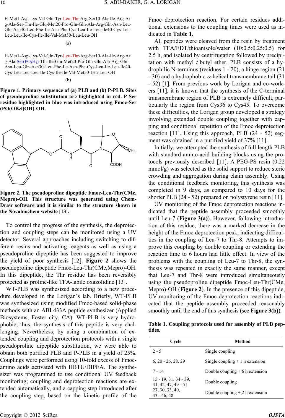 S. ABU-BAKER, G. A. LORIGAN 10 H-Met1-Asp-Lys-Val-Gln-Tyr- Arg-Ser10-Ala-Ile-Arg-Ar g-Ala-Ser-Thr-Ile-Glu-Met20-Pro-Gln-Gln-Ala-Arg-Gln-Asn-Leu- Gln-Asn30-Leu-Phe-Ile-Asn-Phe-Cys-Leu-Ile-Leu-Ile40-Cys-Leu- Leu-Leu-Ile-Cys-Ile-Ile-Val-Met50-Leu-Leu-OH (a) H-Met1-Asp-Lys-Val-Gln-Tyr- Arg-Ser10-Ala-Ile-Arg-Ar g-Ala-Ser(PO 3 H 2 )-Thr-Ile-Glu-Met20-Pro-Gln-Gln-Ala-Arg-Gln- Asn-Leu-Gln-Asn30-Leu-Phe-Ile-Asn-Phe-Cys-Leu-Ile-Leu-Ile40- Cys-Leu-Leu-Leu-Ile-Cys-Ile-Ile-Val-Met50-Leu-Leu-OH (b) Leu-Thr- Leu-Thr- Figure 1. Pr imary sequence of (a) PLB and (b) P-PLB. Site s of pseudoproline substitution are highlighted in red. P-Ser residue highlighted in blue was introduced using Fmoc-Ser (PO(OBzl)OH)-OH. O O N H O N O COOH CH3 Figure 2. The pseudoproline dipeptide F moc -Leu-Thr(CM e, Mepro)-OH. This structure was generated using Chem- Draw software and it is similar to the structure shown in the Novabiochem website [13]. To control the progress of the synthesis, the deprotec- tion and coupling steps can be monitored using a UV detector. Several approaches including switching to dif- ferent resins and activating reagents as well as using a pseudoproline dipeptide has been suggested to improve the yield of poor synthesis [12]. Figure 2 shows the pseudoproline dipeptide Fmoc-Leu-Thr(CMe,Mepro)-OH. In this dipeptide, the Thr residue has been reversibly protected as proline-like TFA-labile oxazolidine [13 ]. WT-PLB was synthesized according to a new proce- dure developed in the Lorigan’s lab. Briefly, WT-PLB was synthesized using modified Fmoc-based solid-phase methods with an ABI 433A peptide synthesizer (Applied Biosystems, Foster city, CA). WT-PLB is very hydro- phobic; thus, the synthesis of this peptide is very chal- lenging. Nevertheless, by using a combination of ex- tended coupling and deprotection protocols with a single pseudoproline dipeptide substitution, we were able to obtain both purified PLB and P-PLB in a yield of 25%. Couplings were performed using 10-fold excess of Fmoc- amino acids activated with HBTU/DIPEA. The synthe- sizer was programmed to use conditional UV feedback monitoring; coupling and deprotection reactions are ex- tended automatically, and a capping step introduced after the coupling step, based on the kinetic profile of the Fmoc deprotection reaction. For certain residues addi- tional extensions to the coupling times were used as in- dicated in Table 1. All peptides were cleaved from the resin by treatment with TFA/EDT/thioanisole/water (10:0.5:0.25:0.5) for 2.5 h, and isolated by centrifugation followed by precipi- tation with methyl t-butyl ether. PLB consists of a hy- drophilic N-terminus (residues 1 - 20), a hinge region (21 - 30) and a hydrophobic -helical transmembrane tail (31 - 52) [1]. From previous work by Lorigan and co-work- ers [11], it is known that the synthesis of the C-terminal transmembrane region of PLB is extremely difficult, par- ticularly the region from Cys36 to Cys45. To overcome these difficulties, the Lorigan gro up developed a strategy involving extended double coupling together with cap- ping and conditional repetition of the Fmoc deprotection reaction [11]. Using this approach, PLB (24 - 52) seg- ment was obtained in a purified yield of 37% [11]. Initially, we attempted the synthesis of full length PLB with standard amino-acid building blocks using the pro- tocols previously described [11]. A PEG-PS resin (0.22 mmol/g) was selected as the solid support to reduce steric crowding and aggregation during chain assembly. Using the conditional feedback monitoring, this synthesis was completed in 9 days, as compared to 10 days for the shorter PLB ( 2 4 - 52) prepare d o n p ol ystyrene resi n [ 1 1]. UV monitoring of the Fmoc deprotection reactions in- dicated that the peptide assembly proceeded smoothly until Leu-7 (Figure 3(a)). However, following introduc- tion of this residue, there was a marked decrease in the height of the Fmoc deprotection peak, indicating difficul- ties in the coupling of Leu-7 to Thr-8. Attempts to im- prove this coupling by double coupling or extending the reaction time to 6 hours had little effect. In view of the problems with the coupling of Leu-7 to Thr-8, the syn- thesis was repeated in exactly the same manner, except that Leu-7 and Thr-8 were introduced simultaneously using the pseudoproline dipeptide Fmoc-Leu-Thr(CMe, Mepro)-OH (Figure 2). In the presence of this dipeptide, UV monitoring of the Fmoc deprotection reactions indi- cated that the peptide assembly proceeded reasonably smoothly until the end of t his synthesis (see Figure 3(b)). Table 1. Coupling protocols used for assembly of PLB pep- tides. Cycle Method 2 - 5 Single coupling 6, 20 - 26, 28, 29 Single coupling + 1 h extension 7 - 14 Double coupling + 6 h ex tension 15 - 19, 31, 34 - 39, 41, 42, 47, 49 - 51 Double coupling 27, 30, 33, 40, 43 - 46, 48 Double coupling + 2 h extension Copyright © 2012 SciRes. OJSTA 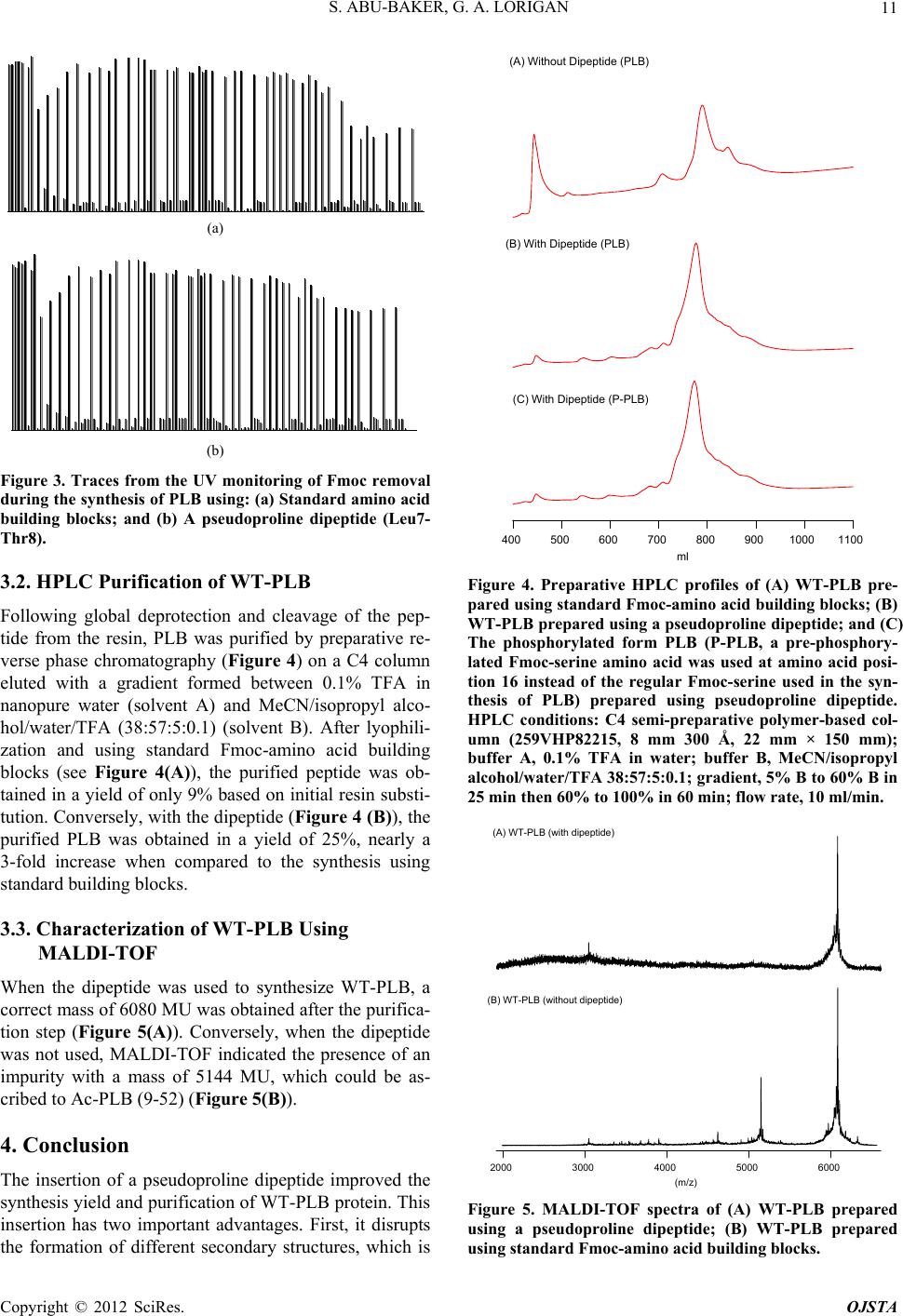 S. ABU-BAKER, G. A. LORIGAN 11 (a) (b) Figure 3. Traces from the UV monitoring of Fmoc removal during the synthesis of PLB using: (a) Standar d amino acid building blocks; and (b) A pseudoproline dipeptide (Leu7- Thr8). 3.2. HPLC Purification of WT-PLB Following global deprotection and cleavage of the pep- tide from the resin, PLB was purified by preparative re- verse phase chromatography (Figure 4) on a C4 column eluted with a gradient formed between 0.1% TFA in nanopure water (solvent A) and MeCN/isopropyl alco- hol/water/TFA (38:57:5:0.1) (solvent B). After lyophili- zation and using standard Fmoc-amino acid building blocks (see Figure 4(A)), the purified peptide was ob- tained in a yield of only 9% based on initial resin substi- tution. Conversely, with the dip eptide (Figure 4 (B)), the purified PLB was obtained in a yield of 25%, nearly a 3-fold increase when compared to the synthesis using standard building blocks. 3.3. Characterization of WT-PLB Using MALDI-TOF When the dipeptide was used to synthesize WT-PLB, a correct mass of 6080 MU was obtained after the purifica- tion step (Figure 5(A)). Conversely, when the dipeptide was not used, MALDI-TOF indicated the presence of an impurity with a mass of 5144 MU, which could be as- cribed to Ac-PLB (9-52) (Figure 5(B)). 4. Conclusion The insertion of a pseudoproline dipeptide improved the synthesis yield and purificatio n of WT-PLB protein. This insertion has two important advantages. First, it disrupts the formation of different secondary structures, which is 11001000900800700600500400 ml (A) Without Dipeptide (PLB) (B) With Dipeptide (PLB) (C) With Dipeptide (P-PLB) Figure 4. Preparative HPLC profiles of (A) WT-PLB pre- pared using standard Fmoc-amino acid building blocks; (B) WT-PLB prepared using a pseudoproline dipe ptide; and (C) The phosphorylated form PLB (P-PLB, a pre-phosphory- lated Fmoc-serine amino acid was used at amino acid posi- tion 16 instead of the regular Fmoc-serine used in the syn- thesis of PLB) prepared using pseudoproline dipeptide. HPLC conditions: C4 semi-preparative polymer-based col- umn (259VHP82215, 8 mm 300 Å, 22 mm × 150 mm); buffer A, 0.1% TFA in water; buffer B, MeCN/isopropyl alcohol/water/TFA 38:57:5:0.1; gradient, 5% B to 60% B in 25 min then 60% to 100% in 60 min; flow rate, 10 ml/min. 60005000400030002000 (m/z) (A) WT-PLB (with dipeptide ) (B) WT-PLB (without dipeptide) Figure 5. MALDI-TOF spectra of (A) WT-PLB prepared using a pseudoproline dipeptide; (B) WT-PLB prepared using standard Fmoc-amino acid building blocks. Copyright © 2012 SciRes. OJSTA  S. ABU-BAKER, G. A. LORIGAN Copyright © 2012 SciRes. OJSTA 12 responsible for poor couplings during the preparation of highly aggregated sequences. Second, it enhances the purities and solubility of crude products leadin g to easier HPLC purification. This technique can be used for simi- lar proteins that show poor synthesis. REFERENCES [1] H. K. B. Simmerman and L. R. Jones, “Phospholamban: Protein Structure, Mechanism of Action, and Role in Car- diac Function,” Physiological Reviews, Vol. 78, No. 4, 1998, pp. 921-947. [2] P. James, M. Inui, M. Tada, M. Chiesi and E. Carafoli, “Nature and Site of Phospholamban Regulation of the Calcium Pump of Sarcoplasmic Reticulum,” Nature, Vol. 342, 1989, pp. 90-92. doi:10.1038/342090a0 [3] M. A. Kirchberger, M. Tada and A. M. Katz, “Phos- pholamban: A Regulatory Protein of the Cardiac Sar- coplasmic Reticulum,” Recent Advances in Studies on Cardiac Structure and Metabolism, Vol. 5, 1975, pp. 103- 115. [4] J. Fuji, A. Ueno, K. Kitano, S. Tanaka, M. Kadoma and M. Tada, “Complete Complementary DNA-Derived Amino Acid Sequence of Canine Cardiac Phospholamban,” Jour- nal of Clinical Investigation, Vol. 79, No. 1, 1987, pp. 301-304. doi:10.1172/JCI112799 [5] Q. Yao, J. L. Bevan, R. F. Weaver and D. J. Bigelow, “Purification of Porcine Phospholamban Expressed in Escherichia Coli,” Protein Expression and Purification, Vol. 8, No. 4, 1996, pp. 463-468. [6] J. G. Collins, E. G. Kranias, A. S. Reeves, L. M. Bilezik- jian and A. Schwartz, “Isolation of Phospholamban and a Second Proteolipid Component from Canine Cardiac Sar- coplasmic Reticulum,” Biochemical and Biophysical Re- search Communications, Vol. 99, No. 3, 1981, pp. 796- 803. doi:10.1016/0006-291X(81)91235-3 [7] E. J. Mayer, E. McKenna, V. M. Garsky, C. J. Burke, H. Mach, C. R. Middaugh, M. Sardana, J. S. Smith and R.G. Johnson Jr., “Biochemical and Biophysical Comparison of Native and Chemically Synthesized Phospholamban and a Monomeric Phospholamban,” Journal of Biologyi- cal Chemistry, Vol. 271, 1996, pp. 1669-1677. doi:10.1074/jbc.271.3.1669 [8] D. J. Hirsh, J. Hammer, W. L. Maloy, J. Blazyk and J. Schaefer, “Secondary Structure and Location of a Ma- gainin Analogue in Synthetic Phospholipid Bilayers,” Biochemistry, Vol. 35, No. 39, 1996, pp. 12733-12741. doi:10.1021/bi961468a [9] A. Mascioni, C. Karim, J. Zamoon, D. D. Thomas and G. Veglia, “Solid-State NMR and Rigid Body Molecular Dynamics to Determine Domain Orientations of Mono- meric Phospholamban,” Journal of the American Chemi- cal Society, Vol. 124, No. 32, 2002, pp. 9392-9393. doi:10.1021/ja026507m [10] E. K. Tiburu, P. C. Dave, K. Damodaran and G. A. Lori- gan, “Investigating the Dynamic Properties of the Trans- membrane Segment of Phospholamban Incorporated into Phospholipid Bilayers Utilizing 2H and 15N Solid-State NMR Spectroscopy,” Biochemistry, Vol. 43, No. 44, 2004, pp. 13899-13909. doi:10.1021/bi0490993 [11] E. K. Tiburu, P. C. Dave, J. F. Vanlerberghe, T. B. Car- don, R. E. Minto and G. A. Lorigan, “An Improved Syn- thetic and Purification Procedure for the Hydrophobic Segment of the Transmembrane Peptide Phospholam- ban,” Analytical Biochemistry, Vol. 318, No. 1, 2003, pp. 146-151. doi:10.1016/S0003-2697(03)00141-6 |