

Paper Menu >>
Journal Menu >>
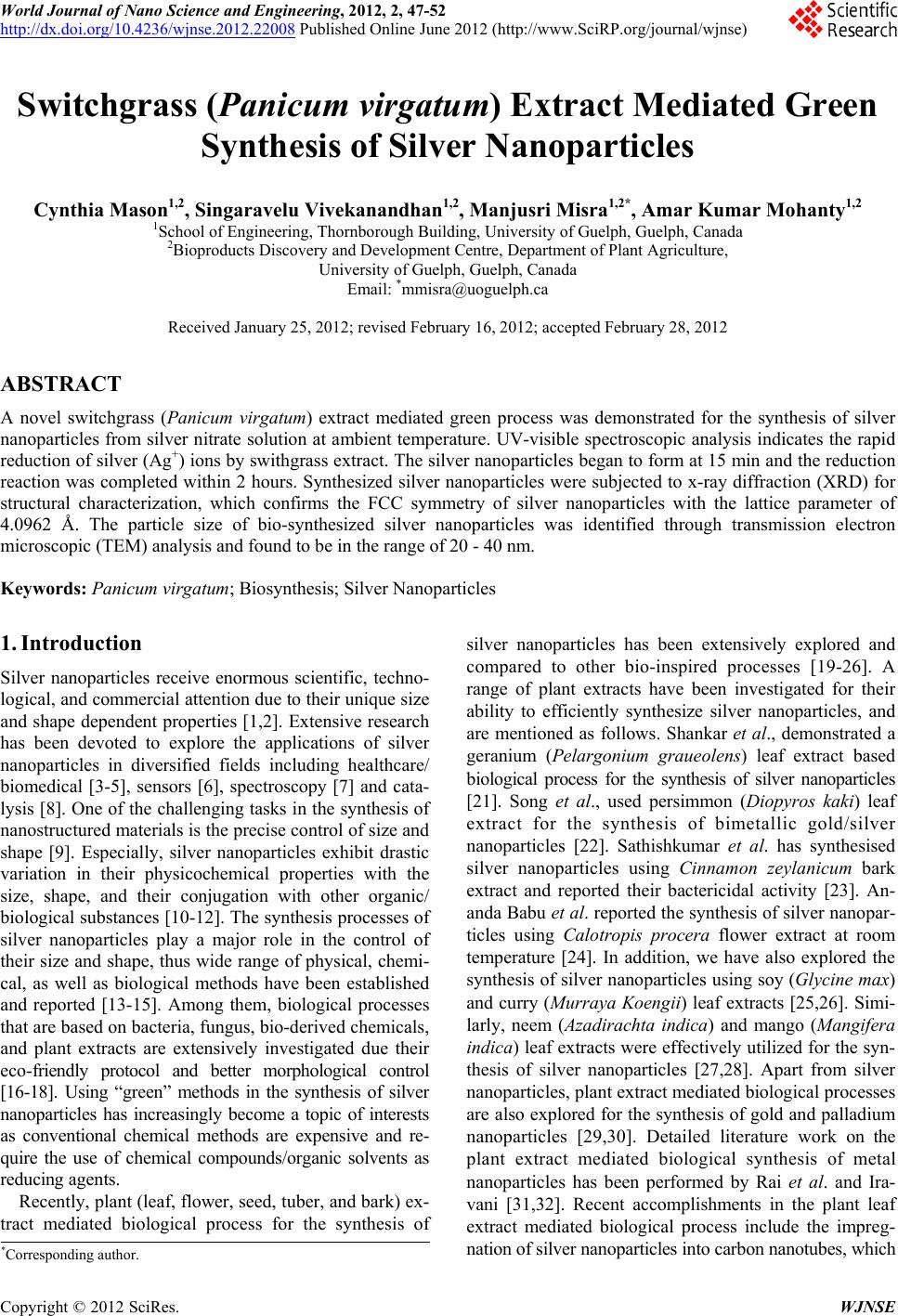 World Journal of Nano Science and Engineering, 2012, 2, 47-52 http://dx.doi.org/10.4236/wjnse.2012.22008 Published Online June 2012 (http://www.SciRP.org/journal/wjnse) Switchgrass (Panicum virgatum) Extract Mediated Green Synthesis of Silver Nanoparticles Cynthia Mason1,2, Singaravelu Vivekanandhan1,2, Manjusri Misra1,2*, Amar Kumar Mohanty1,2 1School of Engineering, Thornborough Building, University of Guelph, Guelph, Canada 2Bioproducts Discovery and Development Centre, Department of Pla nt Agricultu re, University of Guelph, Guelph, Canada Email: *[email protected] Received January 25, 2012; revised February 16, 2012; accepted February 28, 2012 ABSTRACT A novel switchgrass (Panicum virgatum) extract mediated green process was demonstrated for the synthesis of silver nanoparticles from silver nitrate solution at ambient temperature. UV-visible spectroscopic analysis indicates the rapid reduction of silver (Ag+) ions by swithgrass extract. The silver nanoparticles began to form at 15 min and the reduction reaction was completed within 2 hours. Synthesized silver nanoparticles were subjected to x-ray diffraction (XRD) for structural characterization, which confirms the FCC symmetry of silver nanoparticles with the lattice parameter of 4.0962 Å. The particle size of bio-synthesized silver nanoparticles was identified through transmission electron microscopic (TEM) analysis and found to be in the range of 20 - 40 nm. Keywords: Panicum virgatum; Biosynthesis; Silver Nanoparticles 1. Introduction Silver nanoparticles receive enormous scientific, techno- logical, and commercial attention due to their unique size and shape dependent properties [1,2]. Extensive research has been devoted to explore the applications of silver nanoparticles in diversified fields including healthcare/ biomedical [3-5], sensors [6], spectroscopy [7] and cata- lysis [8]. One of the challengin g tasks in the synthesis of nanostructured materials is the precise control of size and shape [9]. Especially, silver nanoparticles exhibit drastic variation in their physicochemical properties with the size, shape, and their conjugation with other organic/ biological substances [10-12]. The synthesis processes of silver nanoparticles play a major role in the control of their size and shape, thus wide range of physical, chemi- cal, as well as biological methods have been established and reported [13-15]. Among them, biological processes that are based on bacteria, fungus, bio-derived chemicals, and plant extracts are extensively investigated due their eco-friendly protocol and better morphological control [16-18]. Using “green” methods in the synthesis of silver nanoparticles has increasingly become a topic of interests as conventional chemical methods are expensive and re- quire the use of chemical compounds/organic solvents as reducing agents. Recently, plant (leaf, flower, seed, tuber, and bark) ex- tract mediated biological process for the synthesis of silver nanoparticles has been extensively explored and compared to other bio-inspired processes [19-26]. A range of plant extracts have been investigated for their ability to efficiently synthesize silver nanoparticles, and are mentioned as follows. Shankar et al., demonstrated a geranium (Pelargonium graueolens) leaf extract based biological process for the synthesis of silver nanoparticles [21]. Song et al., used persimmon (Diopyros kaki) leaf extract for the synthesis of bimetallic gold/silver nanoparticles [22]. Sathishkumar et al. has synthesised silver nanoparticles using Cinnamon zeylanicum bark extract and reported their bactericidal activity [23]. An- anda Babu et al. reported the synthesis of silver nanopar- ticles using Calotropis procera flower extract at room temperature [24]. In addition, we have also explored the synthesis of silver nanoparticles using soy (Glycine max) and curry (Murraya Koengii) leaf extracts [25,26]. Simi- larly, neem (Azadirachta indica) and mango (Mangifera indica) leaf extracts were effectively utilized for the syn- thesis of silver nanoparticles [27,28]. Apart from silver nanoparticles, plant ex tract mediated biological processes are also explored for the synthesis of gold and palladium nanoparticles [29,30]. Detailed literature work on the plant extract mediated biological synthesis of metal nanoparticles has been performed by Rai et al. and Ira- vani [31,32]. Recent accomplishments in the plant leaf extract mediated biological process include the impreg- nation of silver nanopartic l e s into carbon nanotubes, which *Corresponding author. C opyright © 2012 SciRes. WJNSE 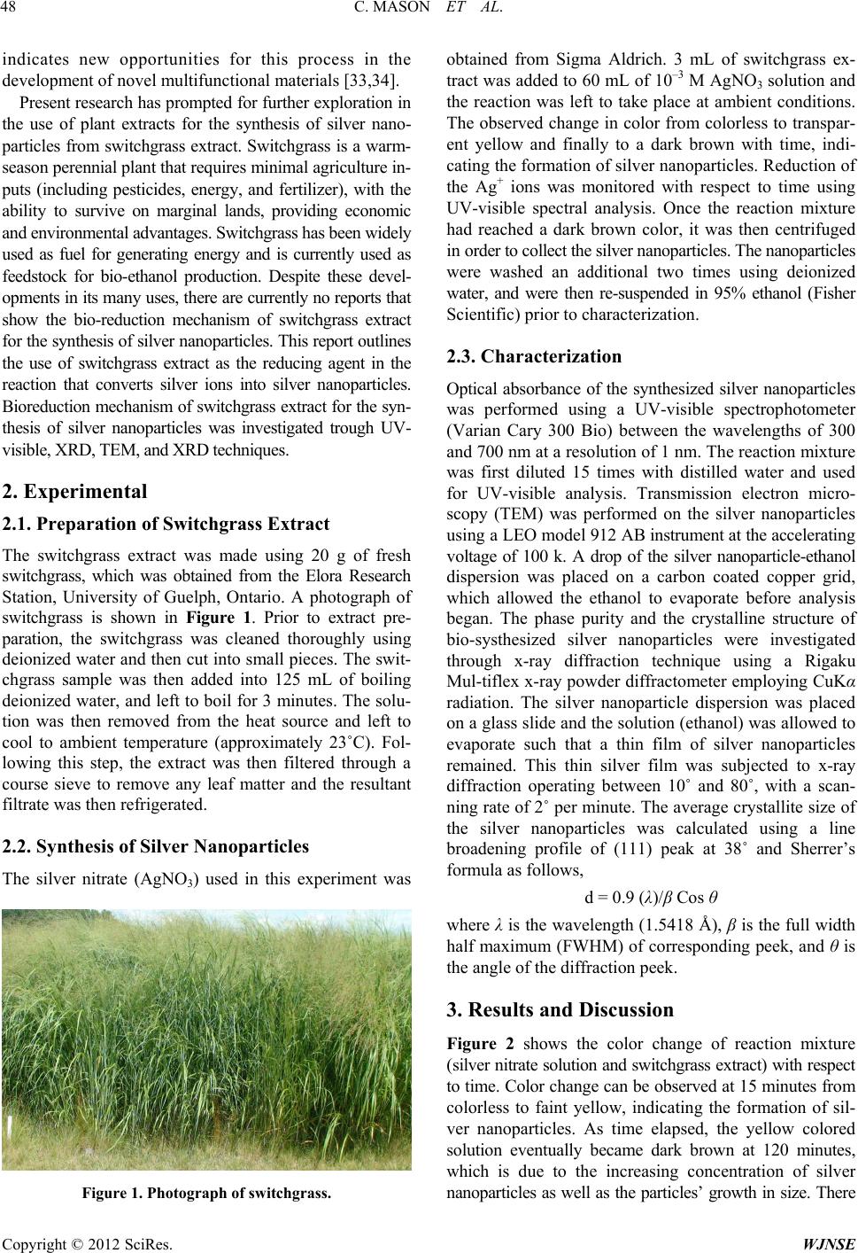 C. MASON ET AL. 48 indicates new opportunities for this process in the development of novel multifunctional materials [33,34]. Present research has prompted for further exploration in the use of plant extracts for the synthesis of silver nano- particles from switchgrass extract. Switchgrass is a war m- season perennial plant that requires minimal agriculture in- puts (including pesticides, energy, and fertilizer), with the ability to survive on marginal lands, providing economic and envi r onm ent al a dv ant a ges. S witc h gr ass ha s be e n wi del y used as fuel for generating energy and is currently used as feedstock for bio-ethanol production. Despite these devel- opments in its ma ny u ses, th ere are currently no r eports that show the bio-reduction mechanism of switchgrass extract for the synthesis of silver nanoparticles. This report outlines the use of switchgrass extract as the reducing agent in the reaction that converts silver ions into silver nanoparticles. Bioreduction mechanism of switchgrass extract for the syn- thesis of silver nanoparticles was investigated trough UV- visible, XRD, TEM, and XRD techniques. 2. Experimental 2.1. Preparation of Switchgrass Extract The switchgrass extract was made using 20 g of fresh switchgrass, which was obtained from the Elora Research Station, University of Guelph, Ontario. A photograph of switchgrass is shown in Figure 1. Prior to extract pre- paration, the switchgrass was cleaned thoroughly using deionized water and then cut into small pieces. The swit- chgrass sample was then added into 125 mL of boiling deionized water, and left to boil for 3 minutes. The so lu- tion was then removed from the heat source and left to cool to ambient temperature (approximately 23˚C). Fol- lowing this step, the extract was then filtered through a course sieve to remove any leaf matter and the resultant filtrate was then refrigerated. 2.2. Synthesis of Silver Nanoparticles The silver nitrate (AgNO3) used in this experiment was Figure 1. Photograph of switchgrass. obtained from Sigma Aldrich. 3 mL of switchgrass ex- tract was added to 60 mL of 10–3 M AgNO3 solution and the reaction was left to take place at ambient conditions. The observed change in color from colorless to transpar- ent yellow and finally to a dark brown with time, indi- cating the formation of silver nanoparticles. Reduction of the Ag+ ions was monitored with respect to time using UV-visible spectral analysis. Once the reaction mixture had reached a dark brown color, it was then centrifuged in order to collect the silver nanoparticles. The nanoparticles were washed an additional two times using deionized water, and were then re-suspended in 95% ethanol (Fisher Scientific) prior to characterization. 2.3. Characterization Optical absorbance of the synthesized silver nanoparticles was performed using a UV-visible spectrophotometer (Varian Cary 300 Bio) between the wavelengths of 300 and 700 nm at a resolution of 1 nm. The reaction mixture was first diluted 15 times with distilled water and used for UV-visible analysis. Transmission electron micro- scopy (TEM) was performed on the silver nanoparticles us in g a LE O mo d el 912 AB instrument at the acceleratin g voltage of 100 k. A drop of the silver nanoparticle-ethanol dispersion was placed on a carbon coated copper grid, which allowed the ethanol to evaporate before analysis began. The phase purity and the crystalline structure of bio-systhesized silver nanoparticles were investigated through x-ray diffraction technique using a Rigaku Mul-tiflex x-ray powder diffractometer employing CuKα radiation. The silver nanoparticle dispersion was placed on a glass slide and the solution (ethanol) was allowed to evaporate such that a thin film of silver nanoparticles remained. This thin silver film was subjected to x-ray diffraction operating between 10˚ and 80˚, with a scan- ning rate of 2˚ per minute. The average crystallite size of the silver nanoparticles was calculated using a line broadening profile of (111) peak at 38˚ and Sherrer’s formula as follows, d = 0.9 (λ)/β Cos θ where λ is the wavelength (1.5418 Å), β is the full width half maximum (FWHM) of corresponding peek, and θ is the angle of the diffra ction pe ek . 3. Results and Discussion Figure 2 shows the color change of reaction mixture ( silver n itrate solu tion and switchg rass extr act) with respect to time. Color change can be observed at 15 minutes from colorless to faint yellow, indicating the formation of sil- ver nanoparticles. As time elapsed, the yellow colored solution eventually became dark brown at 120 minutes, which is due to the increasing concentration of silver nanoparticles as well as the particles’ growth in size. Ther e Copyright © 2012 SciRes. WJNSE  C. MASON ET AL. 49 0 15 30 60 75 90 120 180 (In minutes) Figure 2. Photograph of reaction mixture (switchgrass extract and silver nitrate solution) as a function of time. is no significant change beyond 180 minutes, therefore indicating the completion of the reduction reaction. This was further confirmed by UV-vis spectroscopic analysis. This physical appearance of the reaction mixture turning from yellow to brown is due to the surface plasmon reso- nance (SPR) of the silver nanoparticles, which is consid- ered to be the primary signature of nanoparticle formation. UV-vis spectroscopy is a versatile technique to understand the bioreduction mechanism of silver ions into silver nanoparticles by switchgrass extract. The UV-vis spectra of the reaction mixture recorded as a function of time, is are shown in Figure 3(a). An observed peak at 435 nm is assigned to the surface plasmon resonance band (longitu- dinal vibration) of the silver nanoparticles, which is com- parable with the literature values and exhibits continuous rise in intensity without any ch ange in the peak position as a function of time. During 15 - 60 minutes intervals the absorption peak was weak and broad, which indicates the smaller size of silver nanoparticles. Nucleation occurs between 60 - 90 minutes, which appeared as strong a ab- sorption peak, as shown in Figure 3(a). Figure 3(b) shows the absorbance at λmax (i.e. at 435 nm) as a function of time. From Figure 3(b) it is indentified that the reduction of silver ions to silver nanoparticles occurs quite rapidly, as more than 90% of the bioreduction reaction completes within 90 minutes. Th is is faster than earlier stud ies of the synthesis of silver nanoparticles usi ng biol ogical sources. The crystalline structure of the bio-syntheized silver nanoparticles was investigated by XRD analysis and the obtained x-ray diffraction pattern is shown in Figure 4. The obtained diffraction peaks at 38˚, 44˚, 64˚ and 77˚ are respectively assigned to (111), (200), (311) and (222) plans, which indicates that the synthesized silver nanop ar- ticles are crystallized in face centered cubic (fcc) sym- metry. No additional diffraction peaks were observed other than the characteristic peak of the silver structure that reflects the purity of synthesized silver nanoparticles. The lattice parameter (A) of the bio-synthesized silver nanoparticles was calculated from the diffraction data and was found to be A = 4.0962 Å, which is comparab le with the JCPDS value. Th e calculated crystallite size has been found to be ~10 nm, which is comparable with the particle size as obtained from TEM analysis. (a) (b) Figure 3. (a) UV-vis absorption spectra of reaction mixture (switchgrass and silver nitrate) and (b) Absorbance at λmax (i.e., at 435 nm) as a function of time. Figure 4. XRD pattern of silver nanoparticles synthesized using switchgrass extract. The microscopic structure of the switchgrass extract mediated bio-synthesized silver nanoparticles was inves- tigated by TEM analysis. The obtained TEM images are shown in Figure 5. TEM images confirm the formation of silver nanoparticles with the size range between 20 and 40 nm. In addition, the TEM images show th e shape of the nanoparticles are highly diversified, which includes Copyright © 2012 SciRes. WJNSE  C. MASON ET AL. Copyright © 2012 SciRes. WJNSE 50 (a) (b) (c) (d) Figure 5. (a)-(d) TEM images of silver nanoparticles synthesized using switchgrass extract. 5. Acknowledgements spherical, rod-like, triangular, pentagonal, and hexagonal. It was found that the synthesized silver nanoparticles have the tendency to aggregate and form the agglomerations. The authors would lik e to acknowledge the 2009, Ontario Ministry of Agriculture, Food and Rural Affairs (OMA- FRA)—New Directions & Alternative Renewable Fuels Research Program Project number SR9225 and NSERC NCE AUTO21 project for the financial support to carry out this research. The agglomerated silver nanoparticles assemble to- gether without much physical contact. Thus they can be separated by physical agitation for the further utilization. 4. Conclusion Silver nanoparticles were successfully synthesized using switchgrass extract used at room temperature. Synthesis of silver nanoparticles through this process was fairly rapid, with 90% of silver ion reduction completed within 90 minutes. TEM analysis confirms that the synthesized silver nanoparticles exist between 20 and 40 nm and ex- hibit the tendency to aggregate. XRD analysis indicates the formation of phase pure silver nanoparticles with FCC symmetry. The calculated av erage crystallite size is found to be of 10 nm, which is consistent with TEM analysis. The fundamental understanding of switchgrass mediated biological process for the synthesis of silver nanoparticles will allow the expansion of this p rocess for the synthesis of gold and palladium nanoparticles and their applications. REFERENCES [1] A. Kumar, P. K. Vemula, P. M. Ajayan and G. John, “Sil- ver Nanoparticle Embedded Antimicrobial Paint s Ba s e d o n Vegetable Oil,” Nature Materials, Vol. 7, 2008, pp. 236-241. doi:10.1038/nmat2099 [2] L. S. Nair and C. T. Laurencin, “Silver Nanoparticles: Synthesis and Therapeutic Applications,” Journal of Bio- medical Nanotechnology, Vol. 3, No. 4, 2007, pp. 301-316. doi:10.1166/jbn.2007.041 [3] J. S. Kim, E. Kuk, K. N. Yu, J. H. Kim, S. J. Park, H. J. Lee, S. H. Kim, Y. K. Park, Y. H. Park, C. Y. Hwang, Y. K. Kim, Y. S. Lee, D. H. Jeong and M. H. Cho, “Antim- icrobial Effects of Silver Nanoparticles,” Nanomedicine: Nanotechnology, Biology and Medicine, Vol. 3, No. 1, 2007, pp. 95-101. doi:10.1016/j.nano.2006.12.001  C. MASON ET AL. 51 [4] J. R. Morones, J. L. Elechiguerra, A. Camacho, K. Holt, J. B. Kouri, J. T. Ramírez and M. J. Yacaman, “The Bacteri- cidal Effect of Silver Nanoparticles,” Nanotechnology, Vol. 16, No. 10, 2005, pp. 2346-2353. doi:10.1088/0957-4484/16/10/059 [5] C. M. Jones and E. M. V. Hoek, “A Review of the Anti- bacterial Effects of Silver Nanomaterials and Potential Implications for Human Health and the Environment,” Journal of Nanoparticle Research, Vol. 12, No. 5, 2010, pp. 1531-1551. doi:10.1007/s11051-010-9900-y [6] C. J. Murphy, A. M. Gole, S. E. Huny adi, J. W. Stone, P. N. Sisco, A. Alkilany, B. E. Kinard and P. Hankins, “Chemical Sensing and Imaging with Metallic Nanorods,” Chemical Communications, No. 5, 2008, pp. 544-557. doi:10.1039/b711069c [7] A. J. Haes, C. L. Hay nes, A. D. McFarland, G. C. Schatz, R. P. Van Duyne and S. Zou, “Plasmonic Materials for Surface-Enhanced Sensing and Spectroscopy,” MRS Bulle- tin, Vol. 30, No. 5, 2005, pp. 368-375. doi:10.1557/mrs2005.100 [8] Z. J. Jiang, C. Y. Liu and L. W. Sun, “Catalytic Properties of Silver Nanoparticles Supported on Silica Spheres,” The Journal of Physical Chemistry B, Vol. 109, No. 5, 2005, pp. 1730-1735. doi:10.1021/jp046032g [9] K. L. Kelly, E. Coronado, L. L. Zhao and G. C. Schatz, “The Optical Properties of Metal Nanoparticles: The In- fluence of Size, Shape and Dielectric Environment,” The Journal of Physical Chemistry B, Vol. 107, No. 5, 2003, pp. 668-677. doi:10.1021/jp026731y [10] A. G. Tkachenko, H. Xie, D. Coleman, W. Glomm, J. Ryan, M. F. Anderson, S. Franzen and D. L. Feldheim, “Multifunctional Gold Nanoparticle-Peptide Complexes for Nuclear Targeting,” Journal of the American Chemical Society, Vol. 125, No. 16, 2003, pp. 4700-4701. doi:10.1021/ja0296935 [11] M. Chen, Y. G. Feng, X. Wang, T. C. Li, J. Y. Zhang and D. J. Qian, “Silver Nanoparticles Capped by Oleylamine: Formation, Growth and Self-Organization,” Langmuir, Vol. 23, No. 10, 2007, pp. 5296-5304. doi:10.1021/la700553d [12] C. Aymonier, U. Schlotterbeck, L. Antonietti, P. Zacharias, R. Thomann, J. C. Tiller and S. Mecking, “Hybrids of Sil- ver Nanoparticles with Amphiphilic Hyperbranched Mac- romolecules Exhibiting Antimicrobial Properties,” Chemical Communications, No. 24, 2002, pp. 3018-3019. doi.:10.1039/b208575e [13] J. P. Abid, A. W. Wark, P. F. Brevet and H. H. Girault, “Preparation of Silver Nanoparticles in Solution from a Silver Salt by Laser Irradiation,” Chemical Communica- tions, No. 7, 2002, pp. 792-793. doi:10.1039/b200272h [14] R. Das, S. S. Nath, D. Chakdar, G. Gope and R. Bhat- tacharjee, “Synthesis of Silver Nanoparticles and Their Optical Properties,” Journal of Experimental Nanoscience, Vol. 5, No.4, 2010, pp 357-362. doi:10.1080/17458080903583915 [15] M. Gericke and A. Pinches, “Biological Synthesis of Metal Nanoparticles,” Hydrometallurgy, Vol. 83, No. 1-4, 2006, pp. 132-140. doi:10.1016/j.hydromet.2006.03.019 [16] K. B. Narayanan and N. Sakthivel, “Biological Synthesis of Metal Nanoparticles by Microbes,” Advances in Col- loid and Interface Science, Vol. 156, No. 1-2, 2010, pp. 1-13. doi:10.1016/j.cis.2010.02.001 [17] M. Sastry, A. Ahmad, M. Islam Khan and R. Kumar, “Biosynthesis of Metal Nanoparticles Using Fungi and Actinomycete,” Current Science, Vol. 85, No. 2, 2003, pp. 162-170. [18] S. Kaviya, J. Santhanalakshmi, B. Viswanathan, J. Mu- thumary and K. Srinivasan, “Biosynthesis of Silver Nanopar- ticles Using Citrus Sinensis Peel Extract and Its Antibac- terial Activity,” Spectrochimica Acta Part A: Molecular and Biomolecular Spectroscopy, Vol. 79, No. 3, 2011, pp. 594-598. doi:10.1016/j.saa.2011.03.040 [19] D. Philip, “Green Synthesis of Gold and Silver Nanoparti- cles Using Hibiscus Rosa Sinensis,” Physica E, Vol. 42, No. 5, 2010, pp. 1417-1424. doi:10.1016/j.physe.2009.11.081 [20] V. Kumar, S. C. Yadav and S. K. Yadav, “Syzygium Cumini Leaf and Seed Extract Mediated Biosynthesis of Silver Nanoparticles and Their Characterization,” Journal of Chemical Technology & Biotechnology, Vol. 85, No. 10, 2010, pp. 1301-1309. doi:10.1002/jctb.2427 [21] S. S. Shankar, A. Ahmad and M. Sastry, “Geranium Leaf Assisted Biosynthesis of Silver Nanoparticles,” Biotech- nology Progress, Vol. 19, No. 6, 2003, pp. 1627-1631. doi:10.1021/bp034070w [22] J. Y. Song and B. S. Kim, “Biological Synthesis of Bi- metallic Au/Ag Nanoparticles Using Persimmon (Diopy- ros Kaki) Leaf Extract,” Korean Journal of Chemical En- gineering, Vol. 25, No. 4, 2008, pp. 808-811. doi:10.1007/s11814-008-0133-z [23] M. Sathishkumar, K. Sneha, S. W. Won, C. W. Cho, S. Kim and Y. S. Yun, “Cinnamon Zeylanicum Bark Extract and Powder Mediated Green Synthesi s of Nano-Cry stalline Silver Particles and Its Bactericidal Activity,” Colloids and Surfaces B: Biointerfaces, Vol. 73, No. 2, 2009, pp. 332-338. doi:10.1016/j.colsurfb.2009.06.005 [24] S. A. Babu and H. G. Prabu, “Synthesis of AgNPs Using the Extract of Calotropis Procera Flower at Room Tem- perature,” Materials Letters, Vol. 65, No. 11, 2011, pp. 1675-1677. doi:10.1016/j.matlet.2011.02.071 [25] S. Vivekanandhan, M. Misra and A. K. Mohanty, “Biologi- cal Synthesis of Silver Nanoparticles Using Glycine Max (Soybean) Leaf Extract: An Investigation on Different Soybean Varieties,” Journal of Nanoscience and Nanotech- nology, Vol. 9, No. 12, 2009, pp. 6828-6833. doi:10.1166/jnn.2009.2201 [26] L. Christensen, S. Vivekanandhan, M. Misra and A. K. Mohanty, “Biosynthesis of Silver Nanoparticles Using Murraya Koenigii Leaf: An Investigation on the Effect of Broth Concentration in Reduction Mechanism and Parti- cle Size,” Advanced Materials Letters, Vol. 2, No. 3, 2011, pp. 163-167. doi:10.5185/amlett.2011.4256 [27] S. S. Shankar, A. Rai, A. Ahmad and M. Sastry, “Rapid Synthesis of Au, Ag and Bimetallic Au Core—Ag Shell Nanoparticles Using Neem (Azadirachta indica) Leaf Broth,” Journal of Colloid and Interface Science, Vol. 275, No. 2, 2004, pp. 496-502. Copyright © 2012 SciRes. WJNSE  C. MASON ET AL. Copyright © 2012 SciRes. WJNSE 52 doi:10.1016/j.jcis.2004.03.003 [28] D. Philip, “Mangifera Indica Leaf-Assisted Biosynthesis of Well-Dispersed Silver Nanoparticles,” Spectrochimica Acta Part A: Molecular and Biomolecular Spectroscopy, Vol. 78, No. 1, 2011, pp. 327-331. doi:10.1016/j.saa.2010.10.015 [29] K. B. Narayanan and N. Sakthivel, “Phytosynthesis of Gold Nanoparticles Using Leaf Extract of Coleus Ambo- inicus Lour,” Materials Characterization, Vol. 61, No. 11, 2010, pp. 1232-1238. doi:10.1016/j.matchar.2010.08.003 [30] P. R. Kumar, S. Vivekanandhan, M. Misra, A. K. Mo- hanty and N. Satyanarayana, “Soybean (Glycine max) Leaf Extract Based Green Synthesis of Palladium Nanopar- ticles,” Journal of Biomaterials and Nanobiotechnology, Vol. 3, No. 1, 2012, pp. 14-19. doi:10.4236/jbnb.2011 [31] M. Rai, A. Yadav and A. Cade, “Current Trends in Phy- tosynthesis of Metal Nanoparticles,” Critical Reviews in Biotechnology, Vol. 28, No. 4, 2008, pp. 277-284. doi:10.1080/07388550802368903 [32] S. Iravani, “Green Synthesis of Metal Nanoparticles Using Plants,” Green Chemistry, Vol. 13, No. 10, 2011, pp. 2638-2650. doi:10.1039/c1gc15386b [33] S. Vivekanandhan, D. Tang, M. Misra and A. K. Mohanty, “Novel Glycine Max (Soybean) Leaf Extract Based Bio- logical Process for the Functionalization of Carbon Nano- tubes with Silver Nanoparticles,” Nanoscience and Nano- technology Letters, Vol. 2, No. 3, 2010, pp. 240-243. doi:10.1166/nnl.2010.1087 [34] S. Vivekanandhan, M. Venkateswarlu, D. Carnahan, M. Misra, A. K. Mohanty and N. Satyanarayana, “Function- alization of Single-Walled Carbon Nanotubes with Silver Nanoparticles Using Tecomastans Leaf Extract,” Physica E, Vol. 44, No. 7-8, 2012, pp. 1725-1729. doi:10.1016/j.physe.2011.10.013 |