

Paper Menu >>
Journal Menu >>
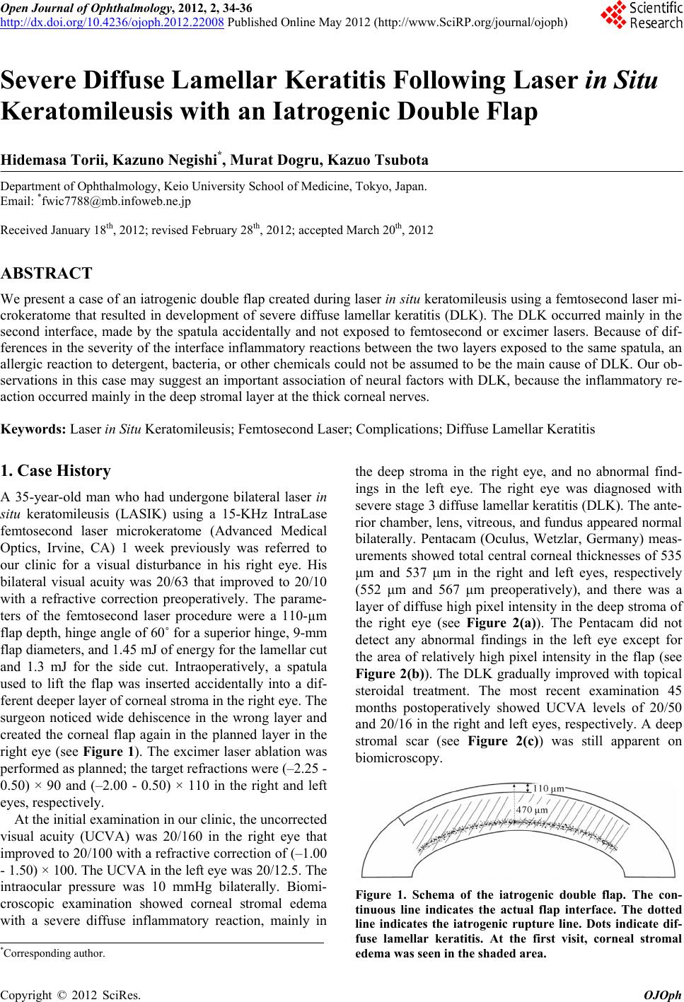 Open Journal of Ophthalmology, 2012, 2, 34-36 http://dx.doi.org/10.4236/ojoph.2012.22008 Published Online May 2012 (http://www.SciRP.org/journal/ojoph) Severe Diffuse Lamellar Keratitis Following Laser in Situ Keratomileusis with an Iatrogenic Double Flap Hidemasa Torii, Kazuno Negishi*, Murat Dogru, Kazuo Tsubota Department of Ophthalmology, Keio University School of Medicine, Tokyo, Japan. Email: *[email protected] Received January 18th, 2012; revised February 28th, 2012; accepted March 20th, 2012 ABSTRACT We present a case of an iatrogenic double flap created during laser in situ keratomileusis using a femtosecond laser mi- crokeratome that resulted in development of severe diffuse lamellar keratitis (DLK). The DLK occurred mainly in the second interface, made by the spatula accidentally and not exposed to femtosecond or excimer lasers. Because of dif- ferences in the severity of the interface inflammatory reactions between the two layers exposed to the same spatula, an allergic reaction to detergent, bacteria, or other chemicals could not be assumed to be the main cause of DLK. Our ob- servations in this case may suggest an important association of neural factors with DLK, because the inflammatory re- action occurred mainly in the deep stromal layer at the thick corneal nerves. Keywords: Laser in Situ Keratomileusis; Femtosecond Laser; Complications; Diffuse Lamellar Keratitis 1. Case History A 35-year-old man who had undergone bilateral laser in situ keratomileusis (LASIK) using a 15-KHz IntraLase femtosecond laser microkeratome (Advanced Medical Optics, Irvine, CA) 1 week previously was referred to our clinic for a visual disturbance in his right eye. His bilateral visual acuity was 20/63 that improved to 20/10 with a refractive correction preoperatively. The parame- ters of the femtosecond laser procedure were a 110-µm flap depth, hinge angle of 60˚ for a superior hinge, 9-mm flap diameters, and 1.45 mJ of energy for the lamellar cut and 1.3 mJ for the side cut. Intraoperatively, a spatula used to lift the flap was inserted accidentally into a dif- ferent deeper layer of corneal stroma in the right eye. The surgeon noticed wide dehiscence in the wrong layer and created the corneal flap again in the planned layer in the right eye (see Figure 1). The excimer laser ablation was performed as planned; the target refractions were (–2.25 - 0.50) × 90 and (–2.00 - 0.50) × 110 in the right and left eyes, respectively. At the initial examination in our clinic, the uncorrected visual acuity (UCVA) was 20/160 in the right eye that improved to 20/100 with a refractive correction of (–1.00 - 1.50) × 100. The UCVA in the left eye was 20/12.5. The intraocular pressure was 10 mmHg bilaterally. Biomi- croscopic examination showed corneal stromal edema with a severe diffuse inflammatory reaction, mainly in the deep stroma in the right eye, and no abnormal find- ings in the left eye. The right eye was diagnosed with severe stage 3 diffuse lamellar keratitis (DLK). The ante- rior chamber, lens, vitreous, and fundus appeared normal bilaterally. Pentacam (Oculus, Wetzlar, Germany) meas- urements showed total central corneal thicknesses of 535 μm and 537 μm in the right and left eyes, respectively (552 μm and 567 μm preoperatively), and there was a layer of diffuse high pixel intensity in the deep stroma of the right eye (see Figure 2(a)). The Pentacam did not detect any abnormal findings in the left eye except for the area of relatively high pixel intensity in the flap (see Figure 2(b)). The DLK gradually improved with topical steroidal treatment. The most recent examination 45 months postoperatively showed UCVA levels of 20/50 and 20/16 in the right and left eyes, respectively. A deep stromal scar (see Figure 2(c)) was still apparent on biomicroscopy. Figure 1. Schema of the iatrogenic double flap. The con- tinuous line indicates the actual flap interface. The dotted line indicates the iatrogenic rupture line. Dots indicate dif- fuse lamellar keratitis. At the first visit, corneal stromal edema was seen in the shaded area. *Corresponding author. Copyright © 2012 SciRes. OJOph 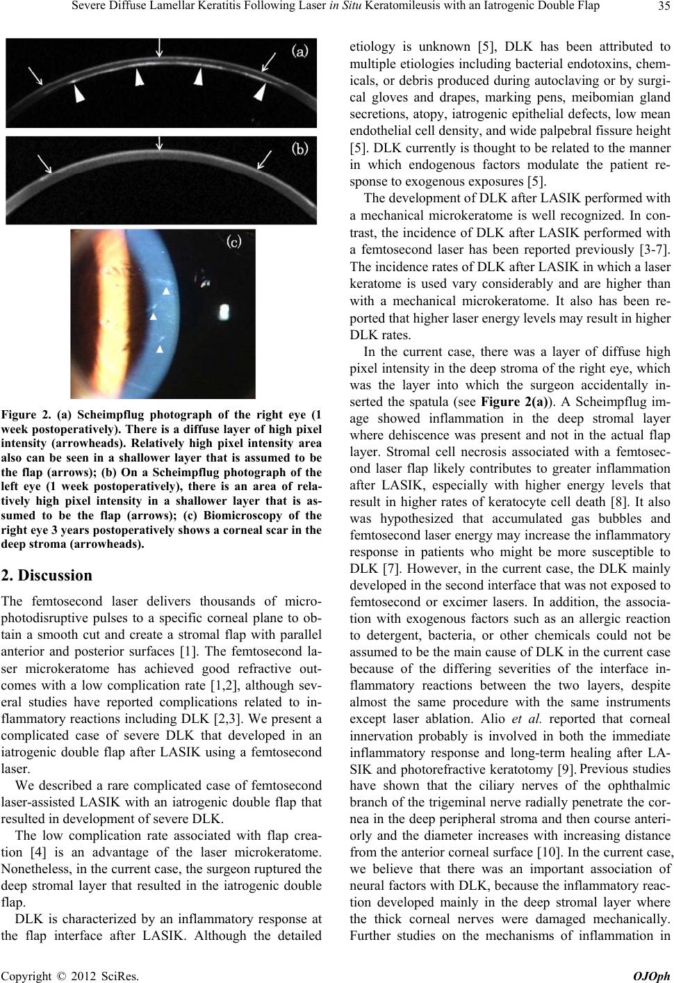 Severe Diffuse Lamellar Keratitis Following Laser in Situ Keratomileusis with an Iatrogenic Double Flap 35 Figure 2. (a) Scheimpflug photograph of the right eye (1 week postoperatively). There is a diffuse layer of high pixel intensity (arrowheads). Relatively high pixel intensity area also can be seen in a shallower layer that is assumed to be the flap (arrows); (b) On a Scheimpflug photograph of the left eye (1 week postoperatively), there is an area of rela- tively high pixel intensity in a shallower layer that is as- sumed to be the flap (arrows); (c) Biomicroscopy of the right eye 3 years postoperatively shows a corneal scar in the deep stroma (arrowheads). 2. Discussion The femtosecond laser delivers thousands of micro- photodisruptive pulses to a specific corneal plane to ob- tain a smooth cut and create a stromal flap with parallel anterior and posterior surfaces [1]. The femtosecond la- ser microkeratome has achieved good refractive out- comes with a low complication rate [1,2], although sev- eral studies have reported complications related to in- flammatory reactions including DLK [2,3]. We present a complicated case of severe DLK that developed in an iatrogenic double flap after LASIK using a femtosecond laser. We described a rare complicated case of femtosecond laser-assisted LASIK with an iatrogenic double flap that resulted in development of severe DLK. The low complication rate associated with flap crea- tion [4] is an advantage of the laser microkeratome. Nonetheless, in the current case, the surgeon ruptured the deep stromal layer that resulted in the iatrogenic double flap. DLK is characterized by an inflammatory response at the flap interface after LASIK. Although the detailed etiology is unknown [5], DLK has been attributed to multiple etiologies including bacterial endotoxins, chem- icals, or debris produced during autoclaving or by surgi- cal gloves and drapes, marking pens, meibomian gland secretions, atopy, iatrogenic epithelial defects, low mean endothelial cell density, and wide palpebral fissure height [5]. DLK currently is thought to be related to the manner in which endogenous factors modulate the patient re- sponse to exogenous exposures [5]. The development of DLK after LASIK performed with a mechanical microkeratome is well recognized. In con- trast, the incidence of DLK after LASIK performed with a femtosecond laser has been reported previously [3-7]. The incidence rates of DLK after LASIK in which a laser keratome is used vary considerably and are higher than with a mechanical microkeratome. It also has been re- ported that higher laser energy levels may result in higher DLK rates. In the current case, there was a layer of diffuse high pixel intensity in the deep stroma of the right eye, which was the layer into which the surgeon accidentally in- serted the spatula (see Figure 2(a)). A Scheimpflug im- age showed inflammation in the deep stromal layer where dehiscence was present and not in the actual flap layer. Stromal cell necrosis associated with a femtosec- ond laser flap likely contributes to greater inflammation after LASIK, especially with higher energy levels that result in higher rates of keratocyte cell death [8]. It also was hypothesized that accumulated gas bubbles and femtosecond laser energy may increase the inflammatory response in patients who might be more susceptible to DLK [7]. However, in the current case, the DLK mainly developed in the second interface that was not exposed to femtosecond or excimer lasers. In addition, the associa- tion with exogenous factors such as an allergic reaction to detergent, bacteria, or other chemicals could not be assumed to be the main cause of DLK in the current case because of the differing severities of the interface in- flammatory reactions between the two layers, despite almost the same procedure with the same instruments except laser ablation. Alio et al. reported that corneal innervation probably is involved in both the immediate inflammatory response and long-term healing after LA- SIK and photorefractive keratotomy [9]. Previous studies have shown that the ciliary nerves of the ophthalmic branch of the trigeminal nerve radially penetrate the cor- nea in the deep peripheral stroma and then course anteri- orly and the diameter increases with increasing distance from the anterior corneal surface [10]. In the current case, we believe that there was an important association of neural factors with DLK, because the inflammatory reac- tion developed mainly in the deep stromal layer where the thick corneal nerves were damaged mechanically. Further studies on the mechanisms of inflammation in Copyright © 2012 SciRes. OJOph  Severe Diffuse Lamellar Keratitis Following Laser in Situ Keratomileusis with an Iatrogenic Double Flap Copyright © 2012 SciRes. OJOph 36 patients with DLK should be performed and hopefully will provide invaluable information. REFERENCES [1] S. G. Slade, “The Use of the Femtosecond Laser in the Customization of Corneal Flaps in Laser in Situ Kera- tomileusis,” Current Opinion in Ophthalmology, Vol. 18, No. 4, 2007, pp. 314-317. doi:10.1097/ICU.0b013e3281bd88a0 [2] H. K. Soong and J. B. Malta, “Femtosecond Lasers in Oph- thalmology,” American Journal of Ophthalmology, Vol. 147, No. 2, 2009, pp. 189-197. [3] J. Javaloy, M. T. Vidal, A. M. Abdelrahman, A. Artola and J. L. Alio, “Confocal Microscopy Comparison of In- tralase Femtosecond Laser and Moria M2 Microkeratome in Lasik,” Journal of Refractive Surgery, Vol. 23, No. 2, 2007, pp. 178-187. [4] M. Moshirfar, J. P. Gardiner, J. A. Schliesser, L. Espan- dar, V. Feiz, M. D. Mifflin and J. C. Chang, “Laser in Situ Keratomileusis Flap Complications Using Mechani- cal Microkeratome versus Femtosecond Laser: Retro- spective Comparison,” Journal of Cataract and Refrac- tive Surgery, Vol. 36, No. 11, 2010, pp. 1925-1933. doi:10.1016/j.jcrs.2010.05.027 [5] C. H. Choe, C. Guss, D. C. Musch, L. M. Niziol and R. M. Shtein, “Incidence of Diffuse Lamellar Keratitis after Lasik with 15 Khz, 30 Khz, and 60 Khz Femtosecond Laser Flap Creation,” Journal of Cataract and Refractive Surgery, Vol. 36, No. 11, 2010, pp. 1912-1918. doi:10.1016/j.jcrs.2010.09.003 [6] P. S. Binder, “Flap Dimensions Created with the Intralase Fs Laser,” Journal of Cataract and Refractive Surgery, Vol. 30, No. 1, 2004, pp. 26-32. doi:10.1016/S0886-3350(03)00578-9 [7] R. Gil-Cazorla, M. A. Teus, L. De Benito-Llopis and I. Fuentes, “Incidence of Diffuse Lamellar Keratitis after Laser in Situ Keratomileusis Associated with the Intralase 15 Khz Femtosecond Laser and Moria M2 Microkera- tome,” Journal of Cataract and Refractive Surgery, Vol. 34, No. 1, 2008, pp. 28-31. doi:10.1016/j.jcrs.2007.08.025 [8] M. V. Netto, R. R. Mohan, F. W. Medeiros, W. J. Dupps Jr., S. Sinha, R. R. Krueger, W. M. Stapleton, M. Ray- born, C. Suto and S. E. Wilson, “Femtosecond Laser and Microkeratome Corneal Flaps: Comparison of Stromal Wound Healing and Inflammation,” Journal of Refractive Surgery, Vol. 23, No. 7, 2007, pp. 667-676. [9] J. L. Alio, J. J. Perez-Santonja, T. Tervo, K. F. Tabbara, M. Vesaluoma, R. J. Smith, B. Maddox and R. K. Ma- loney, “Postoperative Inflammation, Microbial Complica- tions, and Wound Healing Following Laser in Situ Kera- tomileusis,” Journal of Refractive Surgery, Vol. 16, No. 5, 2000, pp. 523-538. [10] J. H. Krachmer, M. J. Mannis and E. J. Holland, “Cor- nea,” Mosby/Elsevier, St. Louis, 2011. |