

Paper Menu >>
Journal Menu >>
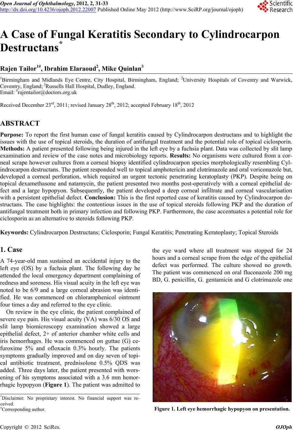 Open Journal of Ophthalmology, 2012, 2, 31-33 http://dx.doi.org/10.4236/ojoph.2012.22007 Published Online May 2012 (http://www.SciRP.org/journal/ojoph) 31 A Case of Fungal Keratitis Secondary to Cylindrocarpon Destructans* Rajen Tailor1#, Ibrahim Elaraoud2, Mike Quinlan3 1Birmingham and Midlands Eye Centre, City Hospital, Birmingham, England; 2University Hospitals of Coventry and Warwick, Coventry, England; 3Russells Hall Hospital, Dudley, England. Email: #[email protected] Received December 23rd, 2011; revised January 28th, 2012; accepted February 18th, 2012 ABSTRACT Purpose: To report the first human case of fungal keratitis caused by Cylindrocarpon destructans and to highlight the issues with the use of topical steroids, the duration of antifu ngal treatment and the potential role of topical ciclosporin. Methods: A patient presented following being inj ured in the left eye by a fuchsia plant. Data was collected by slit lamp examination and review of the case notes and microbiology reports. Results: No organisms were cultured from a cor- neal scrape however cultures from a corneal biopsy identified cylindro carpon species morphologically resembling Cyl- indrocarpon destructans. The patient responded well to topical amphotericin and clotrimazole and oral voriconazole but, developed a corneal perforation, which required an urgent tectonic penetrating keratoplasty (PKP). Despite being on topical dexamethasone and natamycin, the patient presented two months post-operatively with a corneal epithelial de- fect and a large hypopyon. Subsequently, the patient developed a deep corneal infiltrate and corneal vascularisation with a persistent epithelial defect. Conclusion: This is the first reported case of keratitis caused by Cylindrocarpon de- structans. The case highlights: the contentious issues in the use of topical steroids following PKP and the duration of antifungal treatment both in primary infection and following PKP. Furthermore, the case accentuates a potential role for ciclosporin as an alternative to steroids following PKP. Keywords: Cylindrocarpon Destructans; Ciclosporin; Fungal Keratitis; Penetrating Keratoplasty; Topical Steroids 1. Case A 74-year-old man sustained an accidental injury to the left eye (OS) by a fuchsia plant. The following day he attended the local emergency department complaining of redness and soreness. His visual acuity in the left eye was noted to be 6/9 and a large corneal abrasion was identi- fied. He was commenced on chloramphenicol ointment four times a day and referred to the eye clinic. On review in the eye clinic, the patient complained of severe eye pain. His visual acuity (VA) was 6/30 OS and slit lamp biomicroscopy examination showed a large epithelial defect, 2+ of anterior chamber white cells and iris hemorrhages. He was commenced on guttae (G) ce- furoxime 5% and ofloxacin 0.3% hourly. The patients symptoms gradually improved and on day seven of topi- cal antibiotic treatment, prednisolone 0.5% QDS was added. Three days later, the patient presented with wors- ening of his symptoms associated with a 3.6 mm hemor- rhagic hypopyon (Figure 1). The patient was admitted to the eye ward where all treatment was stopped for 24 hours and a corneal scrape from the edge of the epithelial defect was performed. The culture showed no growth. The patient was commenced on oral fluconazole 200 mg BD, G. penicillin, G. gentamicin and G clotrimazole one *Disclaimer: No proprietary interest. No financial support was re- ceived. #Corresponding author. Figure 1. Left eye hemorrhagic hypopyon on presentation. Copyright © 2012 SciRes. OJOph 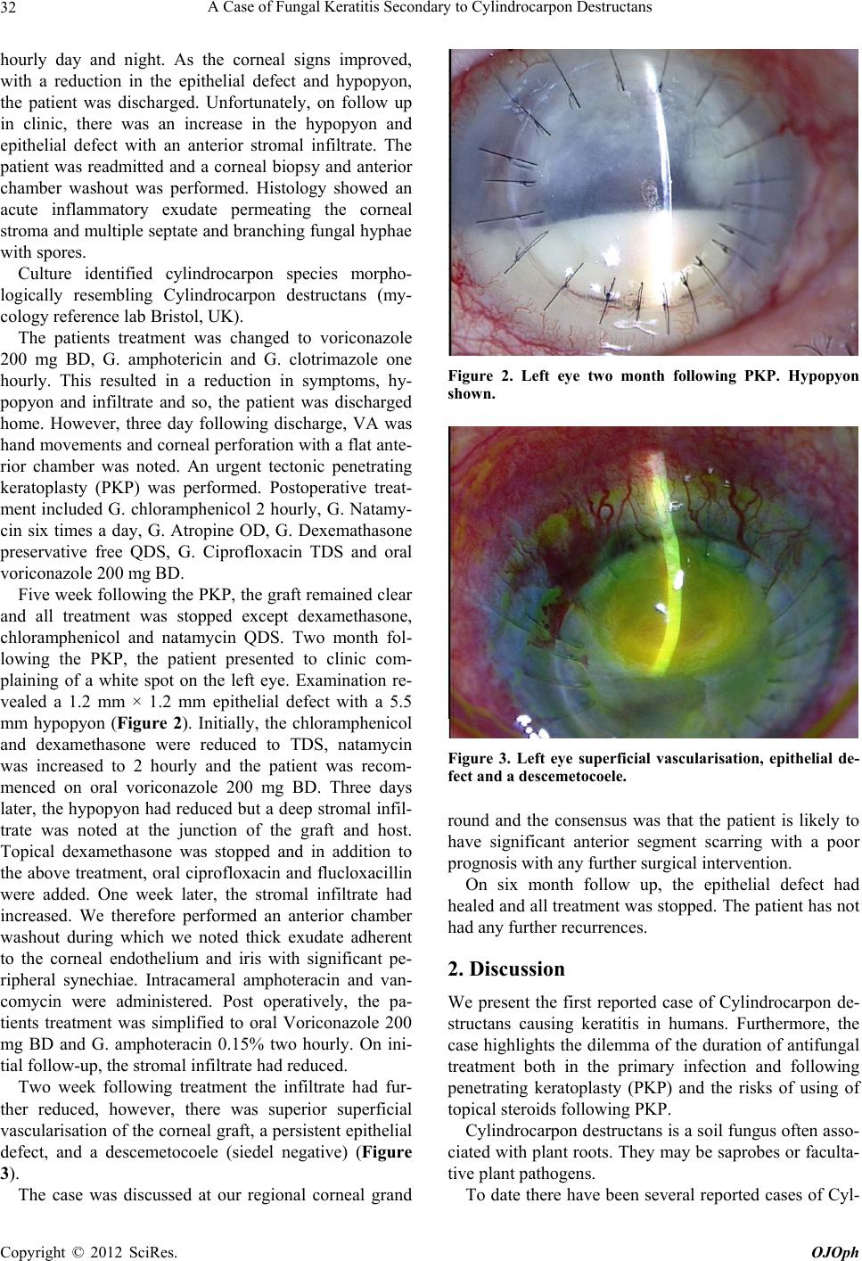 A Case of Fungal Keratitis Secondary to Cylindrocarpon Destructans 32 hourly day and night. As the corneal signs improved, with a reduction in the epithelial defect and hypopyon, the patient was discharged. Unfortunately, on follow up in clinic, there was an increase in the hypopyon and epithelial defect with an anterior stromal infiltrate. The patient was readmitted and a corneal biopsy and anterior chamber washout was performed. Histology showed an acute inflammatory exudate permeating the corneal stroma and multiple septate and br anching fung al hyphae with spores. Culture identified cylindrocarpon species morpho- logically resembling Cylindrocarpon destructans (my- cology reference lab Bristol, UK). The patients treatment was changed to voriconazole 200 mg BD, G. amphotericin and G. clotrimazole one hourly. This resulted in a reduction in symptoms, hy- popyon and infiltrate and so, the patient was discharged home. However, three day following discharge, VA was hand movements and corneal perforation with a flat ante- rior chamber was noted. An urgent tectonic penetrating keratoplasty (PKP) was performed. Postoperative treat- ment included G. chloramphenicol 2 hourly, G. Natamy- cin six times a day, G. Atropine OD, G. Dexemathasone preservative free QDS, G. Ciprofloxacin TDS and oral voriconazole 200 mg BD. Five week following the PKP, the graft remained clear and all treatment was stopped except dexamethasone, chloramphenicol and natamycin QDS. Two month fol- lowing the PKP, the patient presented to clinic com- plaining of a white spot on the left eye. Examination re- vealed a 1.2 mm × 1.2 mm epithelial defect with a 5.5 mm hypopyon (Figure 2). Initially, the chloramphenicol and dexamethasone were reduced to TDS, natamycin was increased to 2 hourly and the patient was recom- menced on oral voriconazole 200 mg BD. Three days later, the hypopyon had reduced but a deep stromal infil- trate was noted at the junction of the graft and host. Topical dexamethasone was stopped and in addition to the above treatment, oral ciprof loxacin and flucloxacillin were added. One week later, the stromal infiltrate had increased. We therefore performed an anterior chamber washout during which we noted thick exudate adherent to the corneal endothelium and iris with significant pe- ripheral synechiae. Intracameral amphoteracin and van- comycin were administered. Post operatively, the pa- tients treatment was simplified to oral Voriconazole 200 mg BD and G. amphoteracin 0.15% two hourly. On ini- tial follow-up, the stromal infiltrate had reduced. Two week following treatment the infiltrate had fur- ther reduced, however, there was superior superficial vascularisation of the corneal graft, a persistent epithelial defect, and a descemetocoele (siedel negative) (Figure 3). The case was discussed at our regional corneal grand Figure 2. Left eye two month following PKP. Hypopyon shown. Figure 3. Left eye superficial vascularisation, epithelial de- fect and a descemet oc oele. round and the consensus was that the patient is likely to have significant anterior segment scarring with a poor prognosis with any further surgical intervention. On six month follow up, the epithelial defect had healed and all treatment was stopped. The patient has not had any further recurrences. 2. Discussion We present the first reported case of Cylindrocarpon de- structans causing keratitis in humans. Furthermore, the case highlights the dilemma of the duration of antifungal treatment both in the primary infection and following penetrating keratoplasty (PKP) and the risks of using of topical steroids following PKP. Cylindrocarpon destructans is a soil fungus often asso- ciated with plant roots. They may be saprobes or faculta- tive plant pathogens. To date there have been several reported cases of Cyl- Copyright © 2012 SciRes. OJOph 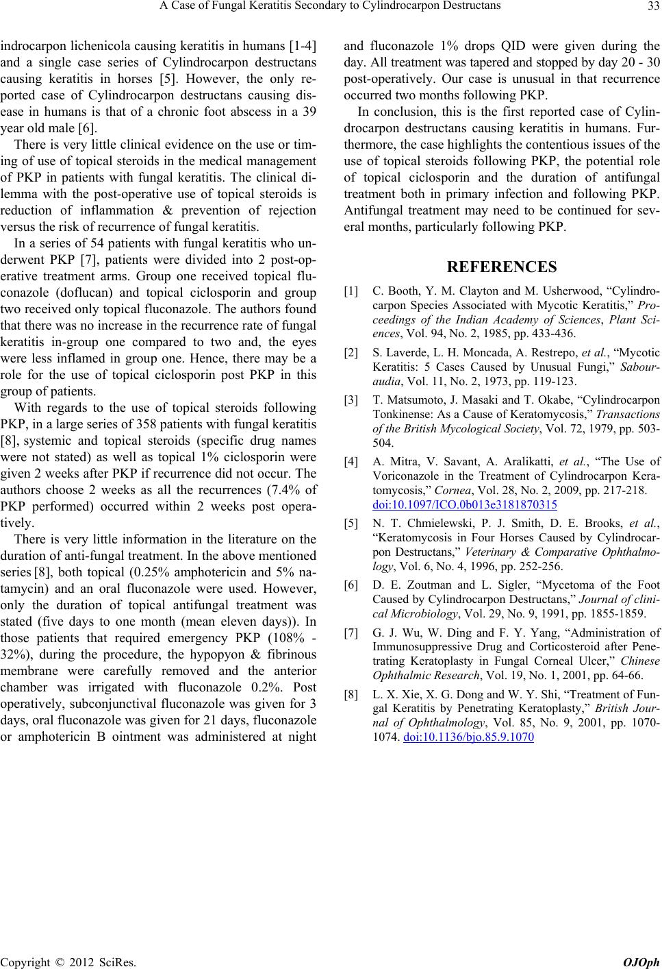 A Case of Fungal Keratitis Secondary to Cylindrocarpon Destructans Copyright © 2012 SciRes. OJOph 33 indrocarpon lichen icola causing keratitis in humans [1-4] and a single case series of Cylindrocarpon destructans causing keratitis in horses [5]. However, the only re- ported case of Cylindrocarpon destructans causing dis- ease in humans is that of a chronic foot abscess in a 39 year old male [6]. There is very little clinical evidence on the use or tim- ing of use of topical steroids in the medical management of PKP in patients with fungal keratitis. The clinical di- lemma with the post-operative use of topical steroids is reduction of inflammation & prevention of rejection versus the risk of recurrence of fungal keratitis. In a series of 54 patients with fungal keratitis who un- derwent PKP [7], patients were divided into 2 post-op- erative treatment arms. Group one received topical flu- conazole (doflucan) and topical ciclosporin and group two received only topical fluconazole. The authors found that there was no increase in the recurrence rate of fungal keratitis in-group one compared to two and, the eyes were less inflamed in group one. Hence, there may be a role for the use of topical ciclosporin post PKP in this group of patients. With regards to the use of topical steroids following PKP, in a large series of 358 patients with fungal keratitis [8], systemic and topical steroids (specific drug names were not stated) as well as topical 1% ciclosporin were given 2 weeks after PKP if recurrence did not occur. The authors choose 2 weeks as all the recurrences (7.4% of PKP performed) occurred within 2 weeks post opera- tively. There is very little information in the literature on the duration of anti-fungal treatment. In the above mentioned series [8], both topical (0.25% amphotericin and 5% na- tamycin) and an oral fluconazole were used. However, only the duration of topical antifungal treatment was stated (five days to one month (mean eleven days)). In those patients that required emergency PKP (108% - 32%), during the procedure, the hypopyon & fibrinous membrane were carefully removed and the anterior chamber was irrigated with fluconazole 0.2%. Post operatively, subconjunctival fluconazole was given for 3 days, oral fluconazole was given for 21 days, fluconazole or amphotericin B ointment was administered at night and fluconazole 1% drops QID were given during the day. All treatment was tapered and stopped by day 20 - 30 post-operatively. Our case is unusual in that recurrence occurred two months following PKP. In conclusion, this is the first reported case of Cylin- drocarpon destructans causing keratitis in humans. Fur- thermore, the case highlights the contentious issues of th e use of topical steroids following PKP, the potential role of topical ciclosporin and the duration of antifungal treatment both in primary infection and following PKP. Antifungal treatment may need to be continued for sev- eral months, particularly following PKP. REFERENCES [1] C. Booth, Y. M. Clayton and M. Usherwood, “Cylindro- carpon Species Associated with Mycotic Keratitis,” Pro- ceedings of the Indian Academy of Sciences, Plant Sci- ences, Vol. 94, No. 2, 1985, pp. 433-436. [2] S. Laverde, L. H. Moncada, A. Restrepo, et al., “Mycotic Keratitis: 5 Cases Caused by Unusual Fungi,” Sabour- audia, Vol. 11, No. 2, 1973, pp. 119-123. [3] T. Matsumoto, J. Masaki and T. Okabe, “Cylindrocarpon Tonkinense: As a Cause of Keratomycosis,” Transactions of the British Mycological Society, Vol. 72, 1979, pp. 503- 504. [4] A. Mitra, V. Savant, A. Aralikatti, et al., “The Use of Voriconazole in the Treatment of Cylindrocarpon Kera- tomycosis,” Cornea, Vol. 28, No. 2, 2009, pp. 217-218. doi:10.1097/ICO.0b013e3181870315 [5] N. T. Chmielewski, P. J. Smith, D. E. Brooks, et al., “Keratomycosis in Four Horses Caused by Cylindrocar- pon Destructans,” Veterinary & Comparative Ophthalmo- logy, Vol. 6, No. 4, 1996, pp. 252-256. [6] D. E. Zoutman and L. Sigler, “Mycetoma of the Foot Caused by Cylindrocarpon Destructans,” Journal of clini- cal Microbiology, Vol. 29, No. 9, 1991, pp. 1855-1859. [7] G. J. Wu, W. Ding and F. Y. Yang, “Administration of Immunosuppressive Drug and Corticosteroid after Pene- trating Keratoplasty in Fungal Corneal Ulcer,” Chinese Ophthalmic Research, Vol. 19, No. 1, 2001, pp. 64-66. [8] L. X. Xie, X. G. Dong and W. Y. Shi, “Treatment of Fun- gal Keratitis by Penetrating Keratoplasty,” British Jour- nal of Ophthalmology, Vol. 85, No. 9, 2001, pp. 1070- 1074. doi:10.1136/bjo.85.9.1070 |