

Paper Menu >>
Journal Menu >>
 Open Journal of Respiratory Diseases, 2012, 2, 60-62 http://dx.doi.org/10.4236/ojrd.2012.22009 Published Online May 2012 (http://www.SciRP.org/journal/ojrd) Investigation of the Anti-Mycobacterial Mechanism of Action of 7-Methyljuglo n e * Veneesha Thaver1,2,3,4, J. J. M. Meyer 3, Riana Cockeran2#, Moloko C. Cholo2, Ronald Anderson2, Namrita Lall3 1Department of Physiology, University of Limpopo, Medical University of South Africa Campus, Pretoria, South Africa 2MRC Unit for Inflammation and Immunity, Department of Immunology, Faculty of Health Sciences, University of Pretoria and Tshwane Academic Division of the National Health Laboratory Service, Pretoria, South Africa 3Department of Plant Science, Faculty of Natural and Agricultural Sciences, University of Pretoria, Pretoria, South Africa 4Department of Physiology, School of Laboratory Medicine and Medical Sciences, University of KwaZulu Natal, Westville Campus, Durban, South Africa Email: #[email protected] Received February 20, 2012; revised March 19, 2012; accepted March 26, 2012 ABSTRACT Objectives: Although the naphthoquinone, 7-methyljuglone (7-MJ), is active against Mycobacterium tuberculosis (MTB) in vitro, neither the cellular site nor mechanism of anti-mycobacterial action of this agent has been identified. The primary objective of the current study was to investigate the mycobacterial outer membrane as a potential target of 7-MJ by measuring the effects of this agent (0.023 - 1.5 mg/L) on microbial ATP levels and uptake of K+. Methods: Bioluminescence and radiometric (uptake of 86Rb+) procedures were used to assay microbial ATP levels and K+ trans- port respectively. Results: Exposure of MTB (strain H37Rv) to 7-MJ for 60 min resulted in dose-related decreases in both microbial ATP levels and uptake of 86Rb+ which achieved statistical significance (P < 0.05) at concentrations of 0.4 and 0.1 mg/L respectively. Conclusions: These observations are compatible with the mycobacterial membrane as being the putative site of action of 7-MJ, targeting microbial energy metabolism and K+ transport. Keywords: Adenosine Triphosphate; Cell Membrane; Energy Metabolism; Potassium 1. Introduction Tuberculosis (TB) remains a major world health problem, in particular since the incidence of multi-drug-resistant tuberculosis has increased in many countries. Conse- quently, discovery of new drugs with novel modes of action represents a major challenge [1]. Euclea natalensis is a shrub or small to medium size tree, which occurs in a variety of habitats, including coastal and inland forests, as well as bushveld in southern Africa. The roots of E. natalensis are used by indigenous people of southern Africa to treat various bacterial infec- tions. Powdered root bark of this species is used as an ingredient in medicines to treat urinary tract infections, venereal diseases and dysmenorrhoea [2]. The Zulus, a South African tribe, use the root bark to treat respiratory diseases such as TB, bronchitis, pleurisy and asthma [3]. Several secondary metabolites such asterpenoids, naph- thoquinones etc. have been isolated from E. natalensis [3]. According to previous studies, the minimum inhibit- tory concentration (MIC) of a naphthoquinone isolated from the roots of the plant, methyljuglone (7-MJ), against a drug-sensitive strain of Mycobacterium tuber- culosis (MTB) was very significant and comparable to some of the existing antituberculosis drugs (MIC: 0.5 mg/L) [4]. The primary objective of the current study was to in- vestigate the mycobacterial outer membrane as a poten- tial target of 7-MJ by measuring the effects of this agent on microbial ATP levels and uptake of potassium (K+). 2. Materials and Methods 2.1. 7-Methyljuglon e (7-MJ) 7-MJ was dissolved in dimethyl sulphoxide (DMSO) and used at a final concentration range of 0.023 - 1.5 mg/L in the assays described below. Appropriate solvent controls (0.15% DMSO) were included in all experiments. *Funding: This study was supported by National Research Foundation and the Medical Research Council, Pretoria, South Africa. Transparency declarations: Nothing to declare. #Corresponding author. 2.2. Mycobacterium tuberculosis MTB strain H37Rv, ATCC 26518 was provided by the C opyright © 2012 SciRes. OJRD 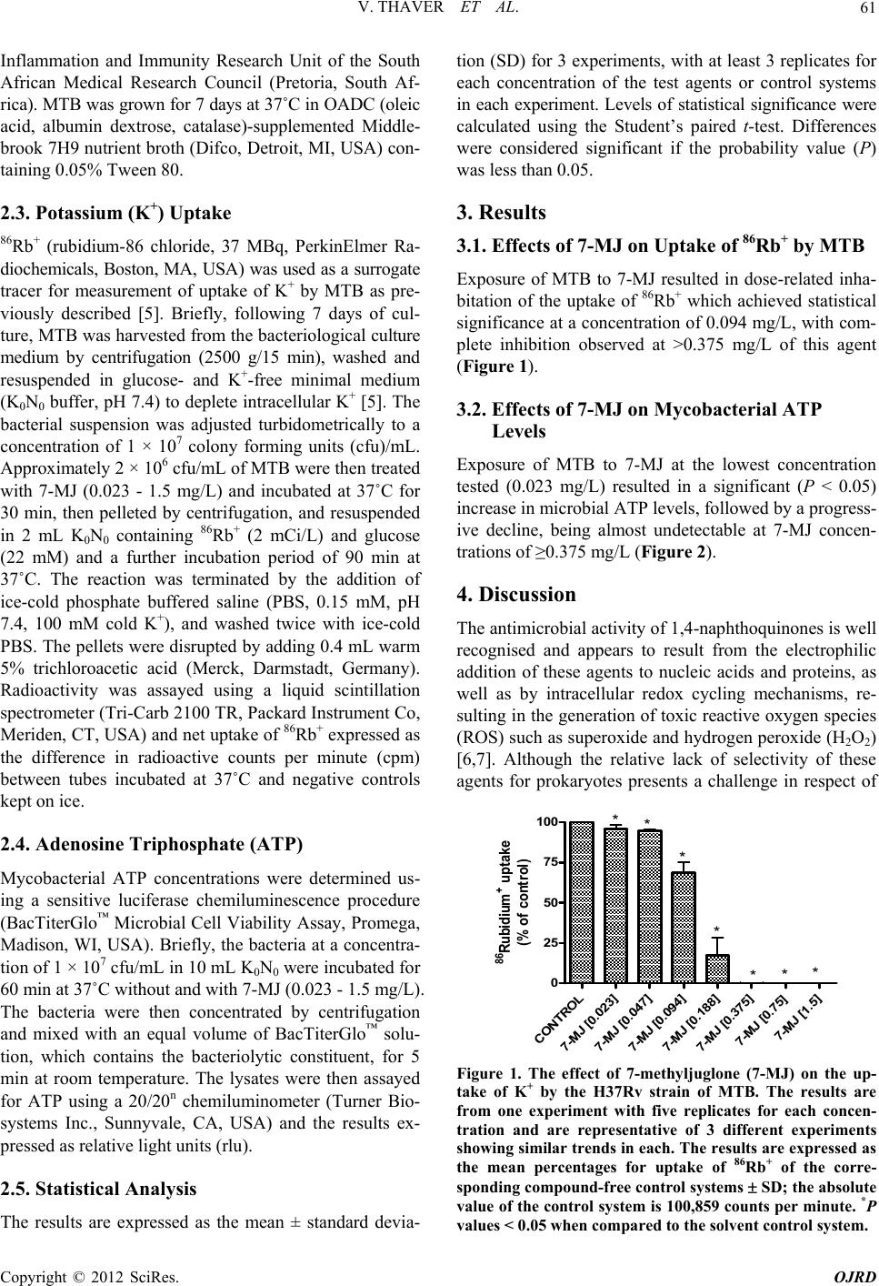 V. THAVER ET AL. 61 Inflammation and Immunity Research Unit of the South African Medical Research Council (Pretoria, South Af- rica). MTB was grown for 7 days at 37˚C in OADC (oleic acid, albumin dextrose, catalase)-supplemented Middle- brook 7H9 nutrient broth (Difco, Detroit, MI, USA) con- taining 0.05% Tween 80. 2.3. Potassium (K+) Uptake 86Rb+ (rubidium-86 chloride, 37 MBq, PerkinElmer Ra- diochemicals, Boston, MA, USA) was used as a surrogate tracer for measurement of uptake of K+ by MTB as pre- viously described [5]. Briefly, following 7 days of cul- ture, MTB was harvested from the bacteriological culture medium by centrifugation (2500 g/15 min), washed and resuspended in glucose- and K+-free minimal medium (K0N0 buffer, pH 7.4) to deplete intracellular K+ [5]. The bacterial suspension was adjusted turbidometrically to a concentration of 1 × 10⁷ colony forming units (cfu)/mL. Approximately 2 × 10⁶ cfu/mL of MTB were then treated with 7-MJ (0.023 - 1.5 mg/L) and incubated at 37˚C for 30 min, then pelleted by centrifugation, and resuspended in 2 mL K0N0 containing 86Rb+ (2 mCi/L) and glucose (22 mM) and a further incubation period of 90 min at 37˚C. The reaction was terminated by the addition of ice-cold phosphate buffered saline (PBS, 0.15 mM, pH 7.4, 100 mM cold K+), and washed twice with ice-cold PBS. The pellets were disrupted by adding 0.4 mL warm 5% trichloroacetic acid (Merck, Darmstadt, Germany). Radioactivity was assayed using a liquid scintillation spectrometer (Tri-Carb 2100 TR, Packard Instrument Co, Meriden, CT, USA) and net uptake of 86Rb+ expressed as the difference in radioactive counts per minute (cpm) between tubes incubated at 37˚C and negative controls kept on ice. 2.4. Adenosine Triphosphate (ATP) Mycobacterial ATP concentrations were determined us- ing a sensitive luciferase chemiluminescence procedure (BacTiterGlo™ Microbial Cell Viability Assay, Promega, Madison, WI, USA). Briefly, the bacteria at a concentra- tion of 1 × 10⁷ cfu/mL in 10 mL K0N0 were incubated for 60 min at 37˚C without and with 7-MJ (0.023 - 1.5 mg/L). The bacteria were then concentrated by centrifugation and mixed with an equal volume of BacTiterGlo™ solu- tion, which contains the bacteriolytic constituent, for 5 min at room temperature. The lysates were then assayed for ATP using a 20/20n chemiluminometer (Turner Bio- systems Inc., Sunnyvale, CA, USA) and the results ex- pressed as relative light units (rlu). 2.5. Statistical Analysis The results are expressed as the mean ± standard devia- tion (SD) for 3 experiments, with at least 3 replicates for each concentration of the test agents or control systems in each experiment. Levels of statistical significance were calculated using the Student’s paired t-test. Differences were considered significant if the probability value (P) was less than 0.05. 3. Results 3.1. Effects of 7-MJ on Uptake of 86Rb+ by MTB Exposure of MTB to 7-MJ resulted in dose-related inha- bitation of the uptake of 86Rb+ which achieved statistical significance at a concentration of 0.094 mg/L, with com- plete inhibition observed at >0.375 mg/L of this agent (Figure 1). 3.2. Effects of 7-MJ on Mycobacterial ATP Levels Exposure of MTB to 7-MJ at the lowest concentration tested (0.023 mg/L) resulted in a significant (P < 0.05) increase in microbial ATP levels, followed by a progress- ive decline, being almost undetectable at 7-MJ concen- trations of ≥0.375 mg/L (Figure 2). 4. Discussion The antimicrobial activity of 1,4-naphthoquinones is well recognised and appears to result from the electrophilic addition of these agents to nucleic acids and proteins, as well as by intracellular redox cycling mechanisms, re- sulting in the generation of toxic reactive oxygen species (ROS) such as superoxide and hydrogen peroxide (H2O2) [6,7]. Although the relative lack of selectivity of these agents for prokaryotes presents a challenge in respect of CONTROL 7-MJ [0.023] 7-MJ [0.047] 7-MJ [0.094] 7-MJ [0.188] 7-MJ [0.375] 7-MJ [0.75] 7-MJ [1.5] 0 25 50 75 100 ** * * *** 86 Rubidium + upt ake (% of control) Figure 1. The effect of 7-methyljuglone (7-MJ) on the up- take of K+ by the H37Rv strain of MTB. The results are from one experiment with five replicates for each concen- tration and are representative of 3 different experiments showing similar trends in each. The results are e xpressed as the mean percentages for uptake of 86Rb+ of the corre- sponding compound-free control systems SD; the absolute value of the control system is 100,859 counts per minute. *P values < 0.05 when compared to the solvent control system. Copyright © 2012 SciRes. OJRD 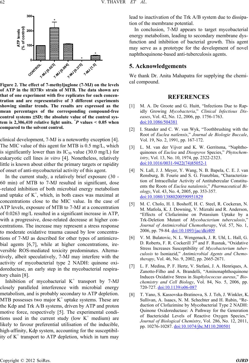 V. THAVER ET AL. Copyright © 2012 SciRes. OJRD 62 CONTROL 7-MJ [0.023] 7-MJ [0.094] 7-MJ [0.375] 7-MJ [1.5] 0 100 200 300 * * * ATP levels ( % of control) Figure 2. The effect of 7-methyljuglone (7-MJ) on the levels of ATP in the H37Rv strain of MTB. The data shown are that of one experiment with five replicates for each concen- tration and are representative of 3 different experiments showing similar trends. The results are expressed as the mean percentages of the corresponding compound-free control systems SD; the absolute value of the control sys- tem is 2,306,410 relative light units. *P values < 0.05 when compared to the solvent control. clinical development, 7-MJ is a noteworthy exception [4]. The MIC value of this agent for MTB is 0.5 mg/L, which is significantly lower than its IC50 value (30.0 mg/L) for eukaryotic cell lines in vitro [4]. Nonetheless, relatively little is known about either the primary targets or rapidity of onset of anti-mycobacterial activity of this agent. In the current study, a relatively brief exposure (30 - 60 min) of MTB to 7-MJ resulted in significant, dose -related inhibition of both microbial energy metabolism and uptake of K+, which, in both cases was maximal at concentrations close to the MIC value. In the case of ATP levels, exposure of MTB to 7-MJ at a concentration of 0.0263 mg/L resulted in a significant increase in ATP, with a progressive, dose-related decrease at higher con- centrations. The increase may represent a stress response to moderate oxidative trauma caused by low concentra- tions of 7-MJ as described for other types of antimicro- bial agents [6,7], while at higher concentrations, ire- versible ROS-mediated toxicity predominates. Alterna- tively, albeit speculatively, 7-MJ may interfere with the activity of mycobacterial type 2 NADH: quinone oxi- doreductase, an early step in the mycobacterial respira- tory chain [8]. Inhibition of mycobacterial K+ transport by 7-MJ closely paralleled interference with microbial energy metabolism, and is probably secondary to ATP depletion. MTB possesses two major K+ uptake systems. These are the Kdp and Trk A/B systems, driven by ATP and proton motive force, respectively [5]. The experimental condi- tions used in the current study (low K+ medium) are likely to favour preferential utilisation of the inducible, high-affinity, Kdp system, accounting for the susceptibil- ity of K+ transport to ATP depletion, which in turn may lead to inactivation of the Trk A/B system due to dissipa- tion of the membrane potential. In conclusion, 7-MJ appears to target mycobacterial energy metabolism, leading to secondary membrane dys- function and inhibition of bacterial growth. This agent may serve as a prototype for the development of novel naphthoquinone-based anti-tuberculosis agents. 5. Acknowledgements We thank Dr. Anita Mahapatra for supplying the chemi- cal compound. REFERENCES [1] M. A. De Groote and G. Huitt, “Infections Due to Rap- idly Growing Mycobacteria,” Clinical Infectious Dis- eases, Vol. 42, No. 12, 2006, pp. 1756-1763. doi:10.1086/504381 [2] I. Stander and C. W. van Wyk, “Toothbrushing with the Root of Euclea natlensis,” Journal de Biologie Buccale, Vol. 19, No. 2, 1991, pp. 167-172. [3] L. M. van der Vijver and K. W. Gerritsma, “Naphtho- quinones of Euclea and Diospyros Species,” Phytochem- istry, Vol. 13, No. 10, 1974, pp. 2322-2323. doi:10.1016/0031-9422(74)85052-1 [4] N. Lall, J. J. Meyer, Y. Wang, N. B. Bapela, C. E. J. van Rensburg, B. Fourie and S. G. Franzblau, “Characteriza- tion of Intracellular Activity of Antitubercular Constitu- ents the Roots of Euclea natalensis,” Pharmaceutical Bi- ology, Vol. 43, No. 4, 2005, pp. 353-357. doi:10.1080/13880200590951829 [5] M. C. Cholo, H. I. Boshoff, H. C. Steel, R. Cockeran, N. M. Matlola, K. J. Downing, V. Mizrahi and R. Anderson, “Effects of Clofazimine on Potassium Uptake by a Trk-Deletion Mutant of Mycobacterium tuberculosis,” Journal of Antimicrobial Chemotherapy, Vol. 57, No. 1, 2006, pp. 79-84. doi:10.1093/jac/dki409 [6] V. M. Bulatovic, N. L. Wengenack, J. R. Uhl, L. Hall, G. D. Roberts, F. R. Cockerill 3rd and F. Rusnak, “Oxidative Stress Increases Susceptibility of Mycobacterium tuber- culosis to Isoniazid,” Antimicrobial Agents and Chemo- therapy, Vol. 46, No. 9, 2002, pp. 2665-2671. [7] L. F. Medina, P. F. Hertz, V. Stefani, J. A. Henriques, A. Zanotto-Filho and A. Brandelli, “Aminonaphthoquinone Induces Oxidative Stress in Staphylococcus aureus,” Bio- chemistry and Cell Biology, Vol. 84, No. 5, 2006, pp. 720-727. doi:10.1139/o06-087 [8] T. Yano, S. Kassovska-Bratinova, S. J. Teh, J. Winkler, K. Sullivan, A. Isaacs, N. M. Schechter and H. Rubin, “Re- duction of Clofazimine by Mycobacterial Type 2 NADH: Quinone Oxidoreductase: A Pathway for the Generation of Bactericidal Levels of Reactive Oxygen Species,” Journal of Biological Chemistry, Vol. 286, No. 12, 2011, pp. 10276-10287. doi:10.1074/jbc.M110.200501 |