

Paper Menu >>
Journal Menu >>
 Open Journal of Respiratory Diseases, 2012, 2, 57-59 http://dx.doi.org/10.4236/ojrd.2012.22008 Published Online May 2012 (http://www.SciRP.org/journal/ojrd) Asthma-Like Tracheo-Br onchial Amyloid osis Fatma Chermiti Ben Abdallah1*, Hanene Smadhi1, Ridha Mahouachi1, Amel Chtourou1, Sofia Taktak1, Ayda Ayadi2, Faouzi El Mezni2, Ali Ben Kheder1 1Pneumology IV Department, Abderrahmen Mami Hospital, Ariana, Tunisia 2Anatomy-Pathology Department, Abderrahmen Mami Hos pital, Ariana, Tunisia Email: *[email protected] Received October 20, 2011; revised November 25, 2011; accepted December 10, 2011 ABSTRACT Localized bronchial Amyloidosis is an uncommon disease of unknown origin. Clinical signs are not specific. Tracheo- bronchial symptoms are the most frequent. We report a case of a 31 year-old man complaining of asthma-like dyspnea. Bronchoscopy was performed because of ineffectiveness of antiasthmatic treatment, showed a submucosal infiltration with stenosis of both right and left upper bronchi and a complete stenosis of intermediate troncus. Multiple biopsies were performed and concluded to Amyloidosis of AL type. Oral corticosteroids were indicated with clinical improve- ment. Keywords: Amyloidosis; Biopsy; Histo logy; Corticosteroids 1. Introduction Tracheobronchial amyloidosis (TBA) is rare and it con- stitutes approximately 1% of benign tumors in these ar- eas. It is characterized by deposition of amyloid within the airway walls, manifesting as submucosal tumors. Symptoms are related to bronchial obstruction leading to a misdiagnosis in some cases. Consequently, the diagno- sis is delayed and made through bronchoscopic biopsy. No consensus exists with regard to its op timal treatment, resulting in different modalities, used to manage this condition. We report a case of localized bronchial amyloidosis diagnosed after a history of asthma-like dyspnea. 2. Case Report A 31-year-old man, non -smoking complained of asthma- like wheezing dyspnea during six months, with a recent worsening during one month. He was treated with in- haled corticosteroids and bronchodilators, but without improvement. He was admitted to our department. His clinical examination showed a respiratory rate of 28/min, a pulse rate of 90/min and blood pressure of 110/80 mmHg. Pulmonary examination revealed diffuses sibi- lant rales. All routine biological examinations were nor- mal. The arterial blood gas levels in room air were as follows: PaCO2 of 38.1, PaO2 of 62.3, and pH of 7.47. Pulmonary function tests showed an obstructive pattern: FVC of 2.33 l (50% of predicted), FEV1 of 1.33 l (34% of predicted) and FEV1/FVC ratio of 57%. Chest X-ray revealed atelectasis in the middle lobe and a right hilar opacity (Figure 1). Bronchodilators, corti- costeroids and antibiotics were administrated for two weeks but without improvement. A Compued tomogram- phy (CT) scan of the chest was performed, showing air- way narrowing caused by circumferential thickening of the bronchial wall and atelectasis in the left upper lobe (Figure 2). Fiberoptic bronchoscopy was then performed reveal- ing severe narrowing of the right and the left main bron- chi with mucosal irregularities and a complete sten osis of intermediary bronchus and the left upper bronchus (Fig- ure 3). Pathological study of bronchial biopsy demon- strated an extensive deposit in the submucosa of a hya- line substance shown to be amyloid. The epithelium over the deposit was intact. No squamous metaplasia was demonstrated. Some giant foreign-body cells were pre- sent in relation to the amyloid and small calcifications were seen. Histopathological analysis of the tissue sam- ple, with Congo red staining, demonstrated apple-green birefringence when viewed under polarized light (Figure 4). The diagnosis of bronchial amyloidosis was made and several investigations were undertaken. The results of urine analysis (for 24-h proteinuria and creatinuria) and protein profiles were within normal limits. An electro- cardiogram, echocardiography and abdominal ultrasound showed no abnormalities. The diagnosis of TBA was then made and the patient *Corresponding author. C opyright © 2012 SciRes. OJRD 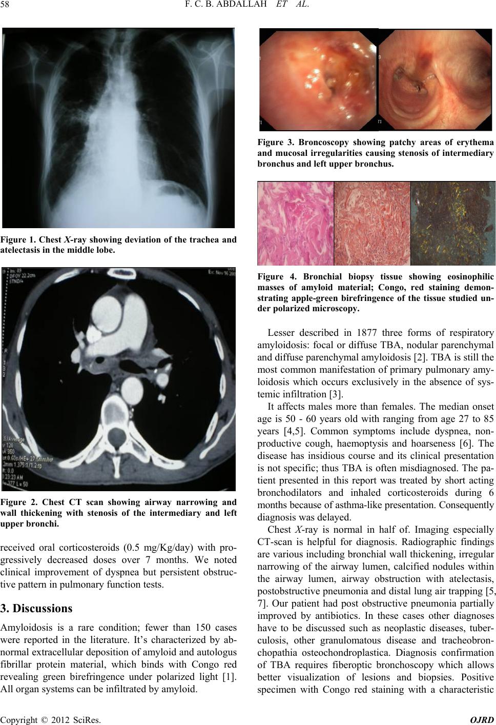 F. C. B. ABDALLAH ET AL. 58 Figure 1. Chest X-ray showing deviation of the trachea and atelectasis in the middle lobe. Figure 2. Chest CT scan showing airway narrowing and wall thickening with stenosis of the intermediary and left upper bronchi. received oral corticosteroids (0.5 mg/Kg/day) with pro- gressively decreased doses over 7 months. We noted clinical improvement of dyspnea but persistent obstruc- tive pattern in pulmonary function tests. 3. Discussions Amyloidosis is a rare condition; fewer than 150 cases were reported in the literature. It’s characterized by ab- normal extracellular deposition of amyloid an d autologus fibrillar protein material, which binds with Congo red revealing green birefringence under polarized light [1]. All organ systems can be infiltrated by amyloid. Figure 3. Broncoscopy showing patchy areas of erythema and mucosal irregularities causing stenosis of intermediary bronchus and left upper br onchus. Figure 4. Bronchial biopsy tissue showing eosinophilic masses of amyloid material; Congo, red staining demon- strating apple-green birefringence of the tissue studied un- der polarized microscopy. Lesser described in 1877 three forms of respiratory amyloidosis: focal or diffuse TBA, nodular parenchymal and diffuse parenchymal amyloidosis [2]. TBA is still th e most common manifestation of primary pulmonary amy- loidosis which occurs exclusively in the absence of sys- temic infiltration [3]. It affects males more than females. The median onset age is 50 - 60 years old with ranging from age 27 to 85 years [4,5]. Common symptoms include dyspnea, non- productive cough, haemoptysis and hoarseness [6]. The disease has insidious course and its clinical presentation is not specific; thus TBA is often misdiagnosed. The pa- tient presented in this report was treated by short acting bronchodilators and inhaled corticosteroids during 6 months because of asthma-like presentation. Consequently diagnosis was delayed. Chest X-ray is normal in half of. Imaging especially CT-scan is helpful for diagnosis. Radiographic findings are various including bronchial wall thickening, irregular narrowing of the airway lumen, calcified nodules within the airway lumen, airway obstruction with atelectasis, postobstructive pneumonia and distal lung air trapping [5, 7]. Our patient had post obstructive pneumonia partially improved by antibiotics. In these cases other diagnoses have to be discussed such as neoplastic diseases, tuber- culosis, other granulomatous disease and tracheobron- chopathia osteochondroplastica. Diagnosis confirmation of TBA requires fiberoptic bronchoscopy which allows better visualization of lesions and biopsies. Positive specimen with Congo red staining with a characteristic Copyright © 2012 SciRes. OJRD  F. C. B. ABDALLAH ET AL. Copyright © 2012 SciRes. OJRD 59 apple green birefringence with polarized light is the proof of amyl oidosis di sease [ 4, 5]. The treatment of localized TBA is essentially symp- tomatic [8]. Strategies depend on the site and extent of the disease. In fact, only observation can be indicated in asymptomatic patients. However, local and or systemic therapies are recommended in patients with severe air- way obstruction [9]. Drugs inhibiting the synthesis, deposition and degradation of amyloid in tissues, espe- cially colchicine, prednisone and melphalan were used but with a limited effect [4,9]. Varied methods of bron- choscopic treatment have been reported such as endo- bronchial Nd-YAG laser, tracheobronchial stents, bal- loon dilation and bronchoscopic ablation of intraluminal amyloid deposits. These methods are associated with a risk of bleeding and recurrences [4]. Surgery may be necessary when airway involvement is extensive [5]. Re- cently, some investigators demonstrated that external beam radiation therapy (EBRT) may provide an objective improvement [10]. Also M. Truong found in his study that EBRT prevents progressive amyloid deposition in 90% of patients with localized airway amyloidosis and it is well tolerated with minimal toxicity [11]. Although TBA is a localized process, it is associated with poorer prognosis. Authors reported that overall sur- vival ranges from 31% to 43% at 4 to 6 years. In absence of curative treatment of TBA, researchers are still con- tinuously trying o ther therapeutic alternatives to improve the prognosis of these patients. 4. Conclusion Diagnosis of TBA has to be considered in patients with asthma-like dyspnea in whom anti-asthmatic treatments are ineffective. The diagnosis can be suggested by radio- graphic findings but it is confirmed essentially through bronchoscopic biopsies, with appropriate Congo red staining of the bronchial tissue samples obtained. At present, there are no therapeutic options proved to be completely successful. However, bronchoscopic man- agement often proves to be temporarily effective, while EBRT still need to be further evaluated. Prognosis of TBA remains poor. REFERENCES [1] H. Gibbaoui, S. Abouchacra and M. Yaman, “A Case of Primary Diffuse Tracheobronchial Amyloidosis,” The Annals of Thoracic Surgery, Vol. 77, No. 5, 2004, pp. 1832-1834. doi:10.1016/S0003-4975(03)00999-8 [2] A. Lesser, “Ein Fall von Enchondroma Osteiodes Mixtum der Lunge Mit Partieller Amyloid Entortung,” Virchows Archiv , Vol. 69, No. 3-4, 1877, pp. 404-409. doi:10.1007/BF02326214 [3] C. Ozer, M. N. Duce, A. Yildiz, F. D. Apaydin, H. Egil- mez and T. Arpaci, “Primary Diffuse Tracheobronchial Amyloidosis: Case Report,” European Journal of Radi- ology, Vol. 44, No. 1, 2002, pp. 37-39. doi:10.1016/S0720-048X(01)00437-5 [4] H. Mao, X. Yang, Z. Wang, X. Chen and Q. Yi, “Asthma- Like Tracheobronchial Amyloidosis: A Case Report and Review of the Literature,” Journal of Nanjing Medical University, Vol. 22, No. 5, 2008, pp. 317-322. doi:10.1016/S1007-4376(08)60088-X [5] K. A. Dahl, K. H. Kernstine, T. L. Vannatta, M. W. Karwal, K. W. Thomas and D. F. Schraith, “Tracheo- bronchial Amyloidosis: A Surgical Disease with Long- Term Consequences,” Journal of Thoracic Cardiovascu- lar Surgery, Vol. 128, No. 5, 2004, pp. 789-792. [6] A. T. Monroe, R. Walia, R. A. Zlotecki and M. A. Jantz, “Tracheobronchial Amyloidosis: A Case Report of Suc- cessful Treatment with External Beam Radiation Ther- apy,” Chest, Vol. 125, No. 2, 2004, pp. 784-789. doi:10.1378/chest.125.2.784 [7] J. W. Santos, A. S. Filho, A. Bertolazzi, G. T. Michel, L. V. Silva, C. R. Melo, V. D. Pedro, D. Spilmann and J. K. Figaro, “Amyloidosis Tracheobronchial Primary,” Jour- nal Brasileiro Pneumologia, Vol. 34, No. 10, 2008, pp. 881-884. doi:10.1590/S1806-37132008001000015 [8] A. K. Brill, K. Woelke, R. Schadlich, C. Weinz and G. Laier- Gr oe neveld, “Tracheobronchial Amyloidosis-Bronchoscopic Diagnosis and Therapy of an Uncommon Disease: A Case Report,” Journal of Physiology and Pharmacology, Vol. 58, No. 5, 2007, pp. 51-55. [9] H. C. Chew, S. Y. Low, P. Eng, T. Agasthian and F. K. Cheah, “Cough and Persistant Wheeze in a Patient with Long-Standing Asthma,” Chest, Vol. 132, No. 2, 2007, pp. 727-731. doi:10.1378/chest.06-2810 [10] M. A. Neben-Wittich, R. L. Foote and S. Kalra, “External Beam Radiation Therapy for Tracheobronchial Amyloi- dosis,” Chest, Vol. 132, No. 1, 2007, pp. 262-267. doi:10.1378/chest.06-3118 [11] M. Truong, L. A. Kachnic, G. A. Grillone and J. L. Berk, “The Role of Radiation Therapy for Progressive Airway Amyloidosis,” International Journal of Radiation On- cology Biology Physics, Vol. 69, No. 3, 2007, p. S122. doi:10.1016/j.ijrobp.2007.07.226 |