

Paper Menu >>
Journal Menu >>
 Journal of Immune Based Therapies, Vaccines and Antimicrobials, 2012, 1, 1-6 http://dx.doi.org/10.4236/jibtva.2012.11001 Published Online April 2012 (http://www.SciRP.org/journal/jibtva) Poly-I:C Decreases Dendritic Cell Viability Independent of PKR Activation Hjalte List Larsen, Anders Elm Pedersen Department of International Health, Immunology and Microbiology, The Panum Institute, University of Copenhagen, Copenhagen, Denmark Email: hjaltel@sund.ku.dk, [email protected] Received March 1, 2012; revised April 2, 2012; accepted April 12, 2012 ABSTRACT Vaccination with tumor-antigen pulsed, monocyte-derived dendritic cells (DCs) has emerged as a promising strategy in cancer immunotherapy. The standard DC maturation cocktail consists of a combination of tumor necrosis factor-α (TNF-α)/interleukin (IL)-1β/IL-6 and prostaglandin E2 (PGE2) for generation of standard DCs (sDCs). In order to im- prove IL-12p70 production and cytotoxic T-lymphocyte (CTL) induction, a novel cocktail composed of TNF-α/IL-1β/ interferon (IFN)-α/IFN-γ and polyinosinic:polycytidylic acid (Poly-I:C) has been introduced to generate so-called α-Type-1 polarized DCs (αDC1s). We and others have previously performed a comprehensive comparison of sDCs and αDC1s. Here we demonstrate that the viability of αDC1s is lowered compared to sDCs and that DC apoptosis is medi- ated by Poly-I:C. We speculated that activation of protein kinase R (PKR) could mediate the observed apoptosis, but despite significantly higher PKR expression in αDC1s compared to sDCs and induction of active threonine (Thr)446 autophosphorylation of PKR in αDC1s, Poly-I:C did not influence total PKR expression or autophosporylation, indi- cating PKR-independent Poly-I:C-induced DC apoptosis. Keywords: Cancer Immunotherapy; Dendritic Cells; Poly-I:C; PKR 1. Introduction In therapeutic cancer vaccination with monocyte-derived DCs [1], maturation is mandatory for proper T-cell stimulation [2]. DCs maturated according to the standard cytokine cocktail consisting of TNF-α/IL-1β/IL-6 and PGE2 [3] show high expression of maturation markers CD80, CD83, CD86, CD134L and CCR7, but low secre- tion of IL-12p70 [4] which may have negative conse- quences for activation of appropriate T cell responses such as TH1 cells and cytotoxic T lymphocytes (CTLs). In the search of cytokine cocktails that increase DC IL-12p70 secretion, DCs maturated according to the α-Type-1 polarizing cocktail containing TNF-α/IL-1β/ Poly-I:C/IFN-α and IFN-γ were developed, as these DCs produce large amounts of IL-12p70 [4,5]. We and others have characterized cytokine secretion and capacity for T cell activation in details elsewhere [4,5] and it has been found that αDC1s may have a reduced capacity for T cell activation under certain conditions [4]. In addition to the criterias for successful DC generation investigated else- where, viability is an important factor. Here, we report a decreased viability of αDC1s and investigate a potential role of PKR mediated mechanisms. The double-stranded RNA (dsRNA)-analog Poly-I:C included in the α-Type-1 polarizing cocktail is a potential inducer of apoptosis, as cytosolic Poly-I:C is capable of activating the dsRNA-activated protein kinase (PKR) [6]. Upon dsRNA/Poly-I:C binding, PKR is autophosphory- lated on multiple sites including Thr446 and Thr451 [7], enabling signal transduction which can induce global translation inhibition and apoptosis [8]. Furthermore, Poly-I:C can be detected by toll-like receptor 3 (TLR3) in the endosomal compartment of DCs [9] and it has been reported that PKR is recruited to a large signaling complex following Poly-I:C binding to TLR3 [10]. We thus speculated that Poly-I:C is the cause of lowered αDC1 viability as compared to sDCs, and that such a Poly- I:C-mediated induction of apoptosis was mediated by PKR activation. In this case, siRNA mediated silencing of PKR-dependent apoptotic pathways could be a future strategy for improved efficacy of therapeutic αDC1s. Here, we report that absence of Poly-I:C from the α-Type-1 polarizing cocktail significantly improved αDC1 viability to levels comparable to sDCs without signifi- cantly decreasing CD80, CD83 and CD86 expression. αDC1s expressed significantly higher amounts of PKR than iDCs and sDCs, with PKR expression being de- pendent on the presence of IFNs in the maturation cock- tail but not on Poly-I:C. We were also unable to detect an C opyright © 2012 SciRes. JIBTVA  H. L. LARSEN ET AL. 2 effect of Poly-I:C on PKR autophosphorylation in mature DCs, suggesting that the observed lowered viability of αDC1s was PKR-independent. 2. Methods 2.1. Generation of Human Monocyte-Derived DCs Human monocyte-derived DCs were generated as previ- ously described with GM-CSF + IL-4 [11] from buffy coats after informed consent from healthy donors at the local department of clinical immunology. Maturation of iDCs was performed on day 7. sDCs were maturated with TNF-α (10 ng/mL), IL-1β (10 ng/mL), IL-6 (10 ng/mL) (all cytokines from Peprotech) and 1 μg/mL PGE2 (Prostin E2, Pfizer). The αDC1 phenotype was induced by TNF-α (10 ng/mL), IL-1β (10 ng/mL), IFN-α (10 ng/mL), IFN-γ (10 ng/mL) (all cytokines from Peprotech) and 20 μg/mL Poly-I:C (Sigma Aldrich). DCs were maturated 48 hours prior to analysis. 2.2. Flow Cytometry Flow cytometry was performed using antibodies against the following surface proteins: CD11c, CD80, CD83 and CD86 (BD-Pharmingen). Isotope control antibodies were obtained from DAKO. Detection of apoptotic DCs were performed using annexin V-FITC apoptosis detection Kit I (BD Pharmingen) according to the manufacturer’s pro- tocol. Data acquisition and analysis was performed on a FACSCalibur flow cytometer (BD) using CellQuest software (BD Biosciences). DCs were identified as the large granular cell population based on forward scatter (FSC)/side scatter (SSC) linear dot plots. 2.3. Western Blotting DCs were lysed in RIPA buffer supplemented with pro- teinase inhibitor cocktail (Sigma Aldrich), PMSF, NaF and Na3VO4 during 20 minutes of centrifugation at 14,000 g, 4˚C. Lysates were treated with SDS and heated for 5 minutes prior to loading onto 4% - 12% Bis-Tris gels (Invitrogen). Electrophoresis was performed in MES running buffer (Invitrogen) for 1 hour at 200 V. Blotting was performed at 30 V for 1 hour onto nitrocellulose membranes (Invitrogen) in transfer buffer (Invitrogen). Membranes were blocked and probed with polyclonal goat anti-pPKR (Thr446) antibody, polyclonal rabbit anti-PKR antibody (both Santa Cruz Biotechnology, Inc.) or goat anti-GAPDH antibody (AbCam) over night at 4˚C. Secondary horse radish peroxidase (HRP)-conjugated polyclonal antibodies (DAKO) were added and the membrane was incubated 1 hour at room temperature. The membrane was washed and visualized using an en- hanced chemiluminescence detection system (ECL plus Western Blotting Detection system, GE Healthcare) ac- cording to the manufacturer’s instructions. Detection of HRP-generated precipitates was performed on a Typhoon Scanner 9410 with Typhoon Scanner software (both from Amersham Biosciences). The same software was used to analyze band intensity, with PKR and pPKR intensity being normalized to GAPDH expression. 3. Results 3.1. DC Phenotype after Maturation Day 7 IL-4 and GM-CSF differentiated monocyte-de- rived DCs were identified as large granular cells ex- pressing the DC marker CD11c (Figures 1(A) and (B)). Maturation using the two established cocktails for gen- eration of sDCs and αDC1s both significantly increased the expression of CD80 (p < 0.05 and p < 0.01 respec- tively) and CD83 (p < 0.05 and p < 0.05 respectively) compared to iDCs, while a stable high expression level of CD86 was maintained from iDCs into mature DCs. CD83 expression in sDC was less pronounced in these DCs from healthy donors as compared to our previous results from cancer patient derived DCs [4]. Removal of individual cytokine components from the α-Type-1 po- larizing cocktail did not induce any significant changes in the expression of the three maturation markers, how- ever absence of Poly-I:C or IFN-α reduced the expres- sion of both CD80 and CD83 compared to expression levels in αDC1s. These observed reductions in CD80 and CD83 expression were not statistical significant but con- sistent in all donors. It was previously published that IL-12p70 secretion was dependent on IFN-γ or Poly-I:C with the largest contribution from IFN-γ [5]. For T cell activation, αDC1s have been shown to be superior for CTL activation [5], whereas assays mostly reflecting CD4+ T cell responses has demonstrated a decreased capacity for T cell activation as compared to sDCs [4]. 3.2. Viability of ex Vivo Generated DCs Annexin V-FITC and propidium iodide (PI) staining re- vealed a tendency toward that sDCs were more viable (mean of all donors: 79.4% living cells) as compared to αDC1s (mean of all donors: 71.7% living cells), a dif- ference that was not statistical significant (p = 0.069) but consistent in all donors. Data from one donor is shown in Figure 2(A) and mean of all donors in Figure 2(B). In order to identify the cause of the viability changes in αDC1s, all single components from the cocktail was sys- tematically excluded (Figure 2(B)). Exclusion of Poly-I:C from the α-type-1 polarizing cocktail significantly im- proved the viability of DCs from a mean of 71.7% (αDC1s) to 79.9% living cells (p = 0.02), thus increasing DC viability to a level comparable to sDCs. The viability Copyright © 2012 SciRes. JIBTVA 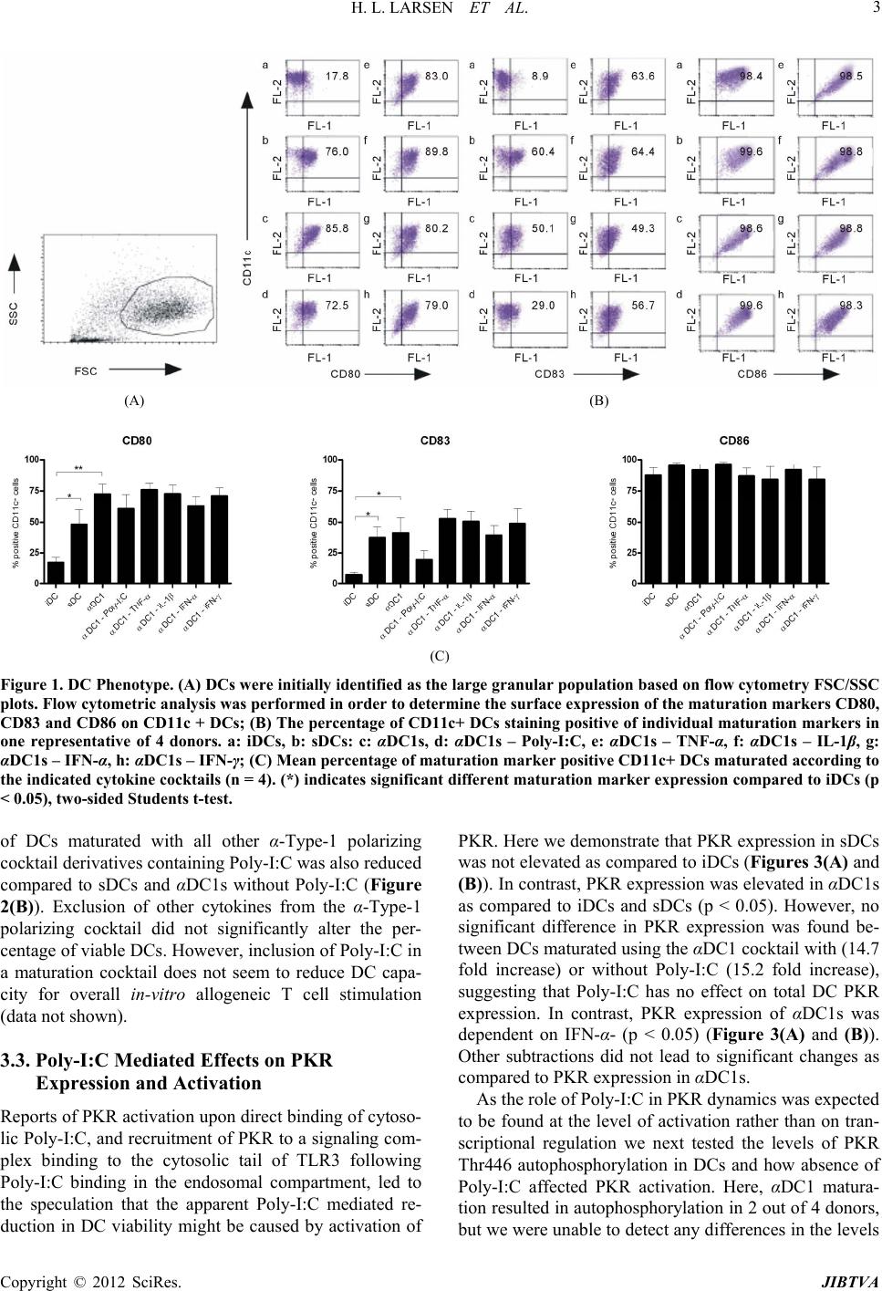 H. L. LARSEN ET AL. Copyright © 2012 SciRes. JIBTVA 3 (A) (B) (C) Figure 1. DC Phenotype. (A) DCs were initially identified as the large granular population based on flow cytometry FSC/SSC plots. Flow cytometric analysis was performed in order to determine the surface expression of the maturation markers CD80, CD83 and CD86 on CD11c + DCs; (B) The percentage of CD11c+ DCs staining positive of individual maturation markers in one representative of 4 donors. a: iDCs, b: sDCs: c: αDC1s, d: αDC1s – Poly-I:C, e: αDC1s – TNF-α, f: αDC1s – IL-1β, g: αDC1s – IFN-α, h: αDC1s – IFN-γ; (C) Mean percentage of maturation marker positive CD11c+ DCs maturated according to the indicated cytokine cocktails (n = 4). (*) indicates significant different maturation marker expression compared to iDCs (p < 0.05), two-sided Students t-test. of DCs maturated with all other α-Type-1 polarizing cocktail derivatives containing Poly-I:C was also reduced compared to sDCs and αDC1s without Poly-I:C (Figure 2(B)). Exclusion of other cytokines from the α-Type-1 polarizing cocktail did not significantly alter the per- centage of viable DCs. However, inclusion of Poly-I:C in a maturation cocktail does not seem to reduce DC capa- city for overall in-vitro allogeneic T cell stimulation (data not shown). 3.3. Poly-I:C Mediated Effects on PKR Expression and Activation Reports of PKR activation upon direct binding of cytoso- lic Poly-I:C, and recruitment of PKR to a signaling com- plex binding to the cytosolic tail of TLR3 following Poly-I:C binding in the endosomal compartment, led to the speculation that the apparent Poly-I:C mediated re- duction in DC viability might be caused by activation of PKR. Here we demonstrate that PKR expression in sDCs was not elevated as compared to iDCs (Figures 3(A) and (B)). In contrast, PKR expression was elevated in αDC1s as compared to iDCs and sDCs (p < 0.05). However, no significant difference in PKR expression was found be- tween DCs maturated using the αDC1 cocktail with (14.7 fold increase) or without Poly-I:C (15.2 fold increase), suggesting that Poly-I:C has no effect on total DC PKR expression. In contrast, PKR expression of αDC1s was dependent on IFN-α- (p < 0.05) (Figure 3(A) and (B)). Other subtractions did not lead to significant changes as compared to PKR expression in αDC1s. As the role of Poly-I:C in PKR dynamics was expected to be found at the level of activation rather than on tran- scriptional regulation we next tested the levels of PKR Thr446 autophosphorylation in DCs and how absence of Poly-I:C affected PKR activation. Here, αDC1 matura- tion resulted in autophosphorylation in 2 out of 4 donors, but we were unable to detect any differences in the levels 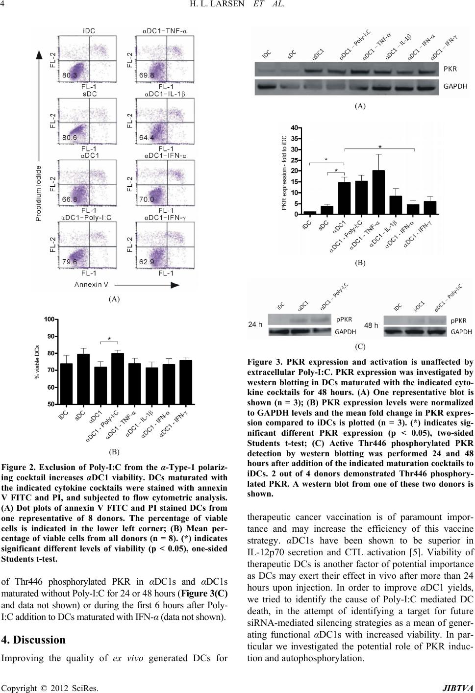 H. L. LARSEN ET AL. 4 (A) (B) Figure 2. Exclusion of Polyα-Type-1 polariz- f Thr446 phosphorylated PKR in αDC1s and αDC1s 4. Discussion uality of ex vivo generated DCs for -I:C from the ing cocktail increases αDC1 viability. DCs maturated with the indicated cytokine cocktails were stained with annexin V FITC and PI, and subjected to flow cytometric analysis. (A) Dot plots of annexin V FITC and PI stained DCs from one representative of 8 donors. The percentage of viable cells is indicated in the lower left corner; (B) Mean per- centage of viable cells from all donors (n = 8). (*) indicates significant different levels of viability (p < 0.05), one-sided Students t-test. o maturated without Poly-I:C for 24 or 48 hours (Figure 3(C) and data not shown) or during the first 6 hours after Poly- I:C addition to DCs maturated with IFN-α (data not shown). Improving the q (A) (B) (C) Figure 3. PKR expression is unaffected by erapeutic cancer vaccination is of paramount impor- and activation extracellular Poly-I:C. PKR expression was investigated by western blotting in DCs maturated with the indicated cyto- kine cocktails for 48 hours. (A) One representative blot is shown (n = 3); (B) PKR expression levels were normalized to GAPDH levels and the mean fold change in PKR expres- sion compared to iDCs is plotted (n = 3). (*) indicates sig- nificant different PKR expression (p < 0.05), two-sided Students t-test; (C) Active Thr446 phosphorylated PKR detection by western blotting was performed 24 and 48 hours after addition of the indicated maturation cocktails to iDCs. 2 out of 4 donors demonstrated Thr446 phosphory- lated PKR. A western blot from one of these two donors is shown. th tance and may increase the efficiency of this vaccine strategy. αDC1s have been shown to be superior in IL-12p70 secretion and CTL activation [5]. Viability of therapeutic DCs is another factor of potential importance as DCs may exert their effect in vivo after more than 24 hours upon injection. In order to improve αDC1 yields, we tried to identify the cause of Poly-I:C mediated DC death, in the attempt of identifying a target for future siRNA-mediated silencing strategies as a mean of gener- ating functional αDC1s with increased viability. In par- ticular we investigated the potential role of PKR induc- tion and autophosphorylation. Copyright © 2012 SciRes. JIBTVA 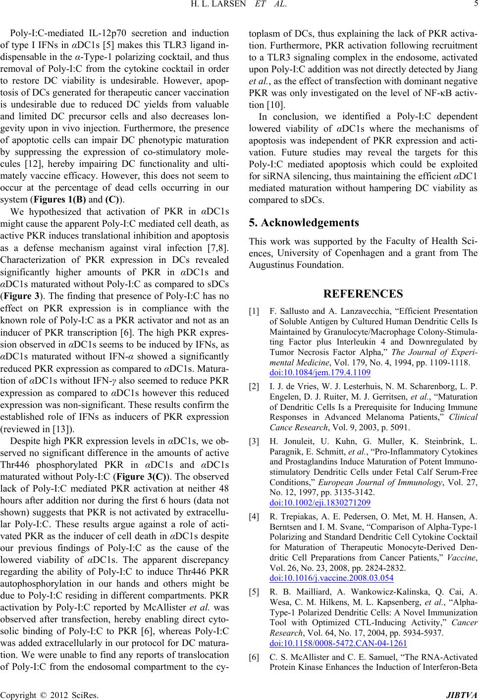 H. L. LARSEN ET AL. 5 Poly-I:C-mediated IL-12p70 secretion and induction of n of PKR in αDC1s m pression levels in αDC1s, we ob- se sion, we identified a Poly-I:C dependent lo 5. Acknowledgements the Faculty of Health Sci- REFERENCES [1] F. Sallusto anicient Presentation type I IFNs in αDC1s [5] makes this TLR3 ligand in- dispensable in the α-Type-1 polarizing cocktail, and thus removal of Poly-I:C from the cytokine cocktail in order to restore DC viability is undesirable. However, apop- tosis of DCs generated for therapeutic cancer vaccination is undesirable due to reduced DC yields from valuable and limited DC precursor cells and also decreases lon- gevity upon in vivo injection. Furthermore, the presence of apoptotic cells can impair DC phenotypic maturation by suppressing the expression of co-stimulatory mole- cules [12], hereby impairing DC functionality and ulti- mately vaccine efficacy. However, this does not seem to occur at the percentage of dead cells occurring in our system (Figures 1(B) and (C)). We hypothesized that activatio ight cause the apparent Poly-I:C mediated cell death, as active PKR induces translational inhibition and apoptosis as a defense mechanism against viral infection [7,8]. Characterization of PKR expression in DCs revealed significantly higher amounts of PKR in αDC1s and αDC1s maturated without Poly-I:C as compared to sDCs (Figure 3). The finding that presence of Poly-I:C has no effect on PKR expression is in compliance with the known role of Poly-I:C as a PKR activator and not as an inducer of PKR transcription [6]. The high PKR expres- sion observed in αDC1s seems to be induced by IFNs, as αDC1s maturated without IFN-α showed a significantly reduced PKR expression as compared to αDC1s. Matura- tion of αDC1s without IFN-γ also seemed to reduce PKR expression as compared to αDC1s however this reduced expression was non-significant. These results confirm the established role of IFNs as inducers of PKR expression (reviewed in [13]). Despite high PKR ex rved no significant difference in the amounts of active Thr446 phosphorylated PKR in αDC1s and αDC1s maturated without Poly-I:C (Figure 3(C)). The observed lack of Poly-I:C mediated PKR activation at neither 48 hours after addition nor during the first 6 hours (data not shown) suggests that PKR is not activated by extracellu- lar Poly-I:C. These results argue against a role of acti- vated PKR as the inducer of cell death in αDC1s despite our previous findings of Poly-I:C as the cause of the lowered viability of αDC1s. The apparent discrepancy regarding the ability of Poly-I:C to induce Thr446 PKR autophosphorylation in our hands and others might be due to Poly-I:C residing in different compartments. PKR activation by Poly-I:C reported by McAllister et al. was observed after transfection, hereby enabling direct cyto- solic binding of Poly-I:C to PKR [6], whereas Poly-I:C was added extracellularly in our protocol for DC matura- tion. We were unable to find any reports of translocation of Poly-I:C from the endosomal compartment to the cy- toplasm of DCs, thus explaining the lack of PKR activa- tion. Furthermore, PKR activation following recruitment to a TLR3 signaling complex in the endosome, activated upon Poly-I:C addition was not directly detected by Jiang et al., as the effect of transfection with dominant negative PKR was only investigated on the level of NF-κB activ- tion [10]. In conclu wered viability of αDC1s where the mechanisms of apoptosis was independent of PKR expression and acti- vation. Future studies may reveal the targets for this Poly-I:C mediated apoptosis which could be exploited for siRNA silencing, thus maintaining the efficient αDC1 mediated maturation without hampering DC viability as compared to sDCs. This work was supported by ences, University of Copenhagen and a grant from The Augustinus Foundation. d A. Lanzavecchia, “Eff of Soluble Antigen by Cultured Human Dendritic Cells Is Maintained by Granulocyte/Macrophage Colony-Stimula- ting Factor plus Interleukin 4 and Downregulated by Tumor Necrosis Factor Alpha,” The Journal of Experi- mental Medicine, Vol. 179, No. 4, 1994, pp. 1109-1118. doi:10.1084/jem.179.4.1109 [2] I. J. de Vries, W. J. Lesterhuis, N. M. Scharenborg, L. P. Steinbrink, L. Engelen, D. J. Ruiter, M. J. Gerritsen, et al., “Maturation of Dendritic Cells Is a Prerequisite for Inducing Immune Responses in Advanced Melanoma Patients,” Clinical Cance Research, Vol. 9, 2003, p. 5091. [3] H. Jonuleit, U. Kuhn, G. Muller, K. Paragnik, E. Schmitt, et al., “Pro-Inflammatory Cytokines and Prostaglandins Induce Maturation of Potent Immuno- stimulatory Dendritic Cells under Fetal Calf Serum-Free Conditions,” European Journal of Immunology, Vol. 27, No. 12, 1997, pp. 3135-3142. doi:10.1002/eji.1830271209 [4] R. Trepiakas, A. E. Pedersen, O. Met, M. H. Hansen, A. Berntsen and I. M. Svane, “Comparison of Alpha-Type-1 Polarizing and Standard Dendritic Cell Cytokine Cocktail for Maturation of Therapeutic Monocyte-Derived Den- dritic Cell Preparations from Cancer Patients,” Vaccine, Vol. 26, No. 23, 2008, pp. 2824-2832. doi:10.1016/j.vaccine.2008.03.054 [5] R. B. Mailliard, A. Wankowicz-Kalinska, Q. Cai, A. Wesa, C. M. Hilkens, M. L. Kapsenberg, et al., “Alpha- Type-1 Polarized Dendritic Cells: A Novel Immunization Tool with Optimized CTL-Inducing Activity,” Cancer Research, Vol. 64, No. 17, 2004, pp. 5934-5937. doi:10.1158/0008-5472.CAN-04-1261 [6] C. S. McAllister and C. E. Samuel, “The RNA-Activated Protein Kinase Enhances the Induction of Interferon-Beta Copyright © 2012 SciRes. JIBTVA  H. L. LARSEN ET AL. Copyright © 2012 SciRes. JIBTVA 6 and Apoptosis Mediated by Cytoplasmic RNA Sensors,” Journal of Biological Chemistry, Vol. 284, No. 3, 2009, pp. 1644-1651. doi:10.1074/jbc.M807888200 [7] F. Zhang, P. R. Romano, T. Nagamura-Inoue, B. Tian, T. E. Dever, M. B. Mathews, et al., “Binding of Double- Stranded RNA to Protein Kinase PKR Is Required for Dimerization and Promotes Critical Autophosphorylation Events in the Activation Loop,” Journal of Biological Chemistry, Vol. 276, No. 27, 2001, pp. 24946-24958. doi:10.1074/jbc.M102108200 [8] D. Scheuner, R. Patel, F. Wang, K. Lee, K. Kumar, J. W et al., “Double-Stranded RNA- u Dependent Protein Kinase , Phosphorylation of the Alpha-Subunit of Eukaryotic Translation Initiation Factor 2 Mediates Apoptosis,” Journal of Biological Chemistry, Vol. 281, No. 30, 2006, pp. 21458-21468. doi:10.1074/jbc.M603784200 [9] L. Alexopoulou, A. C. Holt, R. Medzhitov and R. A. Flavell, “Recognition of Double-Stranded RNA and Activation of NF-kappa B by Toll-Like Receptor 3,” Nature, Vol. 413, No. 6857, 2001, pp. 732-738. doi:10.1038/35099560 [10] Z. Jiang, M. Zamanian-Daryoush, H. Nie, A. M. S R. Williams and X. Li, ilva, B. “Poly(I-C)-Induced Toll-Like Receptor 3 (TLR3)-Mediated Activation of NFkappa B and MAP Kinase Is through an Interleukin-1 Receptor- Associated Kinase (IRAK)-Independent Pathway Employ- ing the Signaling Components TLR3-TRAF6-TAK1- TAB2-PKR,” Journal of Biological Chemistry, Vol. 278, No. 19, 2003, pp. 16713-16719. doi:10.1074/jbc.M300562200 [11] A. E. Pedersen, M. Thorn, M. Gad, M. R. Walter, H. E. Johnsen, E. Gaarsdal, et al., “Phenotypic and Functional Characterization of Clinical Grade Dendritic Cells Generated from Patients with Advanced Breast Cancer for Therapeutic Vaccination,” Scandinavian Journal of Immunology, Vol. 61, No. 2, 2005, pp. 147-156. doi:10.1111/j.0300-9475.2005.01531.x [12] C. A. Williams, R. A. Harry and J. D. McLeod, .x “Apoptotic Cells Induce Dendritic Cell-Mediated Suppres- sion via Interferon-Gamma-Induced IDO,” Immunology, Vol. 124, No. 1, 2008, pp. 89-101. doi:10.1111/j.1365-2567.2007.02743 an, “The dsRNA [13] M. A. Garcia, E. F. Meurs and M. Esteb Protein Kinase PKR: Virus and Cell Control,” Biochimie, Vol. 89, No. 6-7, 2007, pp. 799-811. doi:10.1016/j.biochi.2007.03.001 |