

Paper Menu >>
Journal Menu >>
 Open Journal of Respiratory Diseases, 2012, 2, 31-36 http://dx.doi.org/10.4236/ojrd.2012.22005 Published Online May 2012 (http://www.SciRP.org/journal/ojrd) Should We Measure the FEV1 or the Specific Resistance of the Airways? An Evaluation in Patients with Either COPD, Chronic Dyspnea or Chronic Cough* Kinga Simon1, Viviane De Maertelaer2, André Noseda1 1The Pulmonary Division, CHU Brugmann, Brussels, Belgium 2IRIBHN and Department of Biostatistics and Medical Informatics (SBIM), Université Libre de Bruxelles, Brussels, Belgium Email: [email protected] Received December 27, 2011; revised February 3, 2012; accepted February 13, 2012 ABSTRACT Background: The purpose of this study was to evaluate the relative contribution of measuring the forced expiratory volume in one second (FEV1) or the specific resistance of the airways (sRaw) in adults referred for chronic obstructive pulmonary disease (COPD), chronic dyspnea or chronic cough. Methods: This was a prospective study of 321 subjects referred for lung function testing, in a setting of routine clinical management, for suspicion of COPD (or follow-up of known COPD), chronic dyspnea or chronic cough. The proportions of FEV1 values below the normal range and of sRaw values above the normal range were compared using a Chi-square exact test of Fisher. Results: In the COPD and chronic dyspnea groups, sRaw was as frequently abnormal as FEV1. In the chronic cough group, sRaw was increased in 56.5% of subjects, while FEV1 was decreased in solely 34.8% (p = 0.059). Conclusions: This study suggests that sRaw may be a better tool than FEV1 to detect bronchial obstruction in patients presenting with chronic cough. Keywords: Body Plethysmography; Chronic Cough; Specific Airway Resistance 1. Introduction Spirometry is often considered as the gold standard for determination of obstructive pulmonary disease. This method has however several limitations, which has led to resistance measurements used to clarify lung pathophysi- ology [1]. In the recent years, there has been a progres- sive disinterest in resistance measurements, at least in adults. As an illustration, the 2005 American Thoracic Society and European Respiratory Society statements did not address resistance measurements [2]. In contrast, resistance measurements are still considered to be par- ticularly relevant in pediatric patients [3,4]. The present study was designed to evaluate the relative contribution of measuring the forced expiratory volume in one second (FEV1) or the specific resistance of the airways (sRaw) in adults referred for lung function test- ing (LFT) because of chronic obstructive pulmonary dis- ease (COPD), chronic dyspnea or chronic cough. These indications were chosen because the Belgian authorities recently suggested that the usefulness of assessing airway resistance is not proven in subjects with either COPD, chronic dyspnea or chronic cough [5]. We hypothesized that sRaw may be more sensitive than FEV1 in detecting bronchial obstruction in these groups of patients. The study was performed in a setting of routine clinical man- agement and aimed to evaluate the percentage of subjects with an abnormal test, namely a decreased FEV1 or an increased sRaw. 2. Patients and Methods 2.1. Study Design In the present study, we prospectively evaluated the sRaw and the FEV1 in three groups of subjects (as defined un- derneath) in a setting of routine clinical management. The patients were referred by their physician for LFT because of either COPD (group 1), chronic dyspnea (group 2) or chronic cough (group 3). The study protocol was approved by the Ethics Committee of the CHU Brug- mann and each subject gave written informed consent to the analysis of his (her) lung function data. 2.2. Patients The study was performed at the Pulmonary Division of the CHU Brugmann between 26/10/2009 and 16/07/2010. All males and females between 18 and 70 years old, re- ferred for LFT because of COPD, chronic dyspnea or chronic cough, were eligible provided they gave in- formed consent and were able to perform technically *Conflict of interes t : There is no conflict of interest. C opyright © 2012 SciRes. OJRD  K. SIMON ET AL. 32 acceptable spirometry and plethysmography. Exclusion criteria included age less than 18 or more than 70, being referred for LFT because of another diagnosis, being unable to perform either spirometry or plethysmography, as well as having any contra-indication to LFT (recent heart attack or pneumothorax, recent ocular, abdominal or thoracic surgery, suspicion of tuberculosis, severe claustrophobia). 2.3. Constitution of Groups The classification into three groups was made on basis of the requests for LTF, written by the referring physicians. Subjects were put into group 1 (COPD) when the request mentioned a clinical suspicion of COPD or follow-up of a known COPD. When the request mentioned several respiratory diseases, subjects were included provided COPD was listed as the first diagnosis (e.g. COPD plus bronchectasis). Subjects were put into group 2 (chronic dyspnea) and into group 3 (chronic cough) when the re- quest respectively mentioned chronic breathlessness or chronic cough as the first reason to perform LTF. Pa- tients referred for LFT for other indications (e.g. preop- erative work-up), or for several indications with another diagnosis listed first (e.g. known lung fibrosis and chronic cough) were not included. 2.4. Lung Function Testing A Zan (Waldfenster, Germany) body plethysmograph was used. Patients were instructed in the correct tech- nique while the plethysmography door was open. During the maneuvers, patients were asked to firmly seal their lips around the mouthpiece. Airflow at the mouth was displayed against box pressure and their relationship was computed according to an automated procedure. Signals were digitized over a period of five breaths, with a sam- pling rate of 50 Hz. After correction for the thermal drift induced by the subject’s heat production, a loop was ob- tained and midpoints at flows of +0.5 (point A) and −0.5 l·s−1 (point B) were obtained. The slope of the line be- tween points A and B defined the sRaw. At least two technically satisfactory five breath-loops were obtained and the reported sRaw was an arithmetical mean. Spi- rometry was performed with the door of the box open. The subject was instructed to slowly inspire up to the total lung capacity and to subsequently expire a forced vital capacity. In each subject, two to three technically acceptable curves were obtained. Optimization of the reading of the expiratory curves was performed as rec- ommended by Peslin et al. [6]. The FEV1 was defined as the volume exhaled in one second during the perform- ance of the forced vital capacity maneuver. When a bronchodilation or a provocation test was performed, only the baseline values were retained for analysis. 2.5. Interpretation of Lung Function Data To calculate the predicted values for FEV1, the regres- sion equations of Quanjer et al. [7] were used. An indi- vidual FEV1 value was considered as below the normal range if it was lower than the mean predicted minus 1.64 × standard deviation (SD). The data of Pelzer and Thom- son [8] were used as mean predicted values for sRaw, depending on gender. An individual sRaw value was considered as above the normal range if it was higher than the mean predicted value plus 2 × SD. 2.6. Statistical Analyses The variables recorded in the three groups were com- pared using either an ANOVA procedure (means com- parisons in case of continuous variables) or a Chi-square exact test (proportions comparisons in case of discrete variables). More particularly, the proportions of FEV1 values below the normal range and of sRaw values above the normal range were compared using a Chi-square ex- act test of Fisher. The analysis was made separately in the three groups (COPD, chronic dyspnea, and chronic cough). The statistical software used was SPSS version 17.0. 2.7. Retrospective Review of Medical Charts Twelve months after the study had been completed, medi- cal charts were examined to check for the final cause retained for chronic dyspnea and for chronic cough, in the chronic dyspnea and chronic cough groups. 3. Results During the study period, 321 subjects were included. The allocation into the three groups is listed in Table 1, as well as the mean (SD) values obtained for FEV1 and sRaw. COPD subjects were older than subjects with chronic dyspnea or chronic cough. The proportion of males in the COPD group was higher than in the chronic dyspnea group, and tended to be higher (p = 0.074) than in the chronic cough group. As expected, FEV1 was lower, and sRaw higher, in the COPD group than in the other two groups (Table 2). Individual FEV1 values were plotted against predicted values, separately in males and females. As shown in Figure 1, measured values were predominantly under the identity line in the COPD group, while a large proportion of values were near the identity line in the chronic dysp- nea and chronic cough groups (Figures 2 and 3). The proportions of subjects with FEV1 within versus under the normal range, as well as those of subjects with sRaw within or above the normal range, are shown in Tab le 3. In the chronic dyspnea and COPD groups, sRaw was as frequently abnormal as FEV1 (exact Fisher test, p = 1.000 Copyright © 2012 SciRes. OJRD 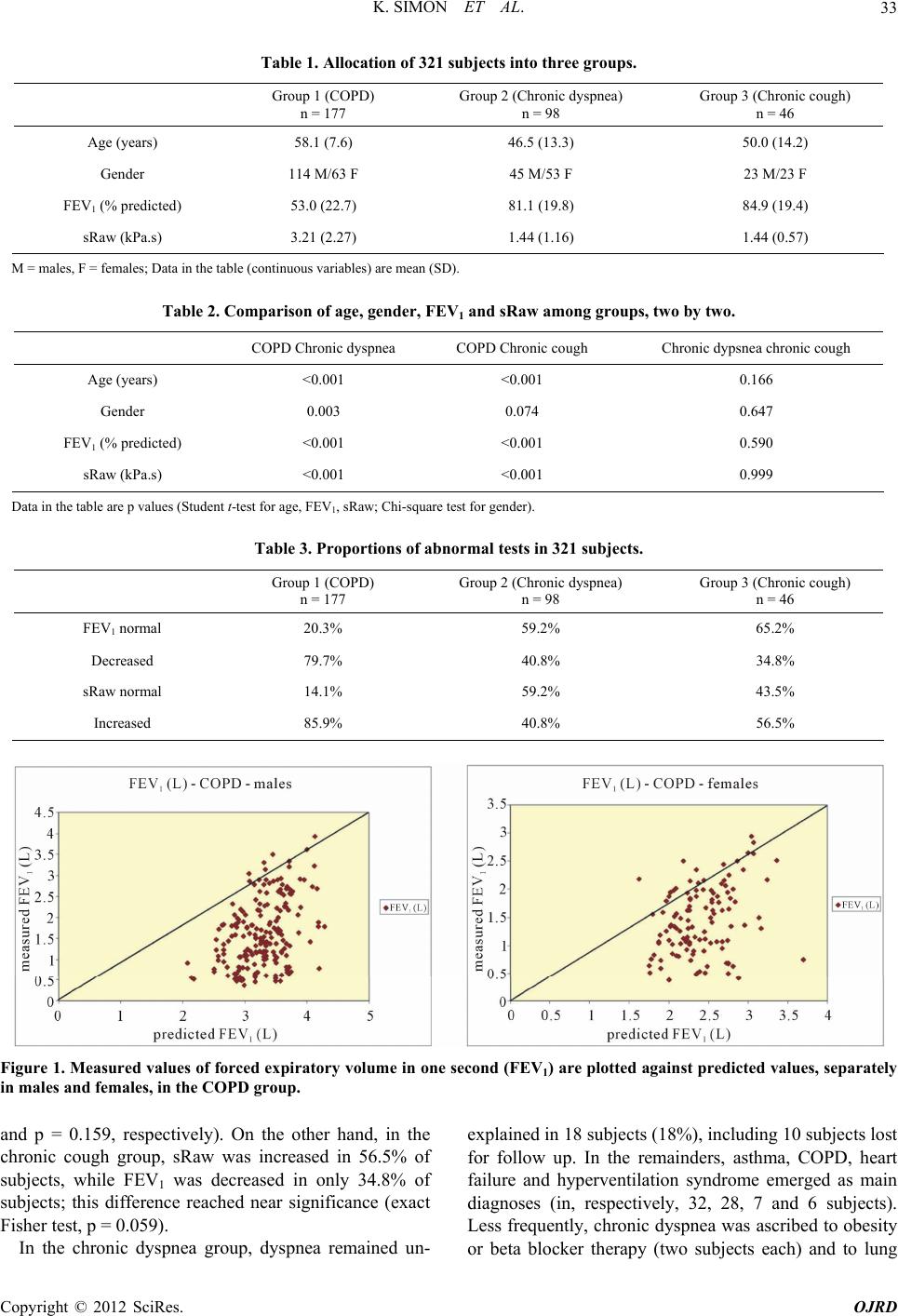 K. SIMON ET AL. Copyright © 2012 SciRes. OJRD 33 Table 1. Allocation of 321 subjects into three groups. Group 1 (COPD) n = 177 Group 2 (Chronic dyspnea) n = 98 Group 3 (Chronic cough) n = 46 Age (years) 58.1 (7.6) 46.5 (13.3) 50.0 (14.2) Gender 114 M/63 F 45 M/53 F 23 M/23 F FEV1 (% predicted) 53.0 (22.7) 81.1 (19.8) 84.9 (19.4) sRaw (kPa.s) 3.21 (2.27) 1.44 (1.16) 1.44 (0.57) M = males, F = females; Data in the table (continuous variables) are mean (SD). Table 2. Comparison of age, gender, FEV1 and sRaw among groups, two by two. COPD Chronic dyspnea COPD Chronic cough Chronic dypsnea chronic cough Age (years) <0.001 <0.001 0.166 Gender 0.003 0.074 0.647 FEV1 (% predicted) <0.001 <0.001 0.590 sRaw (kPa.s) <0.001 <0.001 0.999 Data in the table are p values (Student t-test for age, FEV1, sRaw; Chi-square test for gender). Table 3. Proportions of abnormal tests in 321 subjects. Group 1 (COPD) n = 177 Group 2 (Chronic dyspnea) n = 98 Group 3 (Chronic cough) n = 46 FEV1 normal 20.3% 59.2% 65.2% Decreased 79.7% 40.8% 34.8% sRaw normal 14.1% 59.2% 43.5% Increased 85.9% 40.8% 56.5% Figure 1. Measured values of forced expiratory volume in one second (FEV1) are plotted against predicted values, separately in males and females, in the COPD group. and p = 0.159, respectively). On the other hand, in the chronic cough group, sRaw was increased in 56.5% of subjects, while FEV1 was decreased in only 34.8% of subjects; this difference reached near significance (exact Fisher test, p = 0.059). In the chronic dyspnea group, dyspnea remained un- explained in 18 subjects (18%), including 10 subjects lost for follow up. In the remainders, asthma, COPD, heart failure and hyperventilation syndrome emerged as main diagnoses (in, respectively, 32, 28, 7 and 6 subjects). Less frequently, chronic dyspnea was ascribed to obesity or beta blocker therapy (two subjects each) and to lung 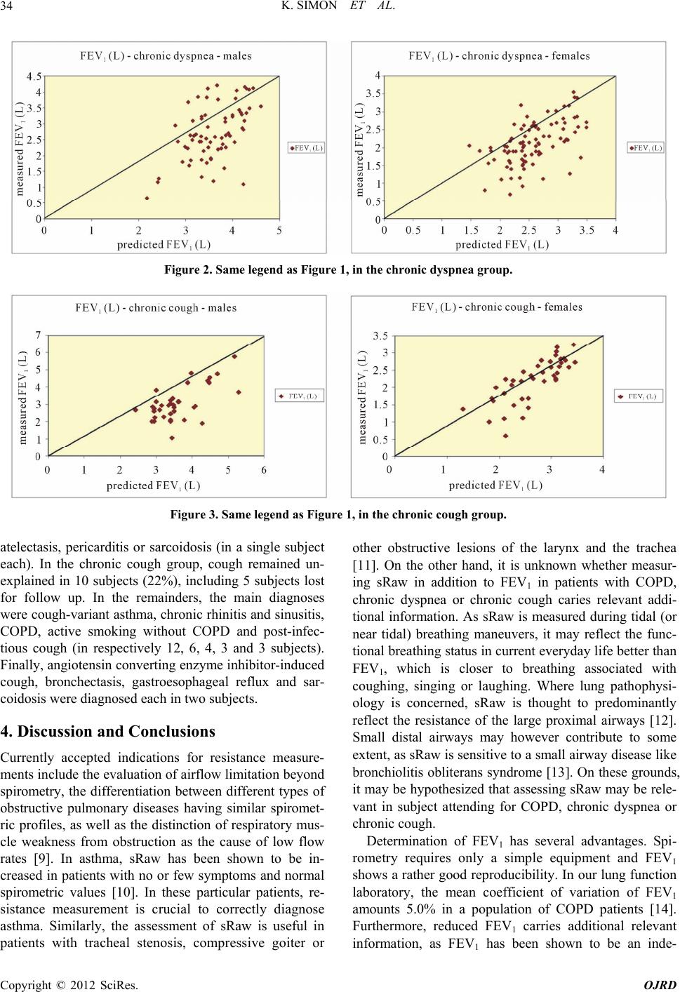 K. SIMON ET AL. 34 Figure 2. Same legend as Figure 1, in the c hr onic dyspnea group. Figure 3. Same legend as Figure 1, in the chronic cough group. atelectasis, pericarditis or sarcoidosis (in a single subject each). In the chronic cough group, cough remained un- explained in 10 subjects (22%), including 5 subjects lost for follow up. In the remainders, the main diagnoses were cough-variant asthma, chronic rhinitis and sinusitis, COPD, active smoking without COPD and post-infec- tious cough (in respectively 12, 6, 4, 3 and 3 subjects). Finally, angiotensin converting enzyme inhibitor-induced cough, bronchectasis, gastroesophageal reflux and sar- coidosis were diagnosed each in two subjects. 4. Discussion and Conclusions Currently accepted indications for resistance measure- ments include the evaluation of airflow limitation beyond spirometry, the differentiation between different types of obstructive pulmonary diseases having similar spiromet- ric profiles, as well as the distinction of respiratory mus- cle weakness from obstruction as the cause of low flow rates [9]. In asthma, sRaw has been shown to be in- creased in patients with no or few symptoms and normal spirometric values [10]. In these particular patients, re- sistance measurement is crucial to correctly diagnose asthma. Similarly, the assessment of sRaw is useful in patients with tracheal stenosis, compressive goiter or other obstructive lesions of the larynx and the trachea [11]. On the other hand, it is unknown whether measur- ing sRaw in addition to FEV1 in patients with COPD, chronic dyspnea or chronic cough caries relevant addi- tional information. As sRaw is measured during tidal (or near tidal) breathing maneuvers, it may reflect the func- tional breathing status in current everyday life better than FEV1, which is closer to breathing associated with coughing, singing or laughing. Where lung pathophysi- ology is concerned, sRaw is thought to predominantly reflect the resistance of the large proximal airways [12]. Small distal airways may however contribute to some extent, as sRaw is sensitive to a small airway disease like bronchiolitis obliterans syndrome [13]. On these grounds, it may be hypothesized that assessing sRaw may be rele- vant in subject attending for COPD, chronic dyspnea or chronic cough. Determination of FEV1 has several advantages. Spi- rometry requires only a simple equipment and FEV1 shows a rather good reproducibility. In our lung function laboratory, the mean coefficient of variation of FEV1 amounts 5.0% in a population of COPD patients [14]. Furthermore, reduced FEV1 carries additional relevant information, as FEV1 has been shown to be an inde- Copyright © 2012 SciRes. OJRD  K. SIMON ET AL. 35 pendent marker for cardiovascular mortality [15]. On the other hand, spirometry is in a large extent dependent on the subject cooperation. As an illustration, as many as 11.5% of subjects are unable to perform correct forced expiratory maneuvers, even in a young population with little or no chronic lung disease [16]. Similarly, measur- ing sRaw via plethysmography has advantages and dis- advantages. The equipment is more sophisticated, more expensive and the reproducibility is somewhat weaker than that of FEV1, with e.g. a mean coefficient of varia- tion of 9.3% in COPD patients in our laboratory [14]. On the other hand, sRaw is more sensitive than FEV1 in a large variety of clinical conditions. Studying the acute effect of physiotherapy on lung function in patients with copious sputum production, Cochrane et al. found a greater change in sRaw than in FEV1 [17]. In patients with heart-lung or bilateral lung transplantation, Bassiri et al found that serial measurements of sRaw were more useful than those of FEV1 for early detection of bron- chiolitis obliterans syndrome [13]. In the present study involving three groups of patients with respectively COPD, chronic dyspnea and chronic cough, we were able to show a larger proportion of sub- jects with increased sRaw than that of subjects with de- creased FEV1 only in chronic coughers. The difference drew near statistical significance. On retrospective ex- amination of medical charts, cough-variant asthma emerged as the most frequent diagnosis retained to ex- plain chronic cough. Cough-variant asthma is known to be a frequent cause of chronic cough, particularly in non- smokers. It may be either a precursor of typical asthma, or persist lifelong as the sole symptom of asthma [18]. Definite diagnosis is usually based on both histamine or metacholine inhalation testing and on resolution of cough with anti-asthmatic therapy [19]. Studies on larger groups of patients are needed to evaluate whether sRaw may be a better tool than FEV1 to detect mild bronchial obstruction in patients presenting with chronic cough. 5. Acknowledgements The authors acknowledge Mrs F. Daimallah, Ch. Jacobs and J. Roobaert for performing lung function testing, as well as Mrs F. Martinez Vadillo for secretarial assis- tance. REFERENCES [1] P. Calverley and N. Koulouris, “Flow Limitation and Dy- namic Hyperinflation: Key Concepts in Modern Respiratory Physiology,” European Respiratory Journal, Vol. 25, No. 1, 2005, pp. 186-199. doi:10.1183/09031936.04.00113204 [2] American Thoracic Society, “Pulmonary Laboratory Man- agement and Procedure Manual,” American Thoracic So- ciety, New York, 2005. [3] H. Bisgaard and K. G. Nielsen, “Plethysmographic Meas- urements of Specific Airway Resistance in Young Chil- dren,” Chest, Vol. 128, No. 1, 2005, 355-362. doi:10.1378/chest.128.1.355 [4] J. Kirkby, S. Stanojevic, L. Welsh, S. Lum, M. Badier, C. Beardsmore, A. Custovic, K. Nielsen, J. Paton and W. Tomalak, “Reference Equations for Specific Airway Re- sistance in Children: The Asthma UK Initiative,” Euro- pean Respiratory Journal, Vol. 36, No. 3, 2010, pp. 622- 629. doi:10.1183/09031936.00135909 [5] A. Van den Bruel, J. Gailly, S. Devriese, F. Vritens and D. Ramaekers, “Pulmonary Function Tests in Adults: Good Clinical Practice,” Belgian Health Care Knowledge Cen- tre, Brussels, 2007, KCE Reports 6. [6] R. Peslin, A. Bohadana, B. Hannhart and P. Jardin, “Com- parison of Various Methods for Reading Maximal Expi- ratory Flow-Volume Curves,” American Review of Res- piratory Disease, Vol. 119, 1979, pp. 271-277. [7] Ph. H. Quanjer, A. Dalhuijsen and B. C. Van Zomeben, “Summary Equations of Reference Values,” Bulletin Eu- ropeen de Physiopathologie Respiratoire, Vol. 19, No. 5, 1983, pp. 45-51. [8] A. M. Pelzer and M. L. Thomson, “Effect of Age, Sex, Stature and Smoking Habits on Human Airway Conduc- tance,” Journal of Applied Physiology, Vol. 21, No. 2, 1966, pp. 469-476. [9] S. Blonshine and M. D. Goldman, “Optimizing Perform- ance of Respiratory Airflow Resistance Measurements,” Chest, Vol. 134, No. 6, 2008, pp. 1304-1309. doi:10.1378/chest.06-2898 [10] D. A. Kaminsky, C. G. Irvin, D. A. Gurka, D. C. Feldsien, E. M. Wagner, M. C. Liu and S. E. Wenzel, “Peripheral Airways Responsiveness to Cool, Dry Air in Normal and Asthmatic Individuals,” American Journal of Respiratory and Critical Care Medicine, Vol. 152, No. 6, 1995, pp. 1784-1790. [11] R. D. Miller and R. E. Hyatt, “Obstructing Lesions of the Larynx and Trachea: Clinical and Physiologic Character- istics,” Mayo Clinic Proceedings, Vol. 44, 1969, pp. 145- 161. [12] P. T. Macklem and T. Mead, “Resistance of Central and Peripheral Airways Measured by Retrograde Catheter,” Journal of Applied Physiology, Vol. 22, No. 3, 1967, pp. 395-401. [13] A. G. Bassiri, R. E. Girgis, R. L. Doyle and J. Theodore, “Detection of Small Airway Dysfunction Using Specific Airway Conductance,” Chest Vol. 111, No. 6, 1997, pp. 1533-1535. doi:10.1378/chest.111.6.1533 [14] A. Noseda, J. Schmerber, T. Prigogine and J. C. Yernault, “Perceived Effect on Shortness of Breath of an Acute In- halation of Saline or Terbutaline: Variability and Sensi- tivity of a Visual Analogue Scale in Patients with Asthma or COPD,” European Respiratory Journal, Vol. 5, 1992, pp. 1043-1053. [15] D. D. Sin, L. Wu and S. F. Man, “The Relationship be- tween Reduced Lung Function and Cardiovascular Mor- tality: A Population-Based Study and a Systematic Re- Copyright © 2012 SciRes. OJRD  K. SIMON ET AL. Copyright © 2012 SciRes. OJRD 36 view of the Literature,” Chest, Vol. 127, No. 6, 2005, pp. 1952-1959. doi:10.1378/chest.127.6.1952 [16] L. W. Ng’Ang’a, T. P. Ernst, M. S. Jaakkola, G. Gerardi, J. H. Hanley and M. R. Becklake, “Spirometric Lung Function: Distribution and Determinants of Test Failure in a Young Adult Population,” American Review of Res- piratory Disease, Vol. 145, No. 1, 1992, pp. 48-52. doi:10.1164/ajrccm/145.1.48 [17] G. M. Cochrane, B. A. Webber and S. W. Clarke, “Ef- fects of Sputum on Pulmonary Function,” British Medical Journal, Vol. 2, No. 6096, 1977, pp. 1181-1183. doi:10.1136/bmj.2.6096.1181 [18] P. V. Dicpinigaitis, “Chronic Cough Due to Asthma. ACCP Evidence-Based Clinical Practice Guidelines,” Chest, Vol. 129, No. 1, 2006, pp. 75S-79S. doi:10.1378/chest.129.1_suppl.75S [19] A. Niimi, “Cough and Asthma,” Current Respiratory Medi - cine Reviews, Vol. 7, No. 1, 2011, pp. 47-54. doi:10.2174/157339811794109327 List of Abbreviations COPD: chronic obstructive pulmonary disease FEV1: forced expiratory volume in one second LFT: lung function testing SD: standard deviation sRaw: specific resistance of the airways |