

Paper Menu >>
Journal Menu >>
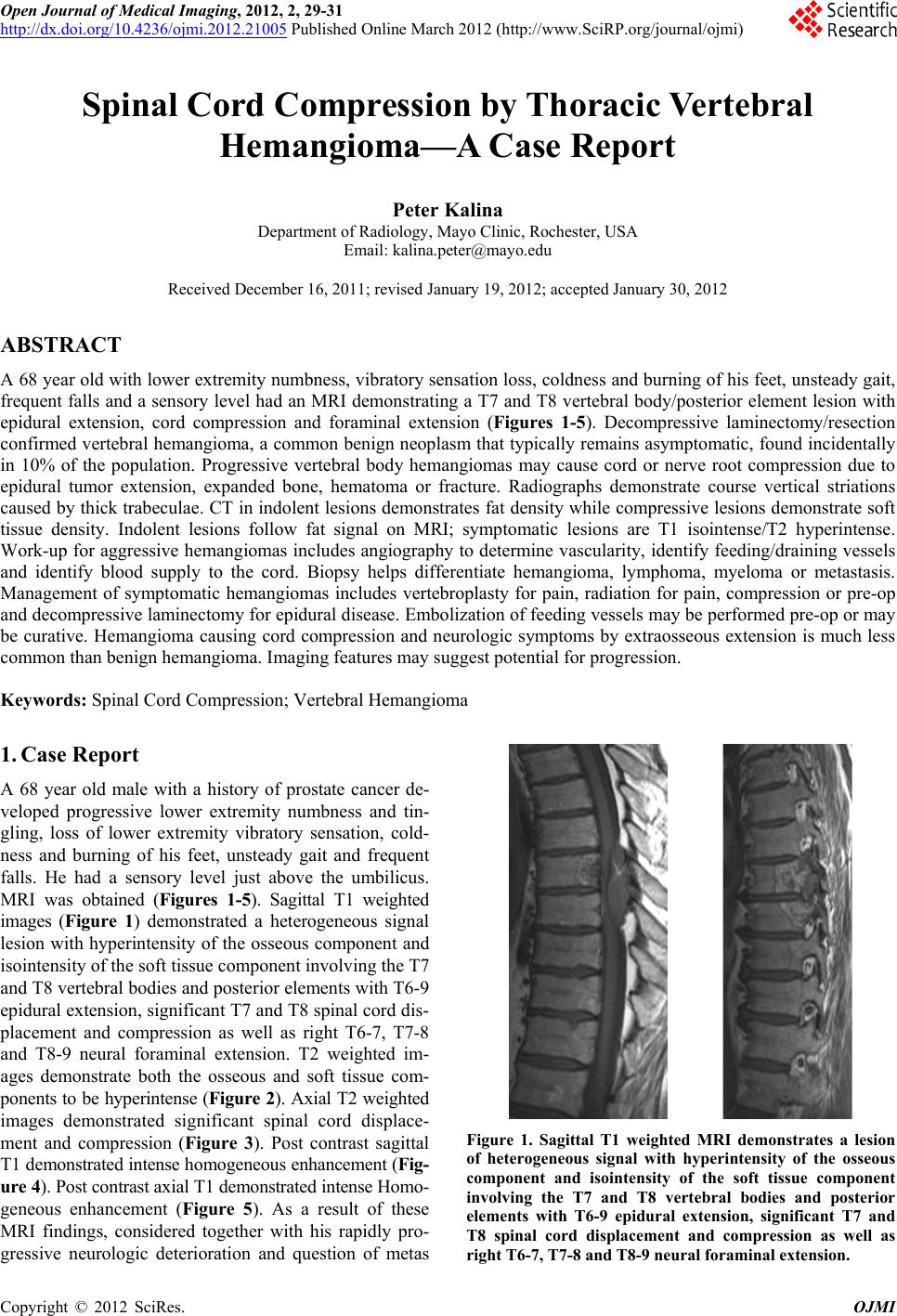 Open Journal of Medical Imaging, 2012, 2, 29-31 http://dx.doi.org/10.4236/ojmi.2012.21005 Published Online March 2012 (http://www.SciRP.org/journal/ojmi) Spinal Cord Compression by Thoracic Vertebral Hemangioma—A Case Report Peter Kalina Department of Radiology, Mayo Clinic, Rochester, USA Email: [email protected] Received December 16, 2011; revised January 19, 2012; accepted January 30, 2012 ABSTRACT A 68 year old with lower extremity numbness, vibratory sensation loss, coldness and burning of his feet, unsteady gait, frequent falls and a sensory level had an MRI demonstrating a T7 and T8 vertebral body/p osterior element lesion with epidural extension, cord compression and foraminal extension (Figures 1-5). Decompressive laminectomy/resection confirmed vertebral hemangioma, a common benign neoplasm that typically remains asymptomatic, found incidentally in 10% of the population. Progressive vertebral body hemangiomas may cause cord or nerve root compression due to epidural tumor extension, expanded bone, hematoma or fracture. Radiographs demonstrate course vertical striations caused by thick trabeculae. CT in indolent lesions demonstrates fat density while compressive lesions demonstrate soft tissue density. Indolent lesions follow fat signal on MRI; symptomatic lesions are T1 isointense/T2 hyperintense. Work-up for aggressive hemangiomas includes angiography to determine vascularity, identify feeding/draining vessels and identify blood supply to the cord. Biopsy helps differentiate hemangioma, lymphoma, myeloma or metastasis. Management of symptomatic hemangiomas includes vertebroplasty for pain, radiation for pain, compression or pre-op and decompressive laminectomy for epidural disease. Embolization of feeding vessels may be performed pre-op or may be curative. Hemangioma causing cord compression and neurologic symptoms by extraosseous extension is much less common than benign hemangioma. Imaging features may suggest potential for progression. Keywords: Spinal Cord Compression; Vertebral Hemangioma 1. Case Report A 68 year old male with a history of prostate cancer de- veloped progressive lower extremity numbness and tin- gling, loss of lower extremity vibratory sensation, cold- ness and burning of his feet, unsteady gait and frequent falls. He had a sensory level just above the umbilicus. MRI was obtained (Figures 1-5). Sagittal T1 weighted images (Figure 1) demonstrated a heterogeneous signal lesion with hyperintensity of the osseous component and isointensity of the soft tissue component involving the T7 and T8 vertebral bodies and posterior elements with T6-9 epidural exten sion, significant T7 an d T8 spinal cord dis- placement and compression as well as right T6-7, T7-8 and T8-9 neural foraminal extension. T2 weighted im- ages demonstrate both the osseous and soft tissue com- ponents to be hyperintense (Figure 2). Axial T2 weighted images demonstrated significant spinal cord displace- ment and compression (Figure 3). Post contrast sagittal T1 demonstrated intense homogeneous enhancement (Fig- ure 4). Post contrast axial T1 demonstrated intense Homo- geneous enhancement (Figure 5). As a result of these MRI findings, considered together with his rapidly pro- gressive neurologic deterioration and question of metas Figure 1. Sagittal T1 weighted MRI demonstrates a lesion of heterogeneous signal with hyperintensity of the osseous component and isointensity of the soft tissue component involving the T7 and T8 vertebral bodies and posterior elements with T6-9 epidural extension, significant T7 and T8 spinal cord displacement and compression as well as right T6-7, T7-8 and T8-9 neural foraminal extension. C opyright © 2012 SciRes. OJMI 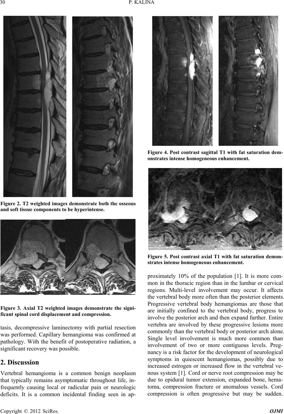 P. KALINA 30 Figure 2. T2 weighted images demonstrate both the osseous and soft tissue components to be hyperintense. Figure 3. Axial T2 weighted images demonstrate the signi- ficant spinal cord displacement and compression. tasis, decompressive laminectomy with partial resection was performed. Capillary hemangioma was confirmed at pathology. With the benefit of postoperative radiation, a significant recovery was possi bl e . 2. Discussion Vertebral hemangioma is a common benign neoplasm that typically remains asymptomatic throughout life, in- frequently causing local or radicular pain or neurologic deficits. It is a common incidental finding seen in ap- Figure 4. Post contrast sagittal T1 with fat saturation dem- onstrates intense homogeneous enhance m e nt. Figure 5. Post contrast axial T1 with fat saturation demon- strates intense homogeneous enhance ment. proximately 10% of the population [1]. It is more com- mon in the thoracic region than in the lumbar or cervical regions. Multi-level involvement may occur. It affects the vertebral body more often than the posterior elements. Progressive vertebral body hemangiomas are those that are initially confined to the vertebral body, progress to involve the posterio r arch and then expand further. Entire vertebra are involved by these progressive lesions more commonly than the vertebral body or posterior arch alone. Single level involvement is much more common than involvement of two or more contiguous levels. Preg- nancy is a risk factor for the development of neurological symptoms in quiescent hemangiomas, possibly due to increased estrogen or increased flow in the vertebral ve- nous system [1]. Cord or nerve root compression may be due to epidural tumor extension, expanded bone, hema- toma, compression fracture or anomalous vessels. Cord compression is often progressive but may be sudden. Copyright © 2012 SciRes. OJMI  P. KALINA Copyright © 2012 SciRes. OJMI 31 Pain often precedes neurological symptoms, most com- monly thoracic myelopathy. Imaging findings have been well characterized [2,3] Radiographs demonstrate course vertical striations caused by thick trabeculae. CT in in- dolent or inactive lesions demonstrates fat density while in symptomatic compressive lesions CT will demonstrate soft tissue density. Indolent lesions tend to follow fat signal on MRI while symptomatic lesions are T1 isoin- tense and T2 hyperintense. Extraosseous tumor contains very little fat although may demonstrate flow voids. Both types typically enhance. Pathology reveals hamartomatous proliferation of endo- thelial vascular tissue with secondary resorption of un- derlying bone. Most trabeculae are atrophic due to the abnormal blood vessels although some become thickened and sclerotic. Cavernous hemangiomas have multiple large thin walled vascular spaces while capillary heman- giomas have multiple capillary channels se parated by reac- tive fibrous tissue [4]. Work-up for aggressive heman- giomas may include angiography to determine vascular- ity, identify feeding an d draining vessels and identify the blood supply to the cord. CT guided biopsy may be war- ranted to differentiate hemangioma, lymphoma, myeloma or metastasis. Management of symptomatic vertebral hemangiomas can be very variable [5]. Vertebroplasty is contemplated for patients with localized pain. Radiation has been utilized for those with pain, pain and compres- sion or pre-operatively. Post-op radiation may reduce the recurrence risk in subtotal tumor removal. Decompres- sive laminectomy is considered for resection of epidural disease. Embolization of feeding vessels may be a pre- operative measure or may be curative. However it is not always necessary or possible if the feeding vessel also supplies the anterior spinal artery. Ethanol embolization has also been utilized. Compressive vertebral hemangioma causing cord compression and neurologic symptoms by extraosseous extension is much less common than benign hemangioma. The CT and MR features may suggest the potential for progresssion. An asymptomatic incidental hemangioma does not need further evaluation unless pain or neurological deficit develops at the appropriate level. REFERENCES [1] Y. Kiroglu, B. Benek, B. Yagci, et al., “Spinal Cord Co m- pression Caused by Vertebral Hemangioma Being Symp- tomatic during Pregnancy,” Surgical Neurology, Vol. 71, No. 4, 2009, pp. 487-492. doi:10.1016/j.surneu.2007.09.025 [2] S. Nassar, F. Hanbali, M. Haddad and M. Fahl, “Thoracic Vertebral Hemangioma with Extradural Extension and Spinal Cord Compression,” Clinical Imaging, Vol. 22, No. 1, 1996, pp. 65-68. doi:10.1016/S0899-7071(97)00068-5 [3] C. R. Templin, J. B. Stambough and J. L. Stambough, “Acute Spinal Cord Compression Caused by Vertebral Hemangioma,” The Spine Journal, Vol. 4, No. 5, 2004, pp. 595-600. doi:10.1016/j.spinee.2003.08.034 [4] J. Rich, T. Donahue and T. Mick, “Symptomatic Expan- sile Vertebral Hemangioma Causing C onus M edullaris Com- pression,” Journal of Manipulative and Physiological Ther- apeutics, Vol. 28, No. 3, 2005, pp. 194-198. doi:10.1016/j.jmpt.2005.02.017 [5] P. Singh, N. Mishra, H. Dash, et al., “Treatment of Ver- tebral Hemangioma with Absolute Alcohol Embolization, Cord Decompression, and Single Level Intstrumentation,” Neurosurgery, Vol. 68, No. 1, 2011, pp. 78-84. doi:10.1227/NEU.0b013e3181fc60e9 |