

Paper Menu >>
Journal Menu >>
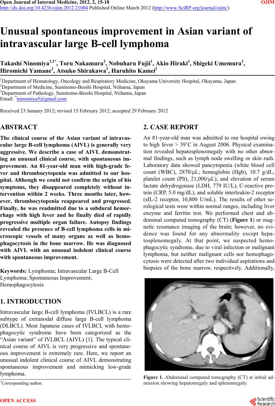 Open Journal of Internal Medicine, 2012, 2, 15-18 OJIM http://dx.doi.org/10.4236/ojim.2012.21004 Published Online March 2012 (http://www.SciRP.org/journal/ojim/) Unusual spontaneous improvement in Asian variant of intravascular large B-cell lymphoma Takashi Ninomiya1,2*, Toru Nakamura2, Nobuharu Fujii1, Akio Hiraki1, Shigeki Umemura2, Hiromichi Yamane2, Atsuko Shirakawa3, Haruhito Kamei2 1Department of Hematology, Oncology and Respiratory Medicine, Okayama University Hospital, Okayama, Japan 2Department of Medicine, Sumitomo-Besshi Hospital, Niihama, Japan 3Department of Pathology, Sumitomo-Besshi Hospital, Niihama, Japan Email: *[email protected] Received 23 January 2012; revised 15 February 2012; accepted 29 February 2012 ABSTRACT The clinical course of the Asian variant of intravas- cular large B-cell lymphoma (AIVL) is generally very aggressive. We describe a case of AIVL demonstrat- ing an unusual clinical course, with spontaneous im- provement. An 81-year-old man with high-grade fe- ver and thrombocytopenia was admitted to our hos- pital. Although we could not confirm the origin of his symptoms, they disappeared completely without in- tervention within 2 weeks. Three months later, how- ever, thrombocytopenia reappeared and progressed. Finally, he was readmitted due to a subdural hemor- rhage with high fever and he finally died of rapidly progressive multiple organ failure. Autopsy findings revealed the presence of B-cell lymphoma cells in mi- croscopic vessels of many organs as well as hemo- phagocytosis in the bone marrow. He was diagnosed with AIVL with an unusual indolent clinical course with spontaneous improvement. Keywords: Lymphoma; Intravascular Large B-Cell Lymphoma; Spontaneous Improvement; Hemophagocytosis 1. INTRODUCTION Intravascular large B-cell lymphoma (IVLBCL) is a rare subtype of extranodal diffuse large B-cell lymphoma (DLBCL). Most Japanese cases of IVLBCL with hemo- phagocytic syndrome have been categorized as the “Asian variant” of IVLBCL (AIVL) [1]. The typical cli- nical course of AIVL is very progressive and spontane- ous improvement is extremely rare. Here, we report an unusual indolent clinical course of AIVL demonstrating spontaneous improvement and mimicking low-grade lymphoma. 2. CASE REPORT An 81-year-old man was admitted to our hospital owing to high fever > 39˚C in August 2006. Physical examina- tion revealed hepatosplenomegaly with no other abnor- mal findings, such as lymph node swelling or skin rash. Laboratory data showed pancytopenia (white blood cell count (WBC), 2870/μL; hemoglobin (Hgb), 10.7 g/dL; platelet count (Plt), 21,000/μL), and elevation of serum lactate dehydrogenase (LDH, 779 IU/L), C-reactive pro- tein (CRP, 5.0 mg/dL), and soluble interleukin-2 receptor (sIL-2 receptor, 10,800 U/mL). The results of other se- rological tests were within normal ranges, including liver enzyme and ferritin test. We performed chest and ab- dominal computed tomography (CT) (Figure 1) or mag- netic resonance imaging of the brain; however, no evi- dence was found for any abnormality except hepa- tosplenomegaly. At that point, we suspected hemo- phagocytic syndrome, due to viral infection or malignant lymphoma, but neither malignant cells nor hemophago- cytosis were detected after two individual aspirations and biopsies of the bone marrow, respectively. Additionally, Figure 1. Abdominal computed tomography (CT) at initial ad- mission showing hepatomegaly and splenomegaly. *Corresponding author. OPEN ACCESS 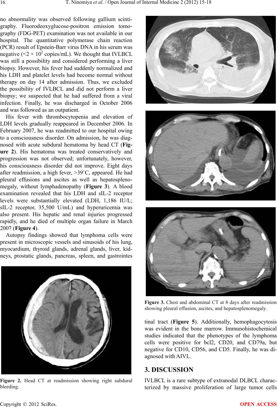 T. Ninomiya et al. / Open Journal of Internal Medicine 2 (2012) 15-18 16 no abnormality was observed following gallium scinti- graphy. Fluorodeoxyglucose-positron emission tomo- graphy (FDG-PET) examination was not available in our hospital. The quantitative polymerase chain reaction (PCR) result of Epstein-Barr virus DNA in his serum was negative (<2 × 10² copies/mL). We thought that IVLBCL was still a possibility and considered performing a liver biopsy. However, his fever had suddenly normalized and his LDH and platelet levels had become normal without therapy on day 14 after admission. Thus, we excluded the possibility of IVLBCL and did not perform a liver biopsy; we suspected that he had suffered from a viral infection. Finally, he was discharged in October 2006 and was followed as an outpatient. His fever with thrombocytopenia and elevation of LDH levels gradually reappeared in December 2006. In February 2007, he was readmitted to our hospital owing to a consciousness disorder. On admission, he was diag- nosed with acute subdural hematoma by head CT (Fig- ure 2). His hematoma was treated conservatively and progression was not observed; unfortunately, however, his consciousness disorder did not improve. Eight days after readmission, a high fever, >39˚C, appeared. He had pleural effusions and ascites as well as hepatospleno- megaly, without lymphadenopathy (Figure 3). A blood examination revealed that his LDH and sIL-2 receptor levels were substantially elevated (LDH, 1,186 IU/L; sIL-2 receptor, 35,500 U/mL) and hyperuricemia was also present. His hepatic and renal injuries progressed rapidly, and he died of multiple organ failure in March 2007 (Figure 4). Autopsy findings showed that lymphoma cells were present in microscopic vessels and sinusoids of his lung, myocardium, thyroid glands, adrenal glands, liver, kid- neys, prostatic glands, pancreas, spleen, and gastrointes Figure 2. Head CT at readmission showing right subdural bleeding. Figure 3. Chest and abdominal CT at 8 days after readmission showing pleural effusion, ascites, and hepatosplenomegaly. tinal tract (Figure 5). Additionally, hemophagocytosis was evident in the bone marrow. Immunohistochemical studies indicated that the phenotypes of the lymphoma cells were positive for bcl2, CD20, and CD79a, but negative for CD10, CD56, and CD5. Finally, he was di- agnosed with AIVL. 3. DISCUSSION IVLBCL is a rare subtype of extranodal DLBCL charac- terized by massive proliferation of large tumor cells Copyright © 2012 SciRes. OPEN ACCESS 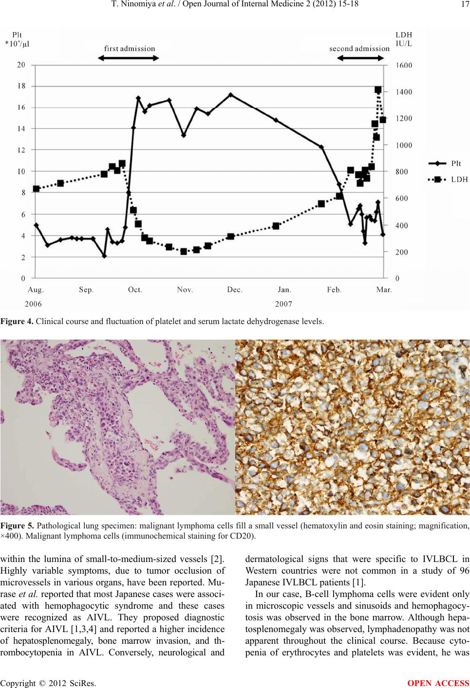 T. Ninomiya et al. / Open Journal of Internal Medicine 2 (2012) 15-18 Copyright © 2012 SciRes. 17 Figure 4. Clinical course and fluctuation of platelet and serum lactate dehydrogenase levels. Figure 5. Pathological lung specimen: malignant lymphoma cells fill a small vessel (hematoxylin and eosin staining; magnification, ×400). Malignant lymphoma cells (immunochemical staining for CD20). within the lumina of small-to-medium-sized vessels [2]. Highly variable symptoms, due to tumor occlusion of microvessels in various organs, have been reported. Mu- rase et al. reported that most Japanese cases were associ- ated with hemophagocytic syndrome and these cases were recognized as AIVL. They proposed diagnostic criteria for AIVL [1,3,4] and reported a higher incidence of hepatosplenomegaly, bone marrow invasion, and th- rombocytopenia in AIVL. Conversely, neurological and dermatological signs that were specific to IVLBCL in Western countries were not common in a study of 96 Japanese IVLBCL patients [1]. In our case, B-cell lymphoma cells were evident only in microscopic vessels and sinusoids and hemophagocy- tosis was observed in the bone marrow. Although hepa- tosplenomegaly was observed, lymphadenopathy was not apparent throughout the clinical course. Because cyto- penia of erythrocytes and platelets was evident, he was OPEN ACCESS  T. Ninomiya et al. / Open Journal of Internal Medicine 2 (2012) 15-18 18 diagnosed with AIVL on the basis of Murase’s criteria for AIVL [2-4]. Although we could not obtain histopa- thological confirmation of malignant lymphoma or he- mophagocytosis on the first admission, his symptoms were suspected to be caused by AIVL. The clinical course of AIVL is typically very aggres- sive. However, a few reports have demonstrated an indo- lent type of IVLBCL. Ishiko et al. reported that a 59- year-old woman with ataxia and gait disturbance after a 3-year history of serum LDH elevation was finally diag- nosed with IVLBCL by brain biopsy [5]. They suggested that there are certain IVLBCL patients with brain in- volvement who present with relatively slow progression, similar to a low-grade lymphoma [5]. Nakao et al. re- ported that a 62-year-old woman with slowly developing paraplegia and a 7-month history of recto-urinary dys- function was diagnosed with IVLBCL by autopsy [6]. In that case, lymphoma cells were only identified in the bone marrow and blood vessels of the cauda equina. Ad- ditionally, Sekine et al. reported that a 71-year-old man with respiratory failure after developing intermittent fe- ver and a high serum LDH for 7 months was diagnosed with IVLBCL by lung biopsy [7]. In that case, lym- phoma cells were identified in the cauda equina and lung. From these different lines of evidence, we suggest that IVLBCL may have a slow progressive clinical course when lymphoma cells are present in limited organs. Al- though we could not show the limited lymphoma cell involvement in a specific organ, our case might be one of indolent AIVL similar to these previous reported cases. In two of three previous cases, a diagnosis of IVLBCL was established in living patients who were treated using CHOP (cyclophosphamide, doxorubicin, vincristine, pred- nisone) with rituximab and showed complete remission. In the other case and in our case, the diagnosis was es- tablished at autopsy. IVLBCL is difficult to diagnose because patients generally show little or no tumor mass formation and more than half of patients are diagnosed postmortem. Recently, some reports have suggested that a random skin biopsy may be efficacious for the early diagnosis of IVLBCL. Asada et al. reported that early diagnosis of IVLBCL was possible by random skin bi- opsy and the early institution of rituximab-based chemo- therapy induced a favorable response [8]. In our case, a liver biopsy or random skin biopsy should have been performed even after the high fever and thrombocyto- penia disappeared. In conclusion, we present a case of AIVL with an un- usual clinical course and spontaneous improvement. We suggest that an indolent clinical course or spontaneous improvement does not exclude AIVL, although most AIVL cases are aggressive and progressive. Importantly, AIVL should be treated immediately with intensive ex- amination for diagnosis, even though the clinical course is apparently indolent. REFERENCES [1] Murase, T., Yamaguchi, M., Suzuki, R., Okamoto, M., Sato, Y., Tamaru, J., Kojima, M., Miura, I., Mori, N., Yoshino, T. and Nakamura, S. (2007) Intravascular large B-cell lymphoma (ivlbcl): A clinicopathologic study of 96 cases with special reference to the immunophenotypic heterogeneity of cd5. Blood, 109, 478-485. doi:10.1182/blood-2006-01-021253 [2] Murase, T. and Nakamura, S. (1999) An Asian variant of intravascular lymphomatosis: An updated review of ma- lignant histiocytosis-like B-cell lymphoma. Leukemia and Lymphoma, 33, 459-473. [3] Murase, T., Nakamura, S., Kawauchi, K., Matsuzaki, H., Sakai, C., Inaba, T., Nasu, K., Tashiro, K., Suchi, T. and Saito, H. (2000) An Asian variant of intravascular large B-cell lymphoma: Clinical, pathological and cytogenetic approaches to diffuse large B-cell lymphoma associated with haemophagocytic syndrome. British Journal of Haematology, 111, 826-834. doi:10.1046/j.1365-2141.2000.02426.x [4] Murase, T., Nakamura, S., Tashiro, K., Suchi, T., Hiraga, J., Hayasaki, N., Kimura, M., Murakami, M., Mizoguchi, Y., Suzuki, T. and Saito, H. (1997) Malignant histiocyto- sis-like B-cell lymphoma, a distinct pathologic variant of intravascular lymphomatosis: A report of five cases and review of the literature. British Journal of Haematology, 99, 656-664. doi:10.1046/j.1365-2141.1997.4623265.x [5] Ishiko, J., Mizuki, M., Yasumi, M., Ujiie, H., Nakamichi, I., Aozasa, K. and Kanakura, Y. (2007) An indolent sub- type of “Intravascular lymphoma”: A case with a 3-year history of ldh elevation. Leukemia and Lymphoma, 48, 1872-1874. doi:10.1080/10428190701493936 [6] Nakao, N., Yoshida, M., Iwata, M., Hashizume, Y. and Sahashi, K. (2008) A case of intravascular malignant lymphomatosis presenting as slowly progressive paraple- gia. Brain Nerve, 60, 181-185. [7] Sekine, A., Hagiwara, E., Okudera, K., Baba, T. and Ogura, T. (2009) A case of slowly progressive intravas- cular lymphoma with respiratory failure caused by diffuse pulmonary vasoconstriction. Nihon Kokyuki Gakkai Zasshi, 47, 924-929. [8] Asada, N., Odawara, J., Kimura, S., Aoki, T., Yamakura, M., Takeuchi, M., Seki, R., Tanaka, A. and Matsue, K. (2007) Use of random skin biopsy for diagnosis of in- travascular large B-cell lymphoma. Mayo Clinic Pro- ceedings, 82, 1525-1527. doi:10.4065/82.12.1525 Copyright © 2012 SciRes. OPEN ACCESS |