

Paper Menu >>
Journal Menu >>
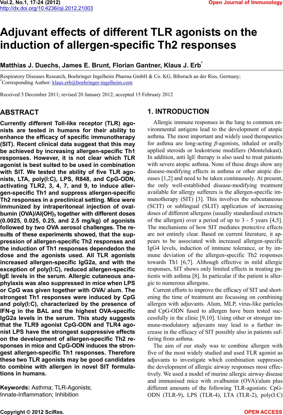 Vol.2, No.1, 17-24 (2012) Open Journal of Immunology http://dx.doi.org/10.4236/oji.2012.21003 Adjuvant effects of different TLR agonists on the induction of allergen-specific Th2 responses Matthias J. Duechs, James E. Brunt, Florian Gantner, Klaus J. Erb* Respiratory Diseases Research, Boehringer Ingelheim Pharma GmbH & Co. KG, Biberach an der Riss, Germany; *Corresponding Author: [email protected] Received 5 December 2011; revised 20 January 2012; accepted 15 February 2012 ABSTRACT Currently different Toll-like receptor (TLR) ago- nists are tested in humans for their ability to enhance the efficacy of specific immunotherapy (SIT). Recent clinical data suggest that this may be achieved by increasing allergen-specific Th1 responses. However, it is not clear which TLR agonist is best suited to be used in combination with SIT. We tested the ability of five TLR ago- nists, LTA, poly(I:C), LPS, R848, and CpG-ODN, activating TLR2, 3, 4, 7, and 9, to induce aller- gen-specific Th1 and suppress allergen-specific Th2 responses in a preclinical setting. Mice were immunized by intraperitoneal injection of oval- bumin (OVA)/Al(OH)3 together with different doses (0.0025, 0.025, 0.25, and 2.5 mg/kg) of agonists followed by two OVA aerosol challenges. The re- sults of these experiments showed, tha t the sup- pression of allergen-specific Th2 responses and the induction of Th1 responses dependedon the dose and the agonists used. All TLR agonists increased allergen-specific IgG2a, and with the exception of poly(I:C), reduced allergen-specific IgE levels in the serum. Allergic cutaneous ana- phylaxis was also suppressed in mice when LPS or CpG was given together with OVA/ alum. The strongest Th1 responses were induced by CpG and poly(I:C), characterized by the presence of IFN-g in the BAL and the highest OVA-specific IgG2a levels in the serum. This study suggests that the TLR9 agonist CpG-ODN and TLR4 ago- nist LPS have the strongest suppressive effects on the development of allergen-specific Th2 re- sponses in mice and CpG-ODN ind uces the stron- gest allergen-specific Th1 responses. Therefore these t wo TLR ago nists may be good candidates to combine with allergen in novel SIT formula- tions in humans. Keywords: Asthma; TLR-Agonists; Innate-Inflammation; Inhibition 1. INTRODUCTION Allergic immune responses in the lung to common en- vironmental antigens lead to the development of atopic asthma. The most important and widely used therapeutics for asthma are long-acting β-agonists, inhaled or orally applied steroids or leukotriene modifiers (Montelukast). In addition, anti IgE therapy is also used to treat patients with severe atopic asthma. None of these drugs show any disease-modifying effects in asthma or other atopic dis- eases [1,2] and need to be taken continuously. At present, the only well-established disease-modifying treatment available for allergy sufferers is the allergen-specific im- munotherapy (SIT) [3]. This involves the subcutaneous (SCIT) or sublingual (SLIT) application of increasing doses of different allergens (usually standardised extracts of the allergen) over a period of up to 3 - 5 years [4,5]. The mechanisms of how SIT mediates protective effects are not entirely clear. Based on current literature, it ap- pears to be associated with increased allergen-specific IgG4 levels, induction of immune tolerance, or by im- mune deviation of the allergen-specific Th2 responses towards Th1 [6,7]. Although effective in mild allergic responses, SIT shows only limited effects in treating pa- tients with asthma [8]. In particular if the patient is aller- gic to numerous allergens. Current efforts to improve the efficacy of SIT and short- ening the time of treatment are focussing on combining allergen with adjuvants. Alum, MLP, virus-like particles and CpG-ODN fused to allergen have been tested suc- cessfully in the clinic [9,10]. Using other or stronger im- mune-modulatory adjuvants may lead to a further in- crease in the efficacy of SIT possibly also in patients suf- fering from asthma. The aim of our study was to combine allergen with five of the most widely studied and used TLR agonist as adjuvants to investigate which combination suppresses the development of allergic airway responses most effec- tively. We used a model of murine allergic airway disease and immunised mice with ovalbumin (OVA)/alum plus different amounts of the following TLR-agonists: CpG- ODN (TLR-9), LPS (TLR-4), LTA (TLR-2), poly(I:C) Copyright © 2012 SciRes. OPEN ACCES S 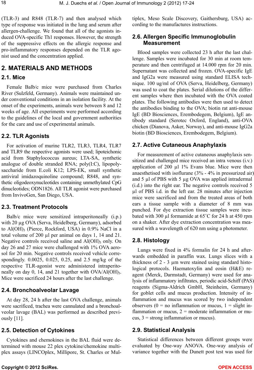 M. J. Duechs et al. / Open Journal of Immunology 2 (2012) 17-24 18 (TLR-3) and R848 (TLR-7) and then analysed which type of response was initiated in the lung and serum after allergen-challenge. We found that all of the agonists in- duced OVA-specific Th1 responses. However, the strength of the suppressive effects on the allergic response and pro-inflammatory responses depended on the TLR ago- nist used and the concentration applied. 2. MATERIALS AND METHODS 2.1. Mice Female Balb/c mice were purchased from Charles River (Sulzfeld, Germany). Animals were maintained un- der conventional conditions in an isolation facility. At the onset of the experiments, animals were between 8 and 12 weeks of age. All experiments were performed according to the guidelines of the local and government authorities for the care and use of experimental animals. 2.2. TLR Agonists For activation of murine TLR2, TLR3, TLR4, TLR7 and TLR9 the respective agonists were used; lipoteichonic acid from Staphylococcus aureus; LTA-SA, synthetic analogue of double stranded RNA; poly(I:C), lipopoly- saccharide from E.coli K12; LPS-EK, small synthetic antiviral imidazoquinoline compound; R848, and syn- thetic oligodeoxynucleotides containing unmethylated CpG dinucleotides; ODN1826. All TLR agonist were purchased from InvivoGen, San Diego, USA. 2.3. Treatment Protocols Balb/c mice were sensitized intraperitoneally (i.p.) with 20 µg OVA (Serva, Heidelberg, Germany), adsorbed to Al(OH)3 (Pierce, Rockford, USA) in 0.9% NaCl in a total volume of 200 µl per animal on days 1, 14 and 21. Negative controls received saline and Al(OH)3 only. On day 26 and 27 mice were challenged with 1% OVA aero- sol for 20 min. Negative controls received vehicle corre- spondingly. 0.0025, 0.025, 0.25, and 2.5 mg/kg of the respective TLR-agonist were administered intraperito- neally on day 0, 14, and 21 together with OVA/Al(OH)3. Mice were sacrificed 24 hours after the last challenge. 2.4. Bronchoalveolar Lavage At day 28, 24 h after the last OVA challenge, animals were sacrificed, trachea were cannulated and a bronchoal- veolar lavage (BAL) was performed as described previ- ously [11]. 2.5. Detection of Cytokines Cytokines and chemokines in the BAL fluid were de- termined with mouse 22 plex cytokine/chemokine multi- plex assays (LINCOplex, Millipore, St. Charles or Mul- tiplex, Meso Scale Discovery, Gaithersburg, USA) ac- cording to the manufactures instructions. 2.6. Allergen Specific Immunoglobulin Measurement Blood samples were collected 23 h after the last chal- lenge. Samples were incubated for 30 min at room tem- perature and then centrifuged at 14.000 rpm for 20 min. Supernatant was collected and frozen. OVA-specific IgE and IgG2a were measured using standard ELISA tech- nique. 100 ug/ml of OVA (Serva, Heidelberg, Germany) was used to coat the plates. Serial dilutions of the differ- ent samples where then incubated with the OVA coated plates. The following antibodies were then used to detect the antibodies binding to the OVA; biotin rat anti-mouse IgE (BD Biosciences, Erembodegem, Belgium), IgE an- tibody standard (Serotec Oxford, England), anti-OVA chicken (Dianova, Asker, Norway), and anti-mouse IgG2a biotin (BD Biosciences, Erembodegem, Belgium). 2.7. Active Cutaneous Anaphy laxis For measurement of active cutaneous anaphylaxis sen- sitized and challenged mice received an intra venous (i.v.) application of 200 µl 1% Evans blue. Mice were then anaesthetised with isoflurane (3% - 4% in pressurized air) and 5 µl of PBS with 5 µg OVA was applied intradermal (i.d.) into the right ear. The negative controls received 5 µl of PBS i.d. in the left ear. 28 minutes after injection mice were sacrificed and from the treated areas of both ears a tissue sample with a diameter of 8 mm was punched. For dye extraction tissue samples were incu- bated with 300 µl formamide at 65˚C for 24 h at 450 rpm on a shaker. After dye extraction concentration was mea- sured with a wavelength of 620 nm using a photometer. 2.8. Histology Lungs were fixed in 4% formalin for 24 h and after- wards embedded in paraffin wax. Lungs slices with a thickness of 2 - 3 µm were stained using standard histo- logical protocols. Haematoxylin and eosin (H&E) re- agent (Merck, Darmstadt, Germany) were used for ana- lysis of inflammatory infiltrates, periodic acid-Schiff (PAS) reagents (Sigma-Aldrich GmbH, Steinheim, Germany) for goblet cells and mucus production. Intensity of in- flammation and mucus was scored by two independent observers (0 = no inflammation or mucus, 1 = slight in- flammation or mucus, 2 = moderate inflammation or mu- cus, 3 = strong inflammation or mucus). 2.9. Statistical Analysis Statistical differences between different groups were evaluated by One-way ANOVA. One-way analysis of variance together with the Dunett post test was used for Copyright © 2012 SciRes. OPEN ACCESS 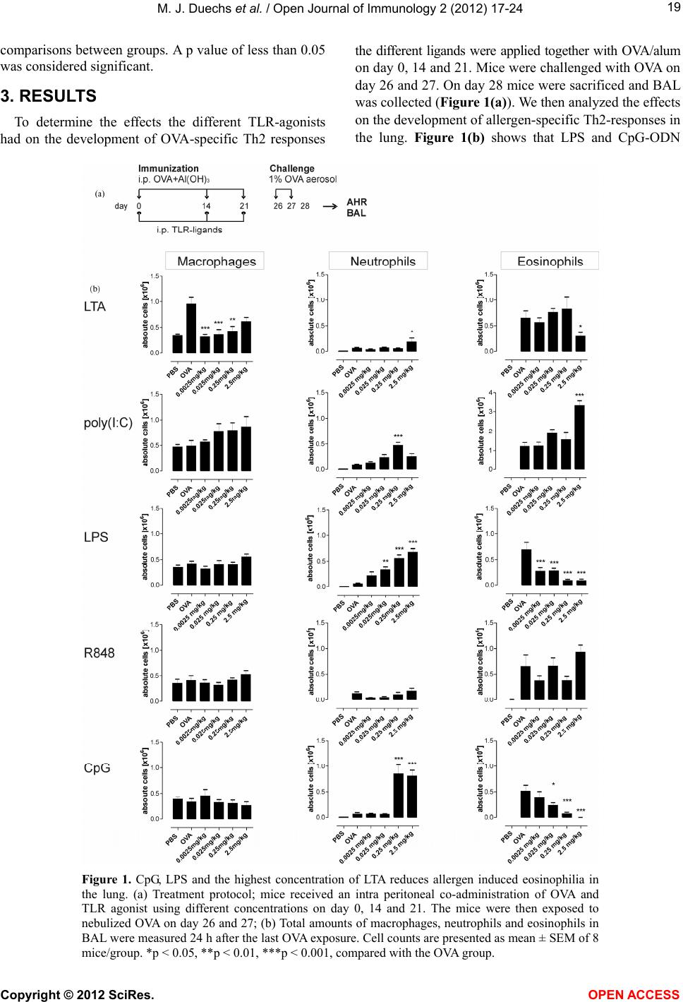 M. J. Duechs et al. / Open Journal of Immunology 2 (2012) 17-24 Copyright © 2012 SciRes. 19 comparisons between groups. A p value of less than 0.05 was considered significant. the different ligands were applied together with OVA/alum on day 0, 14 and 21. Mice were challenged with OVA on day 26 and 27. On day 28 mice were sacrificed and BAL was collected (Figure 1(a)). We then analyzed the effects on the development of allergen-specific Th2-responses in the lung. Figure 1(b) shows that LPS and CpG-ODN 3. RESULTS To determine the effects the different TLR-agonists had on the development of OVA-specific Th2 responses Figure 1. CpG, LPS and the highest concentration of LTA reduces allergen induced eosinophilia in the lung. (a) Treatment protocol; mice received an intra peritoneal co-administration of OVA and TLR agonist using different concentrations on day 0, 14 and 21. The mice were then exposed to nebulized OVA on day 26 and 27; (b) Total amounts of macrophages, neutrophils and eosinophils in BAL were measured 24 h after the last OVA exposure. Cell counts are presented as mean ± SEM of 8 mice/group. *p < 0.05, **p < 0.01, ***p < 0.001, compared with the OVA group. OPEN A CCESS 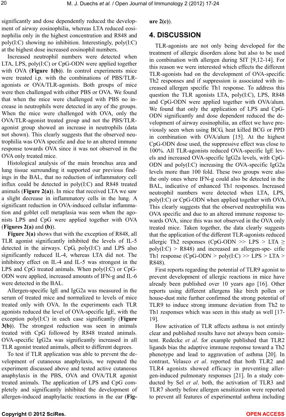 M. J. Duechs et al. / Open Journal of Immunology 2 (2012) 17-24 20 significantly and dose dependently reduced the develop- ment of airway eosinophilia, whereas LTA reduced eosi- nophilia only in the highest concentration and R848 and poly(I:C) showing no inhibition. Interestingly, poly(I:C) at the highest dose increased eosinophil numbers. Increased neutrophil numbers were detected when LTA, LPS, poly(I:C) or CpG-ODN were applied together with OVA (Figure 1(b)). In control experiments mice were treated i.p. with the combinations of PBS/TLR- agonists or OVA/TLR-agonists. Both groups of mice were then challenged with either PBS or OVA. We found that when the mice were challenged with PBS no in- crease in neutrophils were detected in any of the groups. When the mice were challenged with OVA, only the OVA/TLR-agonist treated group and not the PBS/TLR- agonist group showed an increase in neutrophils (data not shown). This clearly suggests that the observed neu- trophilia was OVA specific and due to an altered immune response towards OVA since it was not observed in the OVA only treated mice. Histological analysis of the main bronchus area and lung tissue surrounding it supported our previous find- ings in the BAL, that no reduction of inflammatory cell influx could be detected in poly(I:C) and R848 treated animals (Figure 2(a)). In mice that received LTA we saw a slight decrease in inflammatory cells in the lung. A significant reduction in OVA-induced cellular inflamma- tion and goblet cell metaplasia was seen when the ago- nists LPS and CpG were applied together with OVA (Figures 2( a) and (b)). Figure 3(a) shows that with the exception of R848, all TLR agonist significantly inhibited the levels of IL-5 detected in the airways. CpG, poly(I:C) and LPS also significantly reduced IL-4, whereas LTA did not. The inhibitory effect on IL-4 and IL-5 was strongest in the LPS and CpG treated animals. When poly(I:C) or CpG- ODN were applied, increased amounts of IFN-g and IL-6 were detected in the BAL. Allergen-specific IgE and IgG2a was measured in the serum of treated mice and normalized to levels of mice treated only with OVA. In the experiments each TLR agonists reduced the level of OVA-specific IgE, with the exception poly(I:C) in each case significantly (Figure 3(b)). The strongest reduction was seen in animals treated with CpG followed by R848 treated animals. OVA-specific IgG2a was significantly increased in all TLR agonist treated animals, albeit to different degrees. To test if TLR application was able to prevent the de- velopment of cutaneous anaphylaxis, we repeated the experiment discussed above and tested active cutaneous anaphylaxis in the PBS, OVA and OVA/TLR agonist treated animals. The application of LPS and CpG com- pletely and significantly inhibited the development of allergen-induced anaphylactic reactions in the ear (Fig- ure 2(c)). 4. DISCUSSION TLR-agonists are not only being developed for the treatment of allergic disorders alone but also to be used in combination with allergen during SIT [9,12-14]. For this reason we were interested which effects the different TLR-agonists had on the development of OVA-specific Th2 responses and if suppression is associated with in- creased allergen specific Th1 response. To address this question the TLR agonists LTA, poly(I:C), LPS, R848 and CpG-ODN were applied together with OVA/alum. We found that only the application of LPS and CpG- ODN significantly and dose dependent reduced the de- velopment of airway eosinophilia, an effect we have pre- viously seen when using BCG, heat killed BCG or PPD in combination with OVA/alum [15]. At the highest CpG-ODN dose used, the suppressive effect was close to 100%. All TLR-agonists reduced OVA-specific IgE lev- els and increased OVA-specific IgG2a levels, with CpG- ODN and poly(I:C) increasing the OVA-specific IgG2a levels more than 100 fold. These two groups were also the only ones where IFN-g could also be detected in the BAL, indicative of enhanced Th1 responses. Increased neutrophil numbers were detected when LTA, LPS, poly(I:C) or CpG-ODN when applied together with OVA. This clearly suggests that the observed neutrophilia was OVA specific and due to an altered immune response to- wards OVA, since this was not observed in the OVA only treated mice. Taken together, the data clearly suggests that the application of the different TLR-agonists reduced allergic Th2 responses (CpG-ODN >> LPS > LTA ≥ poly(I:C) > R848) and increased an allergen-spe- cific Th1 response (CpG-ODN > poly(I:C) >> LPS > LTA > R848). First reports regarding the potential of TLR9 agonist to prevent development of allergic reactions in mice have already been published over 10 years ago [16]. Other reports using different allergens like birch pollen or house-dust mite further confirmed the strong potential of TLR9 to induce strong immune deviation from Th2 to Th1 responses which was seen in this study as well [17- 19]. How activation of TLR affects asthma is not entirely clear and published results have not always been consis- tent. Redecke et al. for example published that TLR2 ligands bias the adaptive immune response toward a Th2 phenotype and lead to aggravation of asthma [20]. In contrast, Velasco et al. reported that both TLR2 and TLR4 agonists showed efficacy in preventing aller- gen-induced pulmonary responses [21]. In a study con- ducted by Sel et al. both, the activation of TLR3 and TLR7 shortly before allergen sensitization were reported to prevent all features of experimental asthma including Copyright © 2012 SciRes. OPEN ACCESS 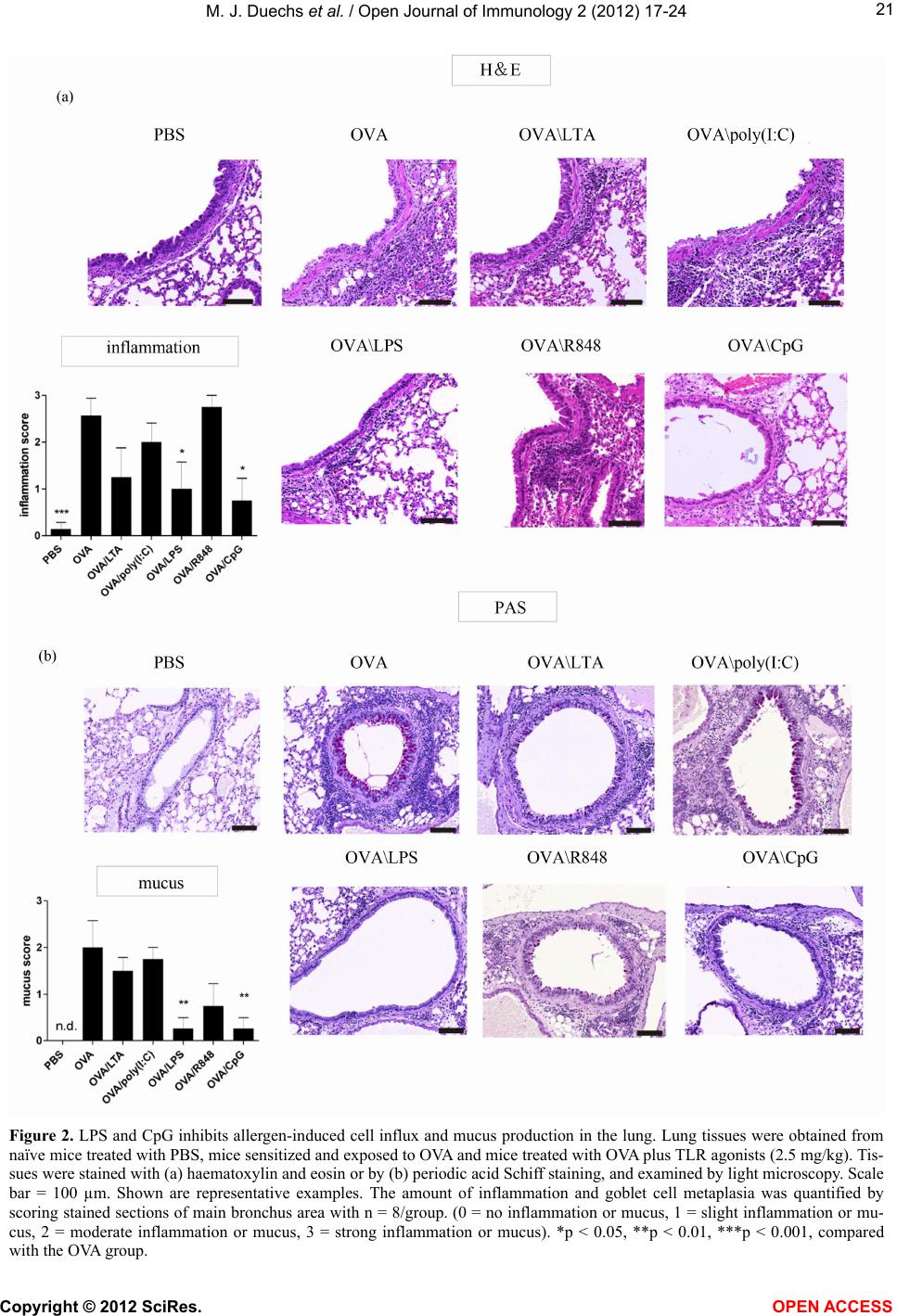 M. J. Duechs et al. / Open Journal of Immunology 2 (2012) 17-24 21 Figure 2. LPS and CpG inhibits allergen-induced cell influx and mucus production in the lung. Lung tissues were obtained from naïve mice treated with PBS, mice sensitized and exposed to OVA and mice treated with OVA plus TLR agonists (2.5 mg/kg). Tis- sues were stained with (a) haematoxylin and eosin or by (b) periodic acid Schiff staining, and examined by light microscopy. Scale bar = 100 µm. Shown are representative examples. The amount of inflammation and goblet cell metaplasia was quantified by scoring stained sections of main bronchus area with n = 8/group. (0 = no inflammation or mucus, 1 = slight inflammation or mu- cus, 2 = moderate inflammation or mucus, 3 = strong inflammation or mucus). *p < 0.05, **p < 0.01, ***p < 0.001, compared with the OVA group. Copyright © 2012 SciRes. OPEN ACCES S 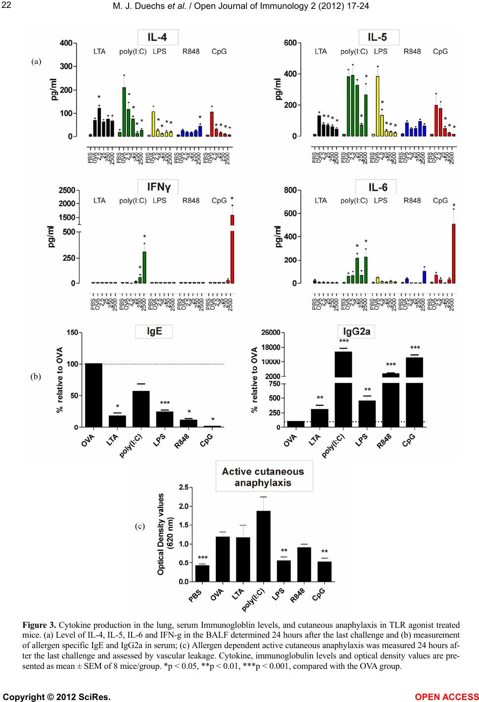 M. J. Duechs et al. / Open Journal of Immunology 2 (2012) 17-24 22 Figure 3. Cytokine production in the lung, serum Immunogloblin levels, and cutaneous anaphylaxis in TLR agonist treated mice. (a) Level of IL-4, IL-5, IL-6 and IFN-g in the BALF determined 24 hours after the last challenge and (b) measurement of allergen specific IgE and IgG2a in serum; (c) Allergen dependent active cutaneous anaphylaxis was measured 24 hours af- ter the last challenge and assessed by vascular leakage. Cytokine, immunoglobulin levels and optical density values are pre- sented as mean ± SEM of 8 mice/group. *p < 0.05, **p < 0.01, ***p < 0.001, compared with the OVA group. Copyright © 2012 SciRes. OPEN ACCESS 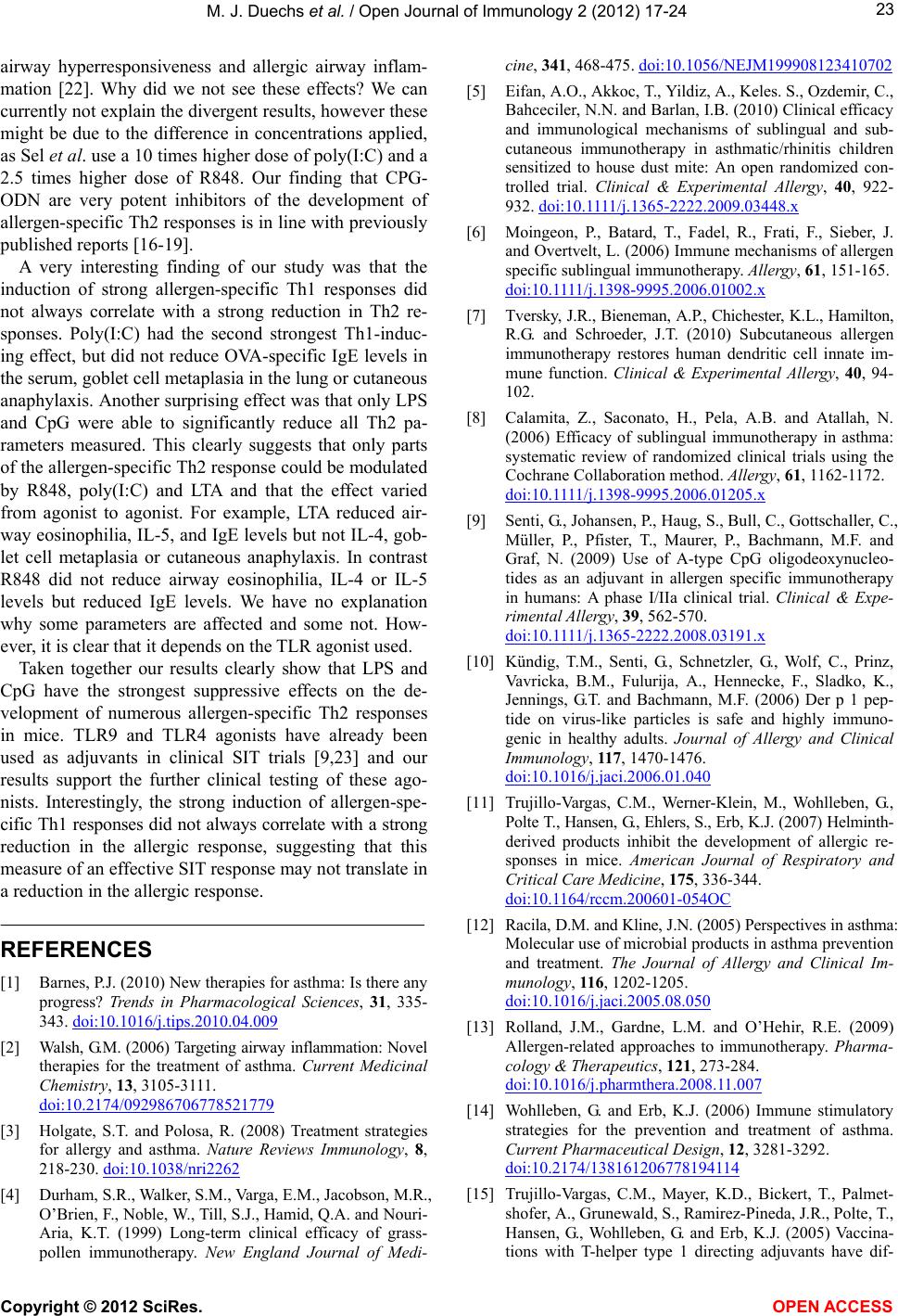 M. J. Duechs et al. / Open Journal of Immunology 2 (2012) 17-24 Copyright © 2012 SciRes. 23 airway hyperresponsiveness and allergic airway inflam- mation [22]. Why did we not see these effects? We can currently not explain the divergent results, however these might be due to the difference in concentrations applied, as Sel et al. use a 10 times higher dose of poly(I:C) and a 2.5 times higher dose of R848. Our finding that CPG- ODN are very potent inhibitors of the development of allergen-specific Th2 responses is in line with previously published reports [16-19]. OPEN A CCESS A very interesting finding of our study was that the induction of strong allergen-specific Th1 responses did not always correlate with a strong reduction in Th2 re- sponses. Poly(I:C) had the second strongest Th1-induc- ing effect, but did not reduce OVA-specific IgE levels in the serum, goblet cell metaplasia in the lung or cutaneous anaphylaxis. Another surprising effect was that only LPS and CpG were able to significantly reduce all Th2 pa- rameters measured. This clearly suggests that only parts of the allergen-specific Th2 response could be modulated by R848, poly(I:C) and LTA and that the effect varied from agonist to agonist. For example, LTA reduced air- way eosinophilia, IL-5, and IgE levels but not IL-4, gob- let cell metaplasia or cutaneous anaphylaxis. In contrast R848 did not reduce airway eosinophilia, IL-4 or IL-5 levels but reduced IgE levels. We have no explanation why some parameters are affected and some not. How- ever, it is clear that it depends on the TLR agonist used. Taken together our results clearly show that LPS and CpG have the strongest suppressive effects on the de- velopment of numerous allergen-specific Th2 responses in mice. TLR9 and TLR4 agonists have already been used as adjuvants in clinical SIT trials [9,23] and our results support the further clinical testing of these ago- nists. Interestingly, the strong induction of allergen-spe- cific Th1 responses did not always correlate with a strong reduction in the allergic response, suggesting that this measure of an effective SIT response may not translate in a reduction in the allergic response. REFERENCES [1] Barnes, P.J. (2010) New therapies for asthma: Is there any progress? Trends in Pharmacological Sciences, 31, 335- 343. doi:10.1016/j.tips.2010.04.009 [2] Walsh, G.M. (2006) Targeting airway inflammation: Novel therapies for the treatment of asthma. Current Medicinal Chemistry, 13, 3105-3111. doi:10.2174/092986706778521779 [3] Holgate, S.T. and Polosa, R. (2008) Treatment strategies for allergy and asthma. Nature Reviews Immunology, 8, 218-230. doi:10.1038/nri2262 [4] Durham, S.R., Walker, S.M., Varga, E.M., Jacobson, M.R., O’Brien, F., Noble, W., Till, S.J., Hamid, Q.A. and Nouri- Aria, K.T. (1999) Long-term clinical efficacy of grass- pollen immunotherapy. New England Journal of Medi- cine, 341, 468-475. doi:10.1056/NEJM199908123410702 [5] Eifan, A.O., Akkoc, T., Yildiz, A., Keles. S., Ozdemir, C., Bahceciler, N.N. and Barlan, I.B. (2010) Clinical efficacy and immunological mechanisms of sublingual and sub- cutaneous immunotherapy in asthmatic/rhinitis children sensitized to house dust mite: An open randomized con- trolled trial. Clinical & Experimental Allergy, 40, 922- 932. doi:10.1111/j.1365-2222.2009.03448.x [6] Moingeon, P., Batard, T., Fadel, R., Frati, F., Sieber, J. and Overtvelt, L. (2006) Immune mechanisms of allergen specific sublingual immunotherapy. Allergy, 61, 151-165. doi:10.1111/j.1398-9995.2006.01002.x [7] Tversky, J.R., Bieneman, A.P., Chichester, K.L., Hamilton, R.G. and Schroeder, J.T. (2010) Subcutaneous allergen immunotherapy restores human dendritic cell innate im- mune function. Clinical & Experimental Allergy, 40, 94- 102. [8] Calamita, Z., Saconato, H., Pela, A.B. and Atallah, N. (2006) Efficacy of sublingual immunotherapy in asthma: systematic review of randomized clinical trials using the Cochrane Collaboration method. Allergy, 61, 1162-1172. doi:10.1111/j.1398-9995.2006.01205.x [9] Senti, G., Johansen, P., Haug, S., Bull, C., Gottschaller, C., Müller, P., Pfister, T., Maurer, P., Bachmann, M.F. and Graf, N. (2009) Use of A-type CpG oligodeoxynucleo- tides as an adjuvant in allergen specific immunotherapy in humans: A phase I/IIa clinical trial. Clinical & Expe- rimental Allergy, 39, 562-570. doi:10.1111/j.1365-2222.2008.03191.x [10] Kündig, T.M., Senti, G., Schnetzler, G., Wolf, C., Prinz, Vavricka, B.M., Fulurija, A., Hennecke, F., Sladko, K., Jennings, G.T. and Bachmann, M.F. (2006) Der p 1 pep- tide on virus-like particles is safe and highly immuno- genic in healthy adults. Journal of Allergy and Clinical Immunology, 117, 1470-1476. doi:10.1016/j.jaci.2006.01.040 [11] Trujillo-Vargas, C.M., Werner-Klein, M., Wohlleben, G., Polte T., Hansen, G., Ehlers, S., Erb, K.J. (2007) Helminth- derived products inhibit the development of allergic re- sponses in mice. American Journal of Respiratory and Critical Care Medicine, 175, 336-344. doi:10.1164/rccm.200601-054OC [12] Racila, D.M. and Kline, J.N. (2005) Perspectives in asthma: Molecular use of microbial products in asthma prevention and treatment. The Journal of Allergy and Clinical Im- munology, 116, 1202-1205. doi:10.1016/j.jaci.2005.08.050 [13] Rolland, J.M., Gardne, L.M. and O’Hehir, R.E. (2009) Allergen-related approaches to immunotherapy. Pharma- cology & Therapeutics, 121, 273-284. doi:10.1016/j.pharmthera.2008.11.007 [14] Wohlleben, G. and Erb, K.J. (2006) Immune stimulatory strategies for the prevention and treatment of asthma. Current Pharmaceutical Design, 12, 3281-3292. doi:10.2174/138161206778194114 [15] Trujillo-Vargas, C.M., Mayer, K.D., Bickert, T., Palmet- shofer, A., Grunewald, S., Ramirez-Pineda, J.R., Polte, T., Hansen, G., Wohlleben, G. and Erb, K.J. (2005) Vaccina- tions with T-helper type 1 directing adjuvants have dif-  M. J. Duechs et al. / Open Journal of Immunology 2 (2012) 17-24 24 ferent suppressive effects on the development of allergen- induced T-helper type 2 responses. Clinical & Experi- mental Allergy, 35, 1003-1013. doi:10.1111/j.1365-2222.2005.02287.x [16] Kline, J.N., Waldschmidt, T.J., Businga, T.R., Lemish, J.E., Weinstock, J.V., Thorne, P.S. and Krieg, A.M. (1998) Cut- ting edge: Modulation of airway inflammation by CpG oligodeoxynucleotides in a murine model of asthma. The Journal of Immunology, 160, 2555-2559. [17] Jahn-Schmid, B., Wiedermann, U., Bohle, B., Repa, A., Kraft, D. and Ebner, C. (1999) Oligodeoxynucleotides containing CpG motifs modulate the allergic TH2 re- sponse of BALB/c mice to Bet v 1, the major birch pollen allergen. Journal of Allergy and Clinical Immunology, 104, 1015-1023. doi:10.1016/S0091-6749(99)70083-7 [18] Serebrisky, D., Teper, A.A., Huang, C.K., Lee, S.Y., Zhang, T.F., Schofield, B.H., Kattan, M., Sampson, H.A. and Li, X.M. (2000) CpG oligodeoxynucleotides can reverse Th2- associated allergic airway responses and alter the B7.1/ B7.2 expression in a murine model of asthma. The Jour- nal of Immunology, 165, 5906-5912. [19] Mo, J.H., Park, S.W., Rhee, C.S., Takabayashi, K., Lee, S.S., Quan, S.H., Kim, I.S., Min, Y.G., Raz, E. and Lee, C.H. (2006) Suppression of allergic response by CpG motif oligodeoxynucleotide house-dust mite conjugate in animal model of allergic rhinitis. American Journal of Rhi- nology, 20, 212-218. [20] Redecke, V., Hacker, H., Datta, S.K., Fermin, A., Pitha, P.M., Broide, D.H. and Raz, E. (2004) Cutting edge: Ac- tivation of Toll-like receptor 2 induces a Th2 immune re- sponse and promotes experimental asthma. The Journal of Immunology, 72, 2739-2743. [21] Velasco, G., Campo, M., Manrique, O.J., Bellou, A., Hong- zheh, H.E., Arestides, R.S.S., Schaub, B., Perkins, D.L. and Finn, P.W. (2005) Toll-like receptor 4 or 2 agonists decrease allergic inflammation. American Journal of Res- piratory Cell and Molecular Biology, 32, 218-224. doi:10.1165/rcmb.2003-0435OC [22] Sel, S., Wegmann, M., Sel, S., Bauer, S., Garn, H., Alber, G. and Renz, H. (2007) Immunomodulatory effects of vi- ral TLR ligands on experimental asthma depend on the additive effects of IL-12 and IL-10. The Journal of Im- munology, 178, 7805-7813. [23] Gawchik, S.M. and Saccar, C.L. (2009) Pollinex quattro tree: Allergy vaccine. Expert Opinion on Biological The- rapy, 9, 377-382. doi:10.1517/14712590802699596 Copyright © 2012 SciRes. OPEN ACCESS |