

Paper Menu >>
Journal Menu >>
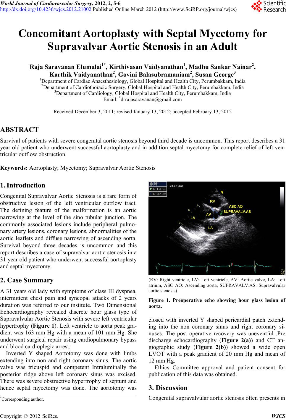 World Journal of Cardiovascular Surgery, 2012, 2, 5-6 http://dx.doi.org/10.4236/wjcs.2012.21002 Published Online March 2012 (http://www.SciRP.org/journal/wjcs) 5 Concomitant Aortoplasty with Septal Myectomy for Supravalvar Aortic Stenosis in an Adult Raja Saravanan Elumalai1*, Kirthivasan Vaidyanathan1, Madhu Sankar Nainar2, Karthik Vaidyanathan2, Govini Balasubramaniam2, Susan George3 1Department of Cardiac Anaesthesiology, Global Hospital and Health City, Perumbakkam, India 2Department of Cardiothor ac ic Surgery, Global Hospital and Health City, Perumbakkam, India 3Department of Cardiology, Global Hospital and Heal th City, Perumbakkam, India Email: *[email protected] Received December 3, 2011; revised January 13, 2012; accepted February 13, 2012 ABSTRACT Survival of patients with severe congenital aortic stenosis beyond third decade is uncommon. This report describes a 31 year old patient who underwent successful aortoplasty and in addition septal myectomy for complete relief of left ven- tricular outflow ob struction. Keywords: Aortoplasty; Myectomy; Supravalvar Aortic Stenosis 1. Introduction Congenital Supravalvar Aortic Stenosis is a rare form of obstructive lesion of the left ventricular outflow tract. The defining feature of the malformation is an aortic narrowing at the level of the sino tubular junction. The commonly associated lesions include peripheral pulmo- nary artery lesions, coronary lesion s, abnormalities of the aortic leaflets and diffuse narrowing of ascending aorta. Survival beyond three decades is uncommon and this report describes a case of supravalva r aortic stenosis in a 31 year old patient who underwent successful aortoplasty and septal myectomy. 2. Case Summary A 31 years old lady with symptoms of class III dyspnea, intermittent chest pain and syncopal attacks of 2 years duration was referred to our institute. Two Dimensional Echocardiography revealed discrete hour glass type of Supravalvular Aortic Stenosis with severe left v entricular hypertrophy (Figure 1). Left ventricle to aorta peak gra- dient was 163 mm Hg with a mean of 101 mm Hg. She underwent surgical repair using cardiopulmonary bypass and blood cardioplegic arrest. Inverted Y shaped Aortotomy was done with limbs extending into non and right coronary sinus. The aortic valve was tricuspid and competent Intraluminally the posterior ridge above left coronary sinus was excised. There was severe obstructive hypertrophy of septum and hence septal myectomy was done. The aortotomy was (RV: Right ventricle, LV: Left ventricle, AV: Aortic valve, LA: Left atrium, ASC AO: Ascending aorta, SUPRAVALV.AS: Supravalvular aortic stenosis) Figure 1. Preoperative echo showing hour glass lesion of aorta. closed with inverted Y shaped pericardial patch extend- ing into the non coronary sinus and right coronary si- nuses. The post operative recovery was uneventful .Pre discharge echocardiography (Figure 2(a)) and CT an- giographic study (Figure 2(b)) showed a wide open LVOT with a peak gradient of 20 mm Hg and mean of 12 mm Hg. Ethics Committee approval and patient consent for publication of this data was obtained. 3. Discussion Congenital supravalvular ao rtic stenosis often presents in *Corresponding a uthor. C opyright © 2012 SciRes. WJCS 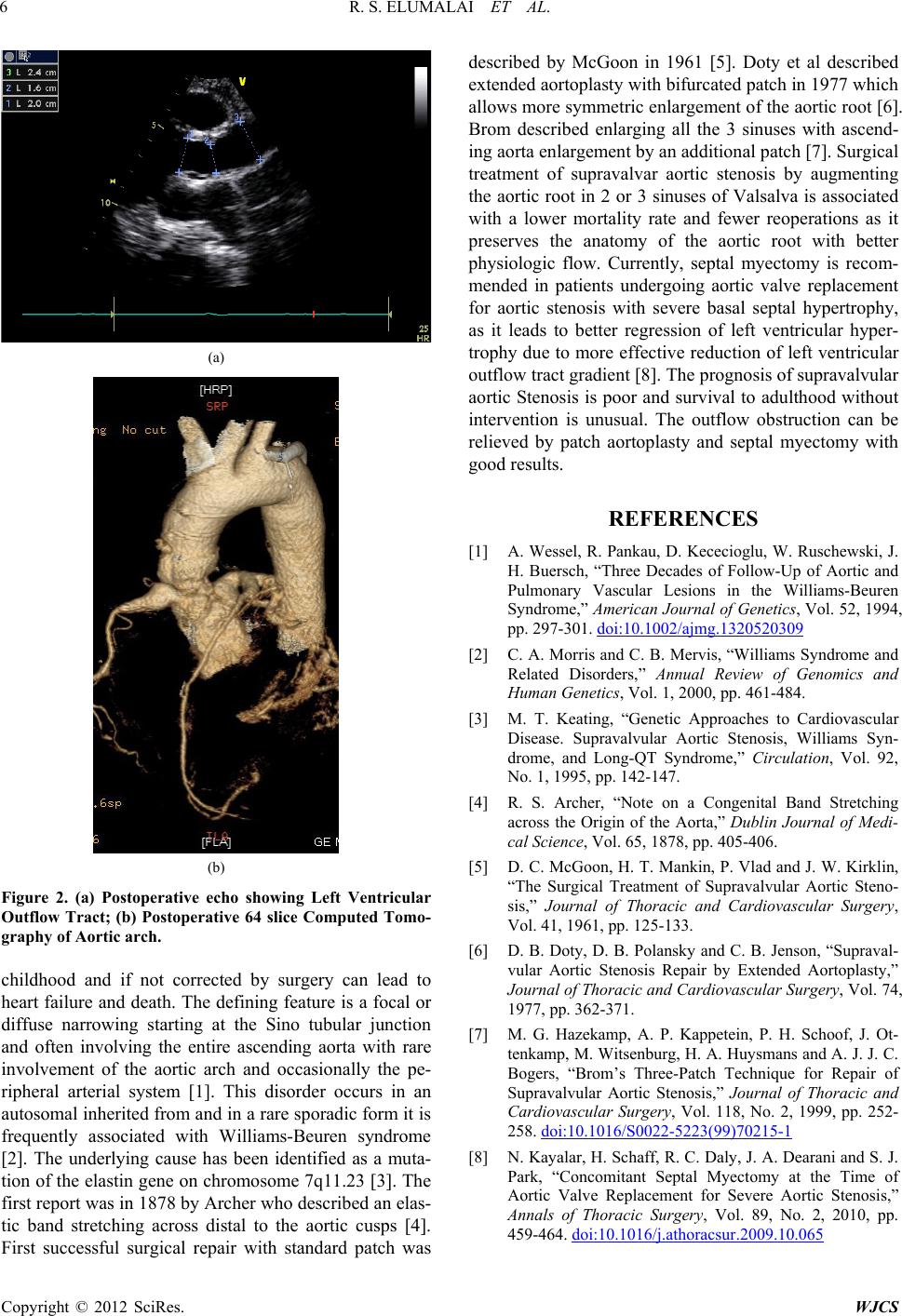 R. S. ELUMALAI ET AL. 6 (a) (b) Figure 2. (a) Postoperative echo showing Left Ventricular Outflow Tract; (b) Postoperative 64 slice Computed Tomo- graphy of Aortic arch. childhood and if not corrected by surgery can lead to heart failure and death. The defining feature is a focal or diffuse narrowing starting at the Sino tubular junction and often involving the entire ascending aorta with rare involvement of the aortic arch and occasionally the pe- ripheral arterial system [1]. This disorder occurs in an autosomal inherited from and in a rare sporadic form it is frequently associated with Williams-Beuren syndrome [2]. The underlying cause has been identified as a muta- tion of the elastin gene on chromosome 7q11.23 [3]. The first repor t was in 187 8 b y Arch er w ho d e scribe d an elas- tic band stretching across distal to the aortic cusps [4]. irst successful surgical repair with standard patch was described by McGoon in 1961 [5]. Doty et al described extended aortoplasty w ith bifur cated patch in 1977 which allows more symmetric enlargement of the ao rtic root [6]. Brom described enlarging all the 3 sinuses with ascend- ing aorta enlarg ement by an additional patch [7]. Surgical treatment of supravalvar aortic stenosis by augmenting the aortic root in 2 or 3 sinuses of Valsalva is associated with a lower mortality rate and fewer reoperations as it preserves the anatomy of the aortic root with better physiologic flow. Currently, septal myectomy is recom- mended in patients undergoing aortic valve replacement for aortic stenosis with severe basal septal hypertrophy, as it leads to better regression of left ventricular hyper- trophy due to more effective reduction of left ventricular outflow tract gradien t [8]. The p rogno sis of suprav alvular aortic Stenosis is poor and survival to adulthood without intervention is unusual. The outflow obstruction can be relieved by patch aortoplasty and septal myectomy with good results. F REFERENCES [1] A. Wessel, R. Pankau, D. Kececioglu, W. Ruschewski, J. H. Buersch, “Three Decades of Follow-Up of Aortic and Pulmonary Vascular Lesions in the Williams-Beuren Syndrome,” American Journal of Genetics, Vol. 52, 1994, pp. 297-301. doi:10.1002/ajmg.1320520309 [2] C. A. Morris and C. B. Mervis, “Williams Syndrome and Related Disorders,” Annual Review of Genomics and Human Genetics, Vol. 1, 2000, pp. 461-484. [3] M. T. Keating, “Genetic Approaches to Cardiovascular Disease. Supravalvular Aortic Stenosis, Williams Syn- drome, and Long-QT Syndrome,” Circulation, Vol. 92, No. 1, 1995, pp. 142-147. [4] R. S. Archer, “Note on a Congenital Band Stretching across the Origin of the Aorta,” Dublin Journal of Medi- cal Science, Vol. 65, 1878, pp. 405-406. [5] D. C. McGoon, H. T. Mankin, P. Vlad and J. W. Kirklin, “The Surgical Treatment of Supravalvular Aortic Steno- sis,” Journal of Thoracic and Cardiovascular Surgery, Vol. 41, 1961, pp. 125-133. [6] D. B. Doty, D. B. Polansky and C. B. Jenson, “Supraval- vular Aortic Stenosis Repair by Extended Aortoplasty,” Journal of Thoracic and Cardiovascular Surgery, Vol. 74, 1977, pp. 362-371. [7] M. G. Hazekamp, A. P. Kappetein, P. H. Schoof, J. Ot- tenkamp, M. Witsenburg, H. A. Huysmans and A. J. J. C. Bogers, “Brom’s Three-Patch Technique for Repair of Supravalvular Aortic Stenosis,” Journal of Thoracic and Cardiovascular Surgery, Vol. 118, No. 2, 1999, pp. 252- 258. doi:10.1016/S0022-5223(99)70215-1 [8] N. Kayalar, H. Schaff, R. C. Daly, J. A. Dearani and S. J. Park, “Concomitant Septal Myectomy at the Time of Aortic Valve Replacement for Severe Aortic Stenosis,” Annals of Thoracic Surgery, Vol. 89, No. 2, 2010, pp. 459-464. doi:10.1016/j.athoracsur.2009.10.065 Copyright © 2012 SciRes. WJCS |