

Paper Menu >>
Journal Menu >>
 Vol.2, No.4, 292-297 (2010) Natural Science http://dx.doi.org/10.4236/ns.2010.24037 Copyright © 2010 SciRes. OPEN ACCESS DNA damage in hemocytes of Schistocerca gregaria (Orthoptera: Acrididae) exposed to contaminated food with cadmium and lead Hesham A. Yousef1*, Amira Afify1, Hany M. Hasan2, Afaf Abdel Meguid1 1Entomology Department, Faculty of Science, Cairo University, Cairo, Egypt;[email protected] 2Agriculture Research Center, Ministry of Agriculture, Cairo, Egypt Received 9 December 2009; Revised 4 January 2010; accepted 5 February 2010. ABSTRACT We measured in a comet assay the damage of DNA in the hemocytes of various stages of the grasshopper Schistocerca gregaria after ex- posing them to various doses of Cd and Pb in the food. The mechanisms of Cd and Pb toxicity for grasshopper are discussed. The accumula- tion of heavy metals and stage of the insect may play important roles in causing the DNA damage. S. gregaria may be considered a valuable bio- indicator for evaluation the genotoxicity of en- vironmental pollutants. Keywords: Comet Assay; Heavy Metals (Cd, Pb); DNA Damage; Schistocerca gregaria 1. INTRODUCTION Heavy metals are among the most problematic causes of water, soil and plant pollution. Genetic and biochemical effects of pollutants on organisms are important in estab- lishing species as bioindicators for environmental haz- ards [1,2]. Heavy metals have been found to induce genotoxic effects in chironomids which are used as a good bioindicator group for aquatic pollution [3]. Ter- restrial insects that develop in the soil are also exposed directly to metal ions present in the soil. Grasshopper species may provide good systems to evaluate the mutagenic effects of some environmental contaminants [4-6]. Cadmium and lead are widespread and dangerous heavy metals that are released into the environment from many sources. Their accumulation in the soils can be- come dangerous to all kinds of organisms, including plants and human life, causing many genotoxic effects [7-9]. They are highly toxic and have been recognized as poison and a probable carcinogen [10]. Clinically, they can adversely affect human health, especially the blood and the renal system [11]. Changes in the cell genome caused by genotoxic agents leading to mutations and possibly tumor forma- tion are some of the lethal or sub-lethal effects induced by a complex mixture of pollutants. Among recently used methods to identify DNA damage is the comet as- say (SCGE-single cell gel electrophoresis). The comet assay provides a rapid, sensitive, and inexpensive method to detect DNA strand breaks in individual eu- karyotic cells [12]. Despite some difficulties in obtaining cell/nuclei suspension, this method has been used to detect and evaluate DNA damage caused by double strand breaks, single strand breaks, alkali labile sites, oxidative base damage, and DNA cross-linking with DNA or protein. It has been successfully applied to cells of various animal groups [13]. Only a few studies have been reported on DNA damage in insects, including D. melanogaster [14], and in the weevil Curculio sikkimen- sis [15], and in grasshoppers Chorthippus brunneus [16]. The aim of the present work was to determine the genotoxic effect of cadmium and lead on the locust S. gregaria and to evaluate its potential as a biomonitor for detecting a heavy-metal polluted environment. 2. MATERIALS AND METHODS 2.1. Colonization of S. Gregaria Locusts were reared in wooden cages at 32 ± 2C°, 50- 60% RH and 16 hrs day light in our Entomology De- partment since about 10 years ago. A daily supply of fresh grass, clover plant was supplied to the locusts. Packed moist sterilized sand in suitable glass containers about 7 cm in diameter and 10 cm deep were prepared for egg-laying. 2.2. Heavy Metals Treatment and Sample Preparation for Alkaline Single Cell Gel (SCG) Assay Living individuals of S. gregaria of the 4th, 5th instars, 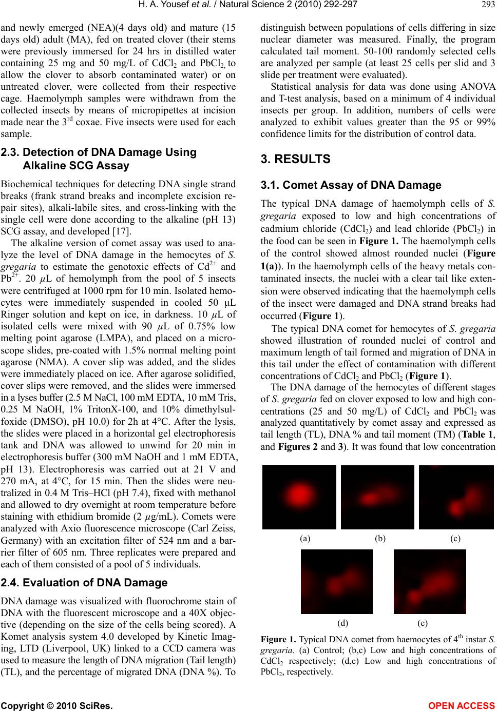 H. A. Yousef et al. / Natural Science 2 (2010) 292-297 Copyright © 2010 SciRes. OPEN ACCESS 293 and newly emerged (NEA)(4 days old) and mature (15 days old) adult (MA), fed on treated clover (their stems were previously immersed for 24 hrs in distilled water containing 25 mg and 50 mg/L of CdCl2 and PbCl2, to allow the clover to absorb contaminated water) or on untreated clover, were collected from their respective cage. Haemolymph samples were withdrawn from the collected insects by means of micropipettes at incision made near the 3rd coxae. Five insects were used for each sample. 2.3. Detection of DNA Damage Using Alkaline SCG Assay Biochemical techniques for detecting DNA single strand breaks (frank strand breaks and incomplete excision re- pair sites), alkali-labile sites, and cross-linking with the single cell were done according to the alkaline (pH 13) SCG assay, and developed [17]. The alkaline version of comet assay was used to ana- lyze the level of DNA damage in the hemocytes of S. gregaria to estimate the genotoxic effects of Cd2+ and Pb2+. 20 µL of hemolymph from the pool of 5 insects were centrifuged at 1000 rpm for 10 min. Isolated hemo- cytes were immediately suspended in cooled 50 μL Ringer solution and kept on ice, in darkness. 10 µL of isolated cells were mixed with 90 µL of 0.75% low melting point agarose (LMPA), and placed on a micro- scope slides, pre-coated with 1.5% normal melting point agarose (NMA). A cover slip was added, and the slides were immediately placed on ice. After agarose solidified, cover slips were removed, and the slides were immersed in a lyses buffer (2.5 M NaCl, 100 mM EDTA, 10 mM Tris, 0.25 M NaOH, 1% TritonX-100, and 10% dimethylsul- foxide (DMSO), pH 10.0) for 2h at 4°C. After the lysis, the slides were placed in a horizontal gel electrophoresis tank and DNA was allowed to unwind for 20 min in electrophoresis buffer (300 mM NaOH and 1 mM EDTA, pH 13). Electrophoresis was carried out at 21 V and 270 mA, at 4°C, for 15 min. Then the slides were neu- tralized in 0.4 M Tris–HCl (pH 7.4), fixed with methanol and allowed to dry overnight at room temperature before staining with ethidium bromide (2 µg/mL). Comets were analyzed with Axio fluorescence microscope (Carl Zeiss, Germany) with an excitation filter of 524 nm and a bar- rier filter of 605 nm. Three replicates were prepared and each of them consisted of a pool of 5 individuals. 2.4. Evaluation of DNA Damage DNA damage was visualized with fluorochrome stain of DNA with the fluorescent microscope and a 40X objec- tive (depending on the size of the cells being scored). A Komet analysis system 4.0 developed by Kinetic Imag- ing, LTD (Liverpool, UK) linked to a CCD camera was used to measure the length of DNA migration (Tail length) (TL), and the percentage of migrated DNA (DNA %). To distinguish between populations of cells differing in size nuclear diameter was measured. Finally, the program calculated tail moment. 50-100 randomly selected cells are analyzed per sample (at least 25 cells per slid and 3 slide per treatment were evaluated). Statistical analysis for data was done using ANOVA and T-test analysis, based on a minimum of 4 individual insects per group. In addition, numbers of cells were analyzed to exhibit values greater than the 95 or 99% confidence limits for the distribution of control data. 3. RESULTS 3.1. Comet Assay of DNA Damage The typical DNA damage of haemolymph cells of S. gregaria exposed to low and high concentrations of cadmium chloride (CdCl2) and lead chloride (PbCl2) in the food can be seen in Figure 1. The haemolymph cells of the control showed almost rounded nuclei (Figure 1(a)). In the haemolymph cells of the heavy metals con- taminated insects, the nuclei with a clear tail like exten- sion were observed indicating that the haemolymph cells of the insect were damaged and DNA strand breaks had occurred (Figure 1). The typical DNA comet for hemocytes of S. gregaria showed illustration of rounded nuclei of control and maximum length of tail formed and migration of DNA in this tail under the effect of contamination with different concentrations of CdCl2 and PbCl2 (Figure 1). The DNA damage of the hemocytes of different stages of S. gregaria fed on clover exposed to low and high con- centrations (25 and 50 mg/L) of CdCl2 and PbCl2 was analyzed quantitatively by comet assay and expressed as tail length (TL), DNA % and tail moment (TM) (Table 1, and Figures 2 and 3). It was found that low concentration (a) (b) (c) (d) (e) Figure 1. Typical DNA comet from haemocytes of 4th instar S. gregaria. (a) Control; (b,c) Low and high concentrations of CdCl2 respectively; (d,e) Low and high concentrations of PbCl2, respectively. 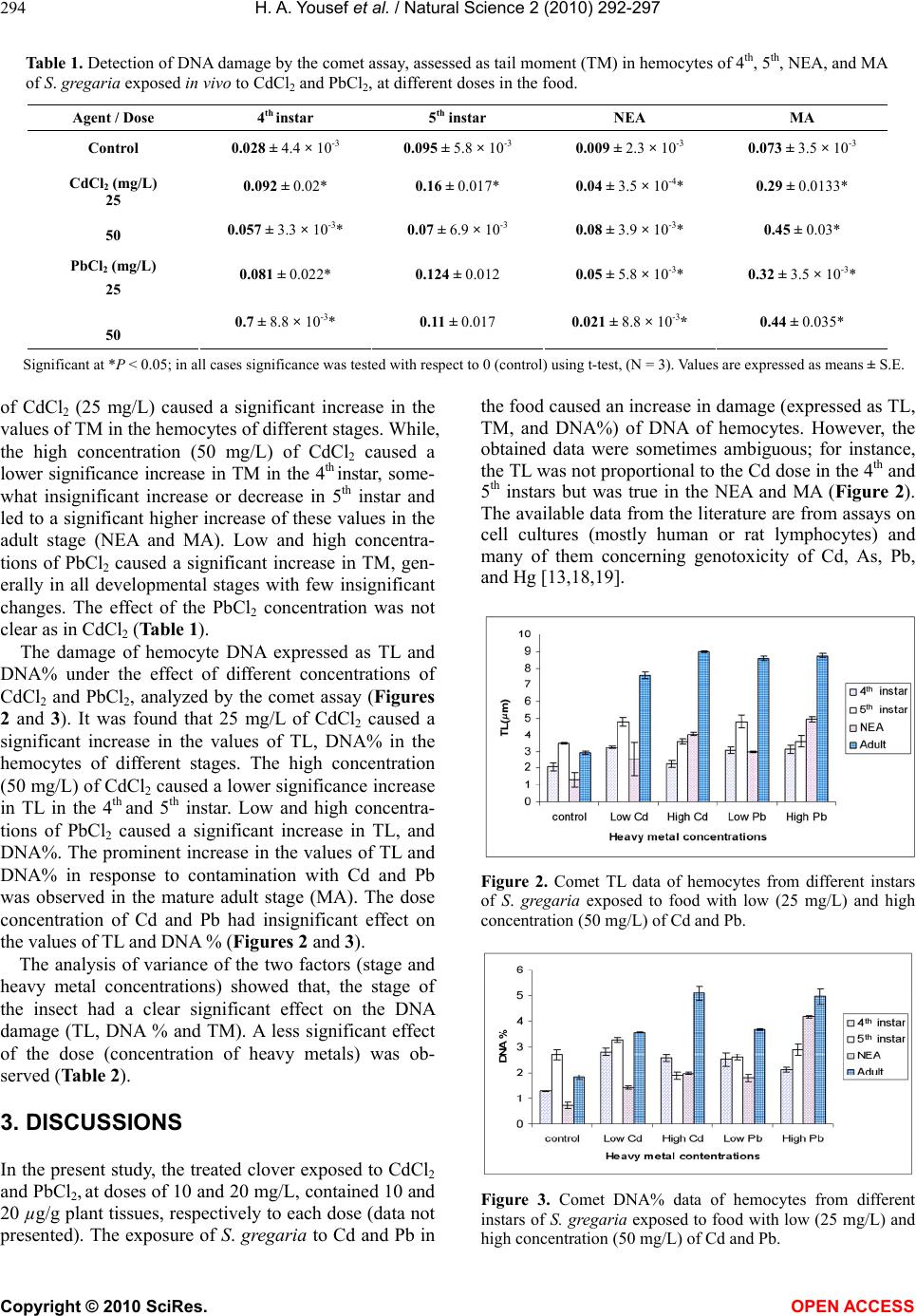 H. A. Yousef et al. / Natural Science 2 (2010) 292-297 Copyright © 2010 SciRes. OPEN ACCESS 294 Table 1. Detection of DNA damage by the comet assay, assessed as tail moment (TM) in hemocytes of 4th, 5th, NEA, and MA of S. gregaria exposed in vivo to CdCl2 and PbCl2, at different doses in the food. Significant at *P < 0.05; in all cases significance was tested with respect to 0 (control) using t-test, (N = 3). Values are expressed as means ± S.E. of CdCl2 (25 mg/L) caused a significant increase in the values of TM in the hemocytes of different stages. While, the high concentration (50 mg/L) of CdCl2 caused a lower significance increase in TM in the 4th instar, some- what insignificant increase or decrease in 5th instar and led to a significant higher increase of these values in the adult stage (NEA and MA). Low and high concentra- tions of PbCl2 caused a significant increase in TM, gen- erally in all developmental stages with few insignificant changes. The effect of the PbCl2 concentration was not clear as in CdCl2 (Table 1). The damage of hemocyte DNA expressed as TL and DNA% under the effect of different concentrations of CdCl2 and PbCl2, analyzed by the comet assay (Figures 2 and 3). It was found that 25 mg/L of CdCl2 caused a significant increase in the values of TL, DNA% in the hemocytes of different stages. The high concentration (50 mg/L) of CdCl2 caused a lower significance increase in TL in the 4th and 5th instar. Low and high concentra- tions of PbCl2 caused a significant increase in TL, and DNA%. The prominent increase in the values of TL and DNA% in response to contamination with Cd and Pb was observed in the mature adult stage (MA). The dose concentration of Cd and Pb had insignificant effect on the values of TL and DNA % (Figures 2 and 3). The analysis of variance of the two factors (stage and heavy metal concentrations) showed that, the stage of the insect had a clear significant effect on the DNA damage (TL, DNA % and TM). A less significant effect of the dose (concentration of heavy metals) was ob- served (Table 2). 3. DISCUSSIONS In the present study, the treated clover exposed to CdCl2 and PbCl2, at doses of 10 and 20 mg/L, contained 10 and 20 µg/g plant tissues, respectively to each dose (data not presented). The exposure of S. gregaria to Cd and Pb in the food caused an increase in damage (expressed as TL, TM, and DNA%) of DNA of hemocytes. However, the obtained data were sometimes ambiguous; for instance, the TL was not proportional to the Cd dose in the 4th and 5th instars but was true in the NEA and MA (Figure 2). The available data from the literature are from assays on cell cultures (mostly human or rat lymphocytes) and many of them concerning genotoxicity of Cd, As, Pb, and Hg [13,18,19]. Figure 2. Comet TL data of hemocytes from different instars of S. gregaria exposed to food with low (25 mg/L) and high concentration (50 mg/L) of Cd and Pb. Figure 3. Comet DNA% data of hemocytes from different instars of S. gregaria exposed to food with low (25 mg/L) and high concentration (50 mg/L) of Cd and Pb. Agent / Dose 4th instar 5th instar NEA MA Control 0.028 ± 4.4 × 10⁻³ 0.095 ± 5.8 × 10⁻³ 0.009 ± 2.3 × 10⁻³ 0.073 ± 3.5 × 10⁻³ 0.092 ± 0.02* 0.16 ± 0.017* 0.04 ± 3.5 × 10-4* 0.29 ± 0.0133* CdCl2 (mg/L) 25 50 0.057 ± 3.3 × 10-3* 0.07 ± 6.9 × 10⁻³ 0.08 ± 3.9 × 10-3* 0.45 ± 0.03* 0.081 ± 0.022* 0.124 ± 0.012 0.05 ± 5.8 × 10-3* 0.32 ± 3.5 × 10⁻³* PbCl2 (mg/L) 25 50 0.7 ± 8.8 × 10-3* 0.11 ± 0.017 0.021 ± 8.8 × 10-3* 0.44 ± 0.035* 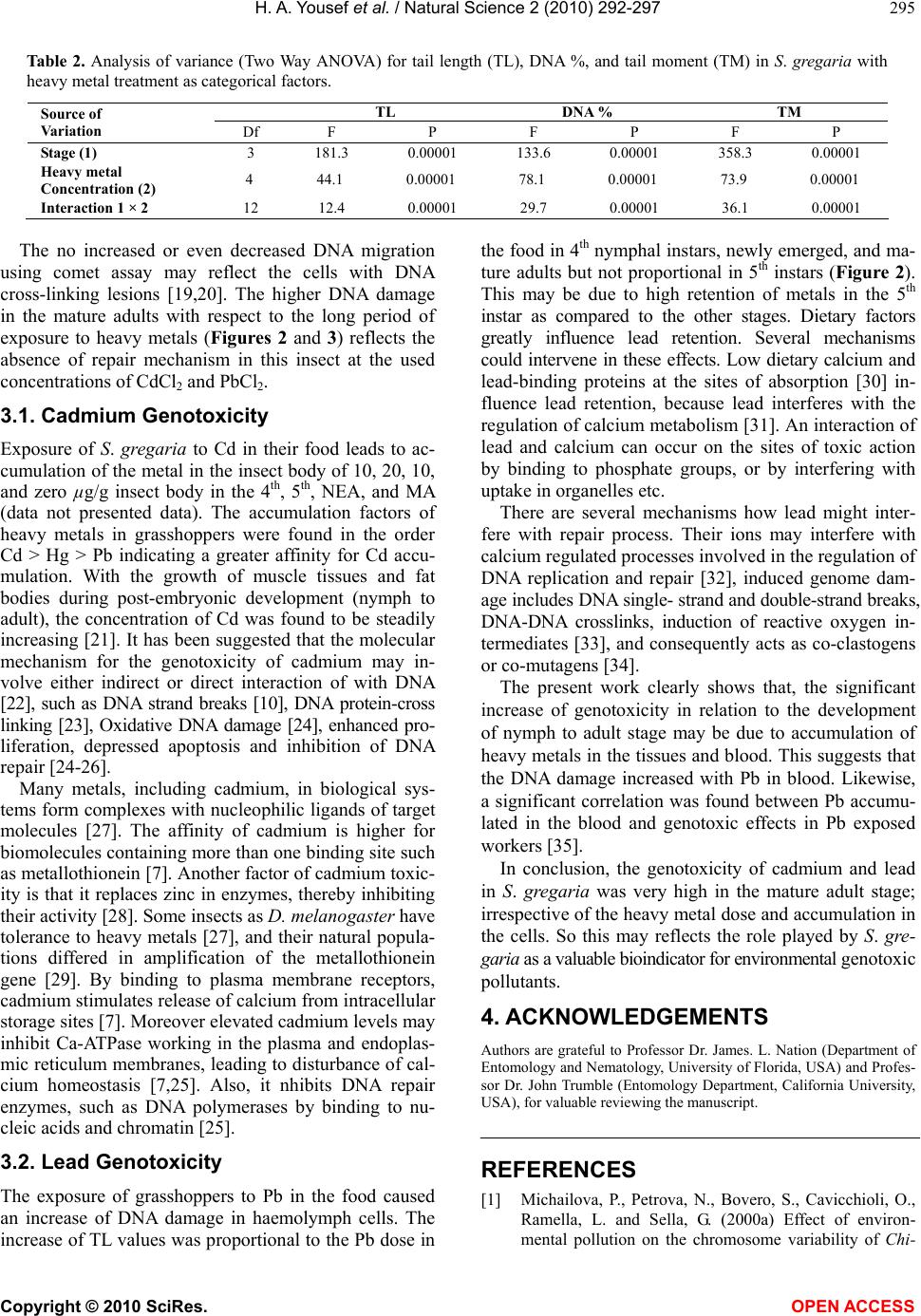 H. A. Yousef et al. / Natural Science 2 (2010) 292-297 Copyright © 2010 SciRes. OPEN ACCESS 295 Table 2. Analysis of variance (Two Way ANOVA) for tail length (TL), DNA %, and tail moment (TM) in S. gregaria with heavy metal treatment as categorical factors. TL DNA % TM Source of Variation Df F P F P F P Stage (1) 3 181.3 0.00001 133.6 0.00001 358.3 0.00001 Heavy metal Concentration (2) 4 44.1 0.00001 78.1 0.00001 73.9 0.00001 Interaction 1 × 2 12 12.4 0.00001 29.7 0.00001 36.1 0.00001 The no increased or even decreased DNA migration using comet assay may reflect the cells with DNA cross-linking lesions [19,20]. The higher DNA damage in the mature adults with respect to the long period of exposure to heavy metals (Figures 2 and 3) reflects the absence of repair mechanism in this insect at the used concentrations of CdCl2 and PbCl2. 3.1. Cadmium Genotoxicity Exposure of S. gregaria to Cd in their food leads to ac- cumulation of the metal in the insect body of 10, 20, 10, and zero µg/g insect body in the 4th, 5th, NEA, and MA (data not presented data). The accumulation factors of heavy metals in grasshoppers were found in the order Cd > Hg > Pb indicating a greater affinity for Cd accu- mulation. With the growth of muscle tissues and fat bodies during post-embryonic development (nymph to adult), the concentration of Cd was found to be steadily increasing [21]. It has been suggested that the molecular mechanism for the genotoxicity of cadmium may in- volve either indirect or direct interaction of with DNA [22], such as DNA strand breaks [10], DNA protein-cross linking [23], Oxidative DNA damage [24], enhanced pro- liferation, depressed apoptosis and inhibition of DNA repair [24-26]. Many metals, including cadmium, in biological sys- tems form complexes with nucleophilic ligands of target molecules [27]. The affinity of cadmium is higher for biomolecules containing more than one binding site such as metallothionein [7]. Another factor of cadmium toxic- ity is that it replaces zinc in enzymes, thereby inhibiting their activity [28]. Some insects as D. melanogaster have tolerance to heavy metals [27], and their natural popula- tions differed in amplification of the metallothionein gene [29]. By binding to plasma membrane receptors, cadmium stimulates release of calcium from intracellular storage sites [7]. Moreover elevated cadmium levels may inhibit Ca-ATPase working in the plasma and endoplas- mic reticulum membranes, leading to disturbance of cal- cium homeostasis [7,25]. Also, it nhibits DNA repair enzymes, such as DNA polymerases by binding to nu- cleic acids and chromatin [25]. 3.2. Lead Genotoxicity The exposure of grasshoppers to Pb in the food caused an increase of DNA damage in haemolymph cells. The increase of TL values was proportional to the Pb dose in the food in 4th nymphal instars, newly emerged, and ma- ture adults but not proportional in 5th instars (Figure 2). This may be due to high retention of metals in the 5th instar as compared to the other stages. Dietary factors greatly influence lead retention. Several mechanisms could intervene in these effects. Low dietary calcium and lead-binding proteins at the sites of absorption [30] in- fluence lead retention, because lead interferes with the regulation of calcium metabolism [31]. An interaction of lead and calcium can occur on the sites of toxic action by binding to phosphate groups, or by interfering with uptake in organelles etc. There are several mechanisms how lead might inter- fere with repair process. Their ions may interfere with calcium regulated processes involved in the regulation of DNA replication and repair [32], induced genome dam- age includes DNA single- strand and double-strand breaks, DNA-DNA crosslinks, induction of reactive oxygen in- termediates [33], and consequently acts as co-clastogens or co-mutagens [34]. The present work clearly shows that, the significant increase of genotoxicity in relation to the development of nymph to adult stage may be due to accumulation of heavy metals in the tissues and blood. This suggests that the DNA damage increased with Pb in blood. Likewise, a significant correlation was found between Pb accumu- lated in the blood and genotoxic effects in Pb exposed workers [35]. In conclusion, the genotoxicity of cadmium and lead in S. gregaria was very high in the mature adult stage; irrespective of the heavy metal dose and accumulation in the cells. So this may reflects the role played by S. gre- garia as a valuable bioindicator for environmental genotoxic pollutants. 4. ACKNOWLEDGEMENTS Authors are grateful to Professor Dr. James. L. Nation (Department of Entomology and Nematology, University of Florida, USA) and Profes- sor Dr. John Trumble (Entomology Department, California University, USA), for valuable reviewing the manuscript. REFERENCES [1] Michailova, P., Petrova, N., Bovero, S., Cavicchioli, O., Ramella, L. and Sella, G. (2000a) Effect of environ- mental pollution on the chromosome variability of Chi-  H. A. Yousef et al. / Natural Science 2 (2010) 292-297 Copyright © 2010 SciRes. OPEN ACCESS 296 ronomus riparius Meigen (1804) (Diptera, Chironomidae) larvae from two Piedmont stations. Genetica, 108(2), 171-180. [2] Michailova, P., Petrova, N., Bovero, S., Sella, G. and Ramella, L. (2000b) Structural and functional rear- rangements in polytene chromosomes of Chironomids (Diptera) as biomarkers for heavy metal pollution in aquatic ecosystems. International Conference on Heavy Metals in the Environment, Michigan University, Michi- gan, 70-79. [3] Michailova, P., Ilkova, J., Petrova, N. and White, K. (2001) Rearrangements in the salivary gland chromo- somes of Chironomus riparius Mg. (Diptera, Chironomi- dae) following exposure to lead. Caryologia, 4, 349-363. [4] Barsiene, J. (1994) Chromosome set changes in molluscs from highly polluted habitats in: A.R. beaumont, editors, genetics and evolution of aquatic organisms. Chapman & Hall, London, 434-447. [5] Barsyte, D. (1999) The analysis of genotoxic damage of industrial pollution in freshwater mollusc tissues. Sum- mery Doctoral Thesis, Institute of Ecology, Vilnius, 1-20. [6] Warchalowska–Sliwa, E., Niklinska, M., Görlich, A., Michailova, P. and Pyza, E. (2005) Heavy metal accu- mulation, heat shock protein expression and cytogenetic changes in Tetrix tenuicornis (L.) (Tetrigidae, Orthoptera) from polluted areas. Environmental Pollution, 133(2), 373-381. [7] Goering, P.L., Waalkes, M.P. and Klaassen, C.D. (1995) Toxicology of cadmium. Goyer, R.A. and Cherian, M.G., Ed., Toxicology of Metals, Springer-Verlag, Berlin, 189- 214. [8] Sanita di Toppi, L. and Gabbrieli, R. (1999) Responses to cadmium in higher plants. Environmental and Experi- mental Botany, 41(2), 105-130. [9] Panda, B.B. and Panda, K.K. (2002) Genotoxicity and mutagenicity of metals in plants. Prasad, K.N.V. and Strzalka, K., Ed., Physiology and BioChemistry of Metal Toxicity and Tolerance in Plants, Kluwer Academic Pub- lishers, Netherlands, 15, 395-414. [10] Waalkes, M. and Misra, R. (1996) Cadmium carcino- genicity and genotoxicity. Chang, L.W., Ed., Toxicology of Metals, CRC Press, Boca Raton, 231-241. [11] Arcadio, P.S. and Gregoria, A.S. (2002) Physical-chemical treatment of water and waste water. CRC Press, Boca Raton, IWA Publishing, London. [12] Rojas, E., Lopez, M.C. and Valverde, M. (1999) Single cell gel electrophoresis assay: Methodology and applica- tions. Journal of Chromatography B: Biomedical Sci- ences and Applications, 722(1-2), 225-254. [13] Woźniak, K. and Blasiak, J. (2003) In vitro genotoxicity of lead acetate: Induction of single and double DNA strand breaks and DNA-protein cross-links. Mutation Research, 535(2), 127-139. [14] Siddique, H.R., Chowdhuri, D.K., Saxena, D.K. and Dhavan, A. (2005) Validation of Drosophila melan- ogaster as an in vivo model for genotoxicity assessment using modified alkaline comet assay. Mutagenesis, 20(4), 285-290. [15] Todoriki, S., Hasan, M., Miyanoshita, A., Imamura, T. and Hayashi, T. (2006) Assessment of electron beam- induced DNA damage in larvae of chestnut weevil. Cur- culio sikkimensis (Coleoptera: Curculionidae) using comet assay. Radiation Physics and Chemistry, 75(2), 292-296. [16] Augustyniak, M., Juchimiuk, J., Przybylowicz, W.J., Mesjasz-Przybylowicz, J., Babczynska, A. and Migula, P. (2006) Zinc-induced DNA damage and the distribution of metals in the brain of grasshoppers by the comet assay and micro-PIXE. Comparative Biochemistry and Physi- ology - Part C, 144(3), 242-251. [17] Singh, N.P., McCoy, M.T., Tice, R.R. and Schneider, E.L. (1998) A simple technique for quantitation of low levels of DNA damage in individual cells. Experimental Cell Research, 175(1), 184-191. [18] Grover, S.A., Coupal, L., Zowall, H., Alexander, C.M., Weiss, T.W. and Gomes, D.R.J. (2001) How cost-effec- tive is the treatment of dyslipidemia in patients with dia- betes but without cardiovascular disease? Diabetes Care, 24(1), 45-50. [19] Mourón, S.A., Golijow, C.D. and Dulout, F.N. (2001) DNA damage by cadmium and arsenic salts assessed by the single cell gel electrophoresis assay. Mutation Re- search, 498(1-2), 47-55. [20] Tice, R., Agurell, E., Anderson, D., Burlinson, B., Hart- mann, A., Kobayashi, H., Miyamae, Y., Rojas, E., Ryu, J.C. and Sasaki, Y. (2000) Single cell gel/comet assay: Guidelines for in vitro and in vivo genetic toxicology testing. Environmental and Molecular Mutagenesis, 35 (3), 206-221. [21] Devkota, B. and Schmidt, G.H. (2000) Accumulation of heavy metals in food plants and grasshoppers from the Taigetos Mountains. Greece Agriculture, Ecosystems and Environment, 78(1), 85-91. [22] Hossain, Z. and Huq, F. (2002) Studies on the interaction between Cd(2+) ions and nucleobases and nucleotides. Journal of Inorganic Biochemistry, 90(3-4), 97-105. [23] Misra, R., Smith, G. and Waalkes, M. (1998) Evaluation of the direct genotoxic potential of cadimium in four dif- ferent rodent cell lines. Toxicology, 126(2), 103-114. [24] Dally, H. and Hartwig, A. (1997) Induction and repair inhibition of oxidative DNA damage by nickel (II) and cadmium (II) in mammalian cells. Carcinogenesis, 18, 1021-1026. [25] Beyersmann, D. and Hechtenberg, S. (1997) Cadmium, gene regulation, and cellular signaling in mammalian cells. Toxicology and Applied Pharmacology, 144(2), 247-261. [26] Waalkes, M., Fox, D., States, C., Patierno, S. and McCabe, M. (2000) Metals and disorder of cell accumulation: Modulation of apoptosis and cell proliferation. Toxico- logical Sciences, 56(2), 255-261. [27] Korsloot, A., van Gestel, C.A.M. and van Straalen, N.M. (2004) Environmental stress and cellular response in ar- thropods. CRC Press, Boca Raton. [28] Hamer, D.H. (1986) Metallothionein. Annual Review of Biochemistry, 55, 931-951. [29] Maroni, G.J., Wise, J., Young, J.E. and Otto, E. (1987) Metallothionein gene duplications and metal tolerance in natural populations of Drosophila melanogaster. Genetics, 117(4), 739-744. [30] Barltop, D. and Meek, F. (1987) Effect of particle size on lead absorption from the gut. Archives of Environmental Health, 34(4), 280-286. [31] Peng, T., Gitelman C.H. and Garner, S.C. (1979) Acute  H. A. Yousef et al. / Natural Science 2 (2010) 292-297 Copyright © 2010 SciRes. OPEN ACCESS 297 lead induced increase in serum calcium in the rat without increased secreation of calcitonin. Proceedings of the So- ciety for Experimental Biology and Medicine, 160(1), 114-117. [32] Johnson, L. and Pellicciari, C.E. (1988) Lead induced changes in the stabilization of the mouse sperm chroma- tin. Toxicology, 51(1), 11-24. [33] Ariza, M.E., Bijur, G.N. and Williams, M.V. (1998) Lead and mercury mutagenesis: Role of H2O2 superoxide dis- mutase and xanthine oxidase. Environmental and Mo- lecular Mutagenesis, 31(4), 352-361. [34] Miadoková, E., Vlčková, V., Jendraššaková, N., Vlček, D. and Šucha, V. (2000) Mutagenic and comutagenic effects of acid-mine water containing heavy metals. Journal of Trace and Microprobe Techniques, 18, 201-207. [35] Duydu, Y., Suzen, H.S., Aydin, A., Cander, O., Uysal, H., Isimer, A. and Vural, N. (2001) Correlation between lead exposure indicators and sister chromatid exchange (SCE) frequencies in lymphocytes from inorganic lead exposed workers. Archives of Environmental Contamination and Toxicology, 41(2), 241-246. |