

Paper Menu >>
Journal Menu >>
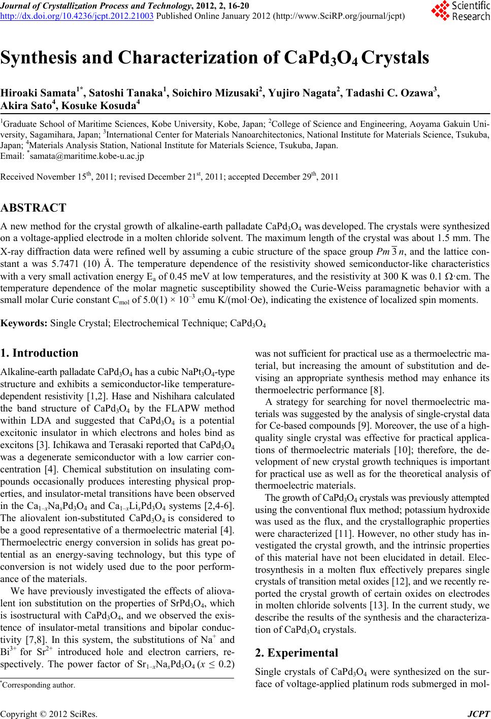 Journal of Crystallization Process and Technology, 2012, 2, 16-20 http://dx.doi.org/10.4236/jcpt.2012.21003 Published Online January 2012 (http://www.SciRP.org/journal/jcpt) Synthesis and Characterization of CaPd3O4 Crystals Hiroaki Samata1*, Satoshi Tanaka1, Soichiro Mizusaki2, Yujiro Nagata2, Tadashi C. Ozawa3, Akira Sato4, Kosuke Kosuda4 1Graduate School of Maritime Sciences, Kobe University, Kobe, Japan; 2College of Science and Engineering, Aoyama Gakuin Uni- versity, Sagamihara, Japan; 3International Center for Materials Nanoarchitectonics, National Institute for Materials Science, Tsukuba, Japan; 4Materials Analysis Station, National Institute for Materials Science, Tsukuba, Japan. Email: *[email protected] Received November 15th, 2011; revised December 21st, 2011; accepted December 29th, 2011 ABSTRACT A new method for the crystal growth of alkaline-earth palladate CaPd3O4 was developed. The crystals were synthesized on a voltage-applied electrode in a molten chloride solvent. The maximum length of the crystal was about 1.5 mm. The X-ray diffraction data were refined well by assuming a cubic structure of the space group Pm 3n, and the lattice con- stant a was 5.7471 (10) Å. The temperature dependence of the resistivity showed semiconductor-like characteristics with a very small activation energy Ea of 0.45 meV at low temperatures, and the resistivity at 300 K was 0.1 ·cm. The temperature dependence of the molar magnetic susceptibility showed the Curie-Weiss paramagnetic behavior with a small molar Curie constant Cmol of 5.0(1) × 10–3 emu K/(mol·Oe), indicating the existence of localized spin moments. Keywords: Single Crystal; Electrochemical Technique; CaPd3O4 1. Introduction Alkaline-earth palladate CaPd3O4 has a cubic NaPt3O4-type structure and exhibits a semiconductor-like temperature- dependent resistivity [1,2]. Hase and Nishihara calculated the band structure of CaPd3O4 by the FLAPW method within LDA and suggested that CaPd3O4 is a potential excitonic insulator in which electrons and holes bind as excitons [3]. Ichikawa and Terasaki reported that CaPd3O4 was a degenerate semiconductor with a low carrier con- centration [4]. Chemical substitution on insulating com- pounds occasionally produces interesting physical prop- erties, and insulator-metal transitions have been observed in the Ca1–xNaxPd3O4 and Ca1–xLixPd3O4 systems [2,4-6]. The aliovalent ion-substituted CaPd3O4 is considered to be a good representative of a thermoelectric material [4]. Thermoelectric energy conversion in solids has great po- tential as an energy-saving technology, but this type of conversion is not widely used due to the poor perform- ance of the materials. We have previously investigated the effects of aliova- lent ion substitution on the properties of SrPd3O4, which is isostructural with CaPd3O4, and we observed the exis- tence of insulator-metal transitions and bipolar conduc- tivity [7,8]. In this system, the substitutions of Na+ and Bi3+ for Sr2+ introduced hole and electron carriers, re- spectively. The power factor of Sr1–xNaxPd3O4 (x ≤ 0.2) was not sufficient for practical use as a thermoelectric ma- terial, but increasing the amount of substitution and de- vising an appropriate synthesis method may enhance its thermoelectric performance [8]. A strategy for searching for novel thermoelectric ma- terials was suggested by the analysis of single-crystal data for Ce-based compounds [9]. Moreover, the use of a high- quality single crystal was effective for practical applica- tions of thermoelectric materials [10]; therefore, the de- velopment of new crystal growth techniques is important for practical use as well as for the theoretical analysis of thermoelectric materials. The growth of CaPd3O4 crystals was previously attempted using the conventional flux method; potassium hydroxide was used as the flux, and the crystallographic properties were characterized [11]. However, no other study has in- vestigated the crystal growth, and the intrinsic properties of this material have not been elucidated in detail. Elec- trosynthesis in a molten flux effectively prepares single crystals of transition metal oxides [12], and we recently re- ported the crystal growth of certain oxides on electrodes in molten chloride solvents [13]. In the current study, we describe the results of the synthesis and the characteriza- tion of CaPd3O4 crystals. 2. Experimental Single crystals of CaPd3O4 were synthesized on the sur- face of voltage-applied platinum rods submerged in mol- *Corresponding author. Copyright © 2012 SciRes. JCPT 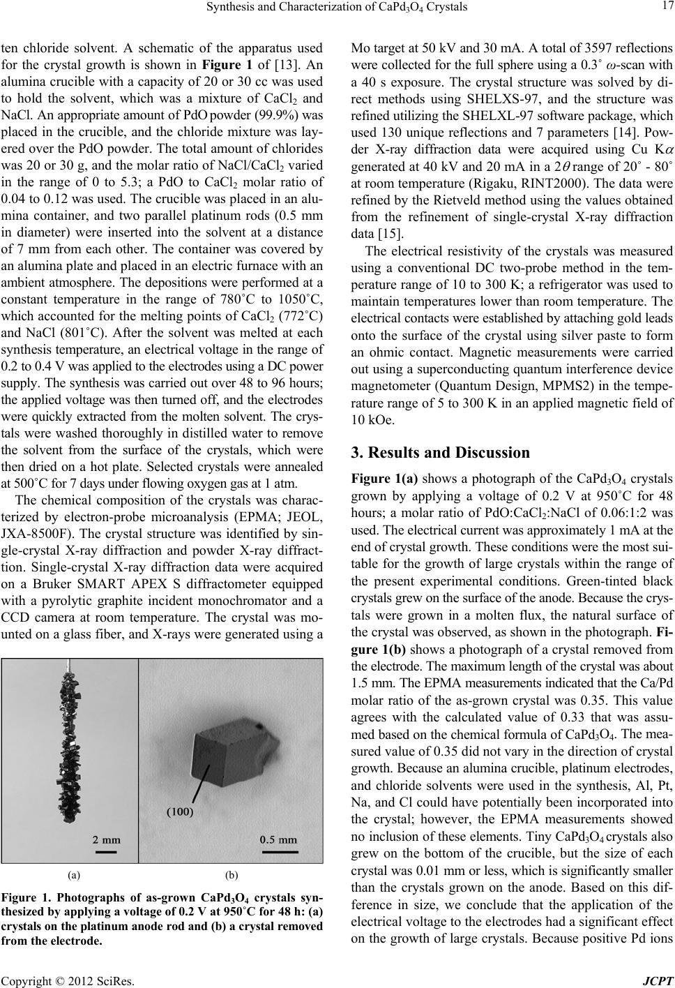 Synthesis and Characterization of CaPd3O4 Crystals 17 ten chloride solvent. A schematic of the apparatus used for the crystal growth is shown in Figure 1 of [13]. An alumina crucible with a capacity of 20 or 30 cc was used to hold the solvent, which was a mixture of CaCl2 and NaCl. An appropriate amount of PdO powder (99.9%) was placed in the crucible, and the chloride mixture was lay- ered over the PdO powder. The total amount of chlorides was 20 or 30 g, and the molar ratio of NaCl/CaCl2 varied in the range of 0 to 5.3; a PdO to CaCl2 molar ratio of 0.04 to 0.12 was used. The crucible was placed in an alu- mina container, and two parallel platinum rods (0.5 mm in diameter) were inserted into the solvent at a distance of 7 mm from each other. The container was covered by an alumina plate and placed in an electric furnace with an ambient atmosphere. The depositions were performed at a constant temperature in the range of 780˚C to 1050˚C, which accounted for the melting points of CaCl2 (772˚C) and NaCl (801˚C). After the solvent was melted at each synthesis temperature, an electrical voltage in the range of 0.2 to 0.4 V was applied to the electrodes using a DC power supply. The synthesis was carried out over 48 to 96 hours; the applied voltage was then turned off, and the electrodes were quickly extracted from the molten solvent. The crys- tals were washed thoroughly in distilled water to remove the solvent from the surface of the crystals, which were then dried on a hot plate. Selected crystals were annealed at 500˚C for 7 days under flowing oxygen gas at 1 atm. The chemical composition of the crystals was charac- terized by electron-probe microanalysis (EPMA; JEOL, JXA-8500F). The crystal structure was identified by sin- gle-crystal X-ray diffraction and powder X-ray diffract- tion. Single-crystal X-ray diffraction data were acquired on a Bruker SMART APEX S diffractometer equipped with a pyrolytic graphite incident monochromator and a CCD camera at room temperature. The crystal was mo- unted on a glass fiber, and X-rays were generated using a (a) (b) Figure 1. Photographs of as-grown CaPd3O4 crystals syn- thesized by applying a voltage of 0.2 V at 950˚C for 48 h: (a) cry stals on the platin um anode rod and ( b) a crystal remove d from the electrode. Mo target at 50 kV and 30 mA. A total of 3597 reflections were collected for the full sphere using a 0.3˚ ω-scan with a 40 s exposure. The crystal structure was solved by di- rect methods using SHELXS-97, and the structure was refined utilizing the SHELXL-97 software package, which used 130 unique reflections and 7 parameters [14]. Pow- der X-ray diffraction data were acquired using Cu K generated at 40 kV and 20 mA in a 2 range of 20˚ - 80˚ at room temperature (Rigaku, RINT2000). The data were refined by the Rietveld method using the values obtained from the refinement of single-crystal X-ray diffraction data [15]. The electrical resistivity of the crystals was measured using a conventional DC two-probe method in the tem- perature range of 10 to 300 K; a refrigerator was used to maintain temperatures lower than room temperature. The electrical contacts were established by attaching gold leads onto the surface of the crystal using silver paste to form an ohmic contact. Magnetic measurements were carried out using a superconducting quantum interference device magnetometer (Quantum Design, MPMS2) in the tempe- rature range of 5 to 300 K in an applied magnetic field of 10 kOe. 3. Results and Discussion Figure 1(a) shows a photograph of the CaPd3O4 crystals grown by applying a voltage of 0.2 V at 950˚C for 48 hours; a molar ratio of PdO:CaCl2:NaCl of 0.06:1:2 was used. The electrical current was approximately 1 mA at the end of crystal growth. These conditions were the most sui- table for the growth of large crystals within the range of the present experimental conditions. Green-tinted black crystals grew on the surface of the anode. Because the crys- tals were grown in a molten flux, the natural surface of the crystal was observed, as shown in the photograph. Fi- gure 1(b) shows a photograph of a crystal removed from the electrode. The maximum length of the crystal was about 1.5 mm. The EPMA measurements indicated that the Ca/Pd molar ratio of the as-grown crystal was 0.35. This value agrees with the calculated value of 0.33 that was assu- med based on the chemical formula of CaPd3O4. The mea- sured value of 0.35 did not vary in the direction of crystal growth. Because an alumina crucible, platinum electrodes, and chloride solvents were used in the synthesis, Al, Pt, Na, and Cl could have potentially been incorporated into the crystal; however, the EPMA measurements showed no inclusion of these elements. Tiny CaPd3O4 crystals also grew on the bottom of the crucible, but the size of each crystal was 0.01 mm or less, which is significantly smaller than the crystals grown on the anode. Based on this dif- ference in size, we conclude that the application of the electrical voltage to the electrodes had a significant effect on the growth of large crystals. Because positive Pd ions Copyright © 2012 SciRes. JCPT 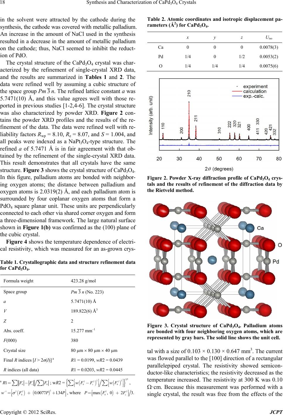 Synthesis and Characterization of CaPd3O4 Crystals 18 in the solvent were attracted by the cathode during the synthesis, the cathode was covered with metallic palladium. An increase in the amount of NaCl used in the synthesis resulted in a decrease in the amount of metallic palladium on the cathode; thus, NaCl seemed to inhibit the reduct- ion of PdO. The crystal structure of the CaPd3O4 crystal was char- acterized by the refinement of single-crystal XRD data, and the results are summarized in Tables 1 and 2. The data were refined well by assuming a cubic structure of the space group Pm 3n. The refined lattice constant a was 5.7471(10) Å, and this value agrees well with those re- ported in previous studies [1-2,4-6]. The crystal structure was also characterized by powder XRD. Figure 2 con- tains the powder XRD profiles and the results of the re- finement of the data. The data were refined well with re- liability factors Rwp = 8.10, Re = 8.07, and S = 1.004, and all peaks were indexed as a NaPt3O4-type structure. The refined a of 5.7471 Å is in fair agreement with that ob- tained by the refinement of the single-crystal XRD data. This result demonstrates that all crystals have the same structure. Figure 3 shows the crystal structure of CaPd3O4. In this figure, palladium atoms are bonded with neighbor- ing oxygen atoms; the distance between palladium and oxygen atoms is 2.0319(2) Å, and each palladium atom is surrounded by four coplanar oxygen atoms that form a PdO4 square planar unit. These units are perpendicularly connected to each other via shared corner oxygen and form a three-dimensional framework. The large natural surface shown in Figure 1(b) was confirmed as the (100) plane of the cubic crystal. Figure 4 shows the temperature dependence of electri- cal resistivity, which was measured for an as-grown crys- Table 1. Crystallographic data and structure refinement data for CaP d3O4. Formula weight 423.28 g/mol Space group Pm3n (No. 223) a 5.7471(10) Å V 189.822(6) Å3 Z 2 Abs. coeff. 15.277 mm–1 F(000) 380 Crystal size 80 m 80 m 40 m Final R indices [I > 2σ(I)] a R1 = 0.0199, wR2 = 0.0439 R indices (all data) R1 = 0.0203, wR2 = 0.0445 a 0c 0 1RFF=- åå F () () ; wR2 = {} 12 22 22 2 00c wF FwF é ùé ù - ê úê ú ë ûë û åå , ( ) ( ) 2 122 0 0.00771.34wF P -éù =+ + êú ëû Table 2. Atomic coordinates and isotropic displacement pa- rameters (Å2) for CaPd3O4. x y z Uiso Ca 0 0 0 0.0078(3) Pd 1/4 0 1/2 0.0053(2) O 1/4 1/4 1/4 0.0075(6) Figure 2. Powder X-ray diffraction profile of CaPd3O4 crys- tals and the results of refinement of the diffraction data by the Rietveld method. Figure 3. Crystal structure of CaPd3O4. Palladium atoms are bonded with four neighboring oxygen atoms, which are represented by gray bars. The solid line shows the unit cell. P, where () 22 0c max, 0 23.PF F é =+ ê ëù ú û tal with a size of 0.103 × 0.130 × 0.647 mm3. The current was flowed parallel to the [100] direction of a rectangular parallelepiped crystal. The resistivity showed semicon- ductor-like characteristics; the resistivity decreased as the temperature increased. The resistivity at 300 K was 0.10 ·cm. Because this measurement was performed with a single crystal, the result was free from the effects of the Copyright © 2012 SciRes. JCPT 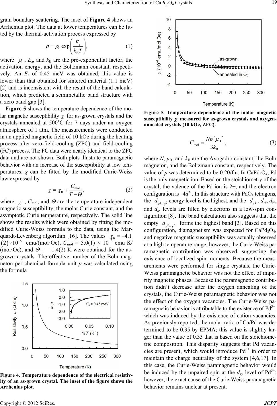 Synthesis and Characterization of CaPd3O4 Crystals 19 grain boundary scattering. The inset of Figure 4 shows an Arrhenius plot. The data at lower temperatures can be fit- ted by the thermal-activation process expressed by a 0 B exp , E kT (1) where 0 , Ea, and kB are the pre-exponential factor, the activation energy, and the Boltzmann constant, respecti- vely. An Ea of 0.45 meV was obtained; this value is lower than that obtained for sintered material (1.1 meV) [2] and is inconsistent with the result of the band calcula- tion, which predicted a semimetallic band structure with a zero band gap [3]. Figure 5 shows the temperature dependence of the mo- lar magnetic susceptibility for as-grown crystals and the crystals annealed at 500˚C for 7 days under an oxygen atmosphere of 1 atm. The measurements were conducted in an applied magnetic field of 10 kOe during the heating process after zero-field-cooling (ZFC) and field-cooling (FC) process. The FC data were nearly identical to the ZFC data and are not shown. Both plots illustrate paramagnetic behavior with an increase of the susceptibility at low tem- peratures; χ can be fitted by the modified Curie-Weiss law expressed by mol 0, C TΘ (2) where 0 , Cmol, and are the temperature-independent magnetic susceptibility, the molar Curie constant, and the asymptotic Curie temperature, respectively. The solid line shows the results which were obtained by fitting the mo- dified Curie-Weiss formula to the data, using the Mar- quardt-Levenberg algorithm [16]. The values 04.1 emu/(mol·Oe), Cmol = 5.0(1) × 03 emu K/ (mol·Oe), and = –1.4(2) K were obtained for the as- grown crystals. The effective number of the Bohr mag- neton per chemical formula unit p was calculated using the formula 215 0 Figure 4. Temperature dependence of the electrical resistiv- ity of an as-grown crystal. The inset of the figure shows the Arrhenius plot. Figure 5. Temperature dependence of the molar magnetic susceptibility measured for as-grow n crystals and oxygen- annealed crystals (10 kOe, ZFC). 22 B mol B , 3 Np Ck (3) where N, μB, and kB are the Avogadro constant, the Bohr magneton, and the Boltzmann constant, respectively. The value of p was determined to be 0.20/f.u. In CaPd3O4, Pd is the only magnetic ion. Based on the stoichiometry of the crystal, the valence of the Pd ion is 2+, and the electron configuration is . In this structure with PdO4 tetragons, the 8 4d 22 x y d-energy level is the highest, and the 2 z d, dxy, dyz, and dzx levels are filled by electrons in a low-spin con- figuration [8]. The band calculation also suggests that the empty 22 x y- forms the highest band [3]. Based on this configuration, diamagnetism was expected for CaPd3O4, and negative magnetic susceptibility was actually observed at a high temperature range; however, the Curie-Weiss pa- ramagnetic contribution was observed, suggesting the existence of localized spin moments. Because the meas- urements were performed for single crystals, the Curie- Weiss paramagnetic behavior was not the effect of impu- rity magnetic phases. Because the paramagnetic contribu- tion didn’t decrease after the oxygen annealing of the crystals, the Curie-Weiss paramagnetic behavior was not the effect of the oxygen vacancies. The Curie-Weiss pa- ramagnetic behavior is attributable to the existence of Pd3+, which was induced by the existence of cation vacancies. As previously reported, the molar ratio of Ca/Pd was de- termined to be 0.35 by EPMA; this value is slightly lar- ger than the value of 0.33 that is based on the stoichiome- tric composition. This disparity suggests that Pd vacan- cies are present, which would introduce Pd3+ in order to maintain the charge neutrality of the system [4,6,17]. In this case, the Curie-Weiss paramagnetic behavior would be induced by the unpaired spin at the dxy level of Pd3+; however, the exact cause of the Curie-Weiss paramagnetic behavior remains unclear at present. d Copyright © 2012 SciRes. JCPT  Synthesis and Characterization of CaPd3O4 Crystals Copyright © 2012 SciRes. JCPT 20 4. Conclusion [7] T. Taniguchi, Y. Nagata, T. C. Ozawa, M. Sato, Y. Noro, T. Uchida and H. Samata, “Insulator-Metal Transition In- duced in Sr1–xNaxPd3O4 for Small Na-Substitutions,” Jour- nal of Alloys and Compounds, Vol. 373, No. 1-2, 2004, pp. 67-72. doi:10.1016/j.jallcom.2003.11.004 Single crystals of alkaline-earth palladate CaPd3O4 were grown by an electrochemical technique that used voltage- applied platinum electrodes inserted into a molten chlo- ride solvent. Crystals with natural surfaces were obtained on the anode, reflecting that the growth proceeded in a liquid phase. The maximum length of the crystal was about 1.5 mm. The crystal structure parameters were refined well by assuming a cubic structure of the space group Pm 3n, and the lattice constant a was 5.7471(10) Å. The resistiv- ity showed semiconductor-like characteristics with a very small activation energy Ea of 0.45 meV at lower tempe- ratures. The temperature dependence of the molar mag- netic susceptibility showed the Curie-Weiss paramagne- tic behavior, indicating the existence of localized spin mo- ments. [8] T. C. Ozawa, A. Matsushita, Y. Hidaka, T. Taniguchi, S. Mizusaki, Y. Nagata, Y. Noro and H. Samata, “Synthesis and Characterization of Electron and Hole Doped Ternary Palladium Oxide: Sr1-xAxPd3O4 (A = Na, Bi),” Journal of Alloys and Compounds, Vol. 448, No. 1-2, 2008, pp. 77- 83. doi:10.1016/j.jallcom.2007.03.137 [9] J. Kitagawa, T. Sasakawa, T. Suemitsu, Y. Echizen and T. Takabatake, “Effects of Valence Fluctuation and Pseu- dogap Formation on Phonon Thermal Conductivity of Ce-Based Compounds with ε-TiNiSi-Type Structure,” Phy- sical Review B, Vol. 66, No. 22, 2002, p. 224304. doi:10.1103/PhysRevB.66.224304 [10] A. Saramat, G. Svensson, A. E. C. Palmqvist, C. Stiewe, E. Mueller, D. Platzek, S. G. K. Williams and D. M. Rowe, “Large Thermoelectric Figure of Merit at High Tem- perature in Czochralski-Grown Clathrate Ba8Ga16Ge30,” Journal of Applied Physics, Vol. 99, No. 2, 2006, p. 023708. doi:10.1063/1.2163979 5. Acknowledgements The work performed at Kobe University and Aoyama Ga- kuin University was supported by Grants-in-Aid for Sci- entific Research from the Ministry of Education, Culture, Sports, Science and Technology of Japan. [11] P. L. Smallwood, M. D. Smith and H.-C. zur Loye, “Flux Synthesis of Alkaline Earth Palladates,” Journal of Crystal Growth, Vol. 216, No. 1-4, 2000, pp. 299-303. doi:10.1016/S0022-0248(00)00432-2 REFERENCES [12] T. N. Nguyen and Hans-Conrad zur Loye, “Electrosyn- thesis in Hydroxide Melts,” Journal of Crystal Growth, Vo. 172, No. 1-2, 1997, pp. 183-189. doi:10.1016/S0022-0248(96)00726-9 [1] R. C. Wnuk, T. R. Touw and B. Post, “The Crystal Struc- ture of CaPd3O4,” IBM Journal of Research and Devel- opment, Vol. 8, No. 2, 1964, pp. 185-186. doi:10.1147/rd.82.0185 [13] H. Samata, Y. Saeki, S. Mizusaki, Y. Nagata, T. C. Oza- wa and A. Sato, “Electrochemical Crystal Growth of Pe- rovskite Ruthenates,” Journal of Crystal Growth, Vol. 311, No. 3, 2009, pp. 623-626. doi:10.1016/j.jcrysgro.2008.09.042 [2] K. Itoh and N. Tsuda, “Metal to Semiconductor Like Transition for Sintered Ca1–xNaxPd3O4,” Solid State Com- munications, Vol. 109, No. 11, 1999, pp. 715-719. doi:10.1016/S0038-1098(98)00549-3 [14] G. M. Sheldrick, SHELXTL, Version 6.10, Bruker AXS Inc., Madison, 1997. [3] I. Hase and Y. Nishihara, “CaPd3O4, as an Excitonic In- sulator,” Physical Review B, Vol. 62, No. 20, 2000, pp. 13426-13429. doi:10.1103/PhysRevB.62.13426 [15] F. Izumi and T. Ikeda, “A Rietveld-Analysis Programm RIETAN-98 and Its Applications to Zeolites,” Materials Science Forum, Vol. 321-324, 2000, pp. 198-205. doi:10.4028/www.scientific.net/MSF.321-324.198 [4] S. Ichikawa and I. Terasaki, “Metal-Insulator Transition in Ca1–xLixPd3O4,” Physical Review B, Vol. 68, 2003, p. 233101. doi:10.1103/PhysRevB.68.233101 [16] W. H. Press, B. P. Flannery, S. A. Teukolsky and W. T. Vet- terling, “Numerical Recipes,” Cambridge University Press, Cambridge, 1986. [5] K. Itoh, Y. Yano and N. Tsuda, “Metal to Insulator Tran- sition for Ca1–xNaxPd3O4,” Journal of the Physical Soci- ety of Japan, Vol. 68, No. 9, 1999, pp. 3022-3026. doi:10.1143/JPSJ.68.3022 [17] S. J. Kim, S. Lemaux, G. Demazeau, J. Y. Kim and J. H. Choy, “LaPdO3: The First PdIII Oxide with the Perovskite Structure,” Journal of the American Chemical Society, Vol. 123, No. 42, 2001, pp. 10413-10414. doi:10.1021/ja016522b [6] Y. Yano, M. Kanazawa, T. Fujii and N. Tsuda, “Magnetic Susceptibility of Ca1-xNaxPd3O4,” Journal of the Physi- cal Society of Japan, Vol. 70, No. 6, 2001, pp. 1772-1176. doi:10.1143/JPSJ.70.1772 |