

Paper Menu >>
Journal Menu >>
 American Journal of Anal yt ical Chemistry, 2011, 2, 32-36 doi:10.4236/ajac.2011.228121 Published Online December 2011 (http://www.SciRP.org/journal/ajac) Copyright © 2011 SciRes. AJAC Analysis of Pesticide Raid® in Feed of Wistar Rat by High-Pressure Liquid Chromatography (HPLC) Albert C. Achudume Institute of Ecology and Environmental Studies, Obafemi Awolowo University, Ile-Ife, Nigeria E-mail: [email protected] Received November 21, 2011; revised December 24, 2011; accepted December 31, 2011 Abstract The distribution of pesticide by-product in tissues of wistar rats were analyzed using high pressure liquid chromatography. The limit of detection of the HPLC was 0.1 µg. Results show bioaccumulation factor of pesticide “Raid®” in lipid, up to three times that of the feed at the first concentration and gradually de- creased as the concentration increased in the muscle > (0.7), brain > (0.5) and liver > (0.3) as indicated in the text. At higher concentration of 961 µg/g, bioaccumulation factor decreased in the lipid to 1.2 and 0.6 in the muscle, 0.03 in the brain and 0.08 in the liver respectively. High Pressure Liquid Chromatography (HPLC) analysis of raid extract suggests the presence of micprothrin and palethrin. The implications are numerous, but simply put that accidental ingestion of chlorinated hydrocarbon as in “Raid®” may involve convulsions, collapse and coma after only brief excitation and ataxia at the onset. Keywords: High Pressure Liquid Chromatography, Pesticide, Raid®, Chlorinated Hydrocarbon, Bioaccumulation 1. Introduction Pesticides have been used to boost food production to a considerable extent and to control vectors of disease [1]. However, these advantages of great economic benefits sometimes come with disadvantages when subjected to critical environmental and human health considerations. The chronic effects of pesticides from food intake on human health are not well defined, but there is increasing evidence of carcinogenetic [2], teratogenic [3] and ge- netotoxic [4], as well as disruption of hormonal functions [5]. Analysis of these pesticides and their residues h ad in the past aided objective re-evaluation and reassessment of these substances on a benefit-risk analysis basis and their subsequent withdrawal from use when found to be hazardous to human health and the environment. Residues of pesticides and heavy metals enter the food system. This occurs more often than one might think. Additionally, residues of pesticides found in the air in- side the home and on floors and other interior surfaces contribute to non-occupational pesticide exposure [6]. For example, any potentially harmful substances that have no set tolerance thresh may not necessarily be harmful. This means that if there is any toxin in food supply or water, such food cannot be ban unless it exists in quantities over th e tolerance threshold no matter what. There are few studies of compliance to pesticide use [7,8]. The quality and sophiscation of analysis have grown and very minute quantities of these pesticides and their residues can be analyzed with high degree of specify, precision and accuracy. Therefore, it is important and utmost necessary to develop methods for analysis of pes- ticides residues in environmental samples. In the light of the above, and in view of pesticide related adverse reac- tions, the study, presents chromatographic and absorp- tion spectra from a common pesticide Raid® in tissu e of rat as it affects basal metabolism using high pressure liquid chromatograph. 2. Materials and Methodology 2.1. Test Facility Tests with Wistar rats were conducted in a room tem- perature exposure facility (23˚C). Thirty-five animals weighing between 180 - 210 were caged in perforated aluminum chambers (38 × 55 × 35 cm). Five animals were placed in each of the chambers containing sawdust shaves, and a light: dark cycle of approximately 12 h.  A. C. ACHUDUME33 Water was given ad libitum, temperature was maintained at 24˚C ± 1˚C. Air temperature was ±2˚C of the water temperature. Protocols describing the use of animals in accordance with National Research Council (NRC) [8] and World Medical Association (WMA) [9] guide on the care and use of laboratory animals. 2.2. Test procedures Wistar rats were obtained from the animal breeding fa- cility of College of Health Sciences, Obafemi Awolowo University, Ile-Ife, Nigeria. Technical grade “RAID” (Johnson Wax, Nigeria), was dissolved in corn oil and with (in a rotary mixer) 2:98 (w/w) the co mmercial feed, Purina 5000, for 10 min: the control diet contained corn oil in the same proportion. Animals were fed raid-spiked commercial feed at measured concentrations of 25.0 ± 2.4, 54.0 ± 9.2, 108.0 ± 12.5, 216.0 ± 14.6, and 430.0 ± 20.2 and 961.0 ± 80.6 µg/g “Raid” for 10 days, increas- ing 5 - 10 g/day and ending with 96 g/rat. Two rats from different chambers receiving the same concentration of raid in the food were selected and tissue samples were dissected out (lipid, muscle, brain and liver) at test ter- mination. The symptom-li mited sensation tests were per- formed after an overnight fast [10]. Mean values were calculated from the two rats sampled from each concen- tration of insecticide-raid. 2.3. Test Extraction In the toxicity persistence test, “Raid” was extracted with 1:1 diethyl ether-petroleum ether [11]. This compound was identified and quantified by high-pressure liquid chromatography (HPLC), using UV1-202nm@01, Base (Amersham Pharmacia Biotech), serial number 90042, mobile phase was Methanol: H2O (1:1). The limit of de- tection of the HPLC was 0.1 µg .The dimension of the column C18, 4.6 × 250 mm and particle size 5 micron. Flow rate was 1 ml/min, the injection volume 100 µl and a wavelength of 202 nm was used for detection. The values were quantified by standard calibration curve. 2.4. Analytical Procedures The brain, muscle and liver homogenate fractions were prepared by the method described by [12] and resus- pended in 0.15 M KCl to an appropriate final concentra- tion of 20 mg of protein per millimeter (approximately) 0.5 g of tissue. Liver tissue lipid s were extracted by ch lo- roform/methanol ratio (2:1) following the method of Folch [13]. Fractions of each tissue homogenate were used for the determination of Glucose-6-phosphatase (G-6-Pase) and Lactate dehydrogenase respectively, to test for inhibition of metabolic enzymes required to aid digestion. Glucose-6-phospatase was determined using aliquot of each homogenate (20%), incubated with 1 ml of P-nitrophenyl phosphate (NPP) for 15 min and the reaction was stopped by addition of 2 ml of 0.2 N NaOH; absorbance was measured at 450 nm as described by [14]. Lactate dehydrogenase was determined by the method of [15]. Protein was determined by [16] method. Experi- ments were conducted in a completely randomized man- ner with five replicates. These were repeated and mean data of two experiments is presented. Data were sub- jected to one-way analysis of variance and significance of treatments from control was tested at 5% level of sig- nificance followed by Dunnet’s test. Further, data were also subjected to determination of correlation coefficient between raid concentration and the observed response. The statistical analysis was performed using SPSS soft- ware version 10.0. 3. Results and Discussion Bioaccumulation factor of pesticide “Raid®” was ob- served in lipid, up to three times that of the feed at the first concentration and gradually decreased as the con- centration increased (Table 1) while accumulation factor in the muscle (0.7), brain (0.5) and liver (0.3) was about the indicated number times that of the feed. At higher concentration of 961 µg/g, bioaccumulation factor de- creased in the lipid to 1.2 and 0.6 in the muscle, 0.03 in the brain and 0.08 in the liver. Using the mean of insecti- cide in feed, the tissues accumulate the insecticide in ascending order: brain < liver < muscle < lipid. The ef- fects increased with increasing insecticide concentration in the feed an d showed a significant deference compared to control (P < 0.05) at a concentration of 430 µg/g. Table 2 indicates the estimated detectable levels of toxicity in rat tissues exposed to the insecticide “Raid®”. Tissue insecticide concentration increased in lipid, mus- cle, brain and liver, respectively. The insecticide accu- mulation in these organs may therefore indicate that the active ingredients are also absorbed resulting in the de- creased enzymes formation, which should accelerate immediate metabolic processes, but instead, remained complexed in the tissue organs. Additionally, the reten- tion time (within 10 min.) may be increased because of it easy solubility and lower excretion rate leading to bio- accumulation. The brain shows insignificant decreases in the enzymes Glucose-6-phosphatase and Lactic acid de- hydrogenase, while significant decreases were noticeable in the muscle and liver. The tissues concentrations were the sums of the two components. High Pressure Liquid Chromatography (HPLC) analy- sis of raid extract suggests the presence of micprothrin Copyright © 2011 SciRes. AJAC  A. C. ACHUDUME Copyright © 2011 SciRes. AJAC 34 Table 1. Tissue total raid concentrations and bioaccumulation factors (BAF) in Wister Rats exposed to insecticide “Raid”. Mean ± SD insecticide “Raid” in feed (µg/g) Raid concentration in Wister rats (µg/g) a and bioaccumulation factor (BAF)b Lipid Muscle Brain Liver 0.00 - - - - 25.0 ± 2.4 72.5 17.5 12.5 7.5 (2.9) (0.7) (0.5) (0.3) 54.0 ± 9.2 86.4 21.7 16.4 9.4 (1.6) (0.4) (0.3) (0.2) 108.2 ± 12.5 172.8 30.4 19.5 10.8 (1.6) (0.3) (0.2) (0.10) 216.2 ± 14.6 280.8 45.8 22.9 19.8 (1.3) (0.2) (0.1) (0.09) 430.0 ± 20.6 324.0 86.4 25.8 37.3 (0.8) (0.2) (0.06) (0.09) 961.2 ± 70.5 1153.2 576.6 28.8 76.9 (1.2) (0.6) (0.03) (0.08) aMean raid concentration at test termination (~steady state value); bBAF = bioaccumulation factor ‘Raid’ in tissue (µg/g)/feed raid concentration (µg/g). Table 2. Index of toxicity in rat tissues exposed to insecticide “Raid”. Indicators Brain IU/L Muscle IU/L Liver IU/L Control 0.28 ± 0.02 1.15 ± 0.20 2.54 ± 0.1 Glucose-6-phospatase 0.25 ± 0.05 0.35 ± 0.09* 0.15 ± 0.03* Control 0.22 ± 0.04 0.10 ± 0.01 1.94 ± 0.02 Lactate dehydrogenase 0.19 ± 0.08 0.15 ± 0.11* 0.07 ± 0.08* *Significantly different P < 0.05 from control. and palethrin. Figure 1(a) shows the chromatographic profile of the raid insecticides. Figure 1(b) shows the chromatographic profile of standards (0.025%). The high- est peak obtained from “Raid®” extract is most likely micprothrin. This peak possessed a retention time (9.25) min. For the identification of raid, co-chromatography with authentic standards in different chromatographic conditions was used. Two different peaks were eluted within 10 min and were well resolved (Figure 1(a)), but unidentified peak was eluted within a retention time of early eluting of 2 min. The HPLC analysis failed to de- tect the unidentified component in “Raid®”. The reason is not clear, but raid may have undergone a structural change because of combination, hence different absorb- ance property [17]. The fine tuning of the mobile phase composition depended on the choice of the internal stan- dard. Most internal standard compounds were rejected because of interfering components. Only micprothrin and palethrin had acceptable chromatographic characteristics. The implications are numerous, but simply put that ac- cidental ingestion of chlorinated hydrocarbon as in “Raid®” may involve convulsions, collapse and coma after only brief excitation and ataxia at the onset. Other workers have contrasted neuritic outgrowth of chlorpyrifos, diazinon and organophosphate pesticides including parathion and concluded that all have adverse effects on brain development and systemic toxicity [18]. In recent study, Walz, [19], shows that glyphosate, the active ingredient in Roundup, causes birth defects in frogs and chicken embryos at far lower levels than used in agricultural and garden applications. The malforma- tions primarily affected the skull, face, midline and spi- nal cord. Other independent scientific research has also found that glyphosate causes endocrine disruption, de- velopmental and reproductive toxicity, DNA damage, neurotoxicity and cancer [3,18]. Many of these effects 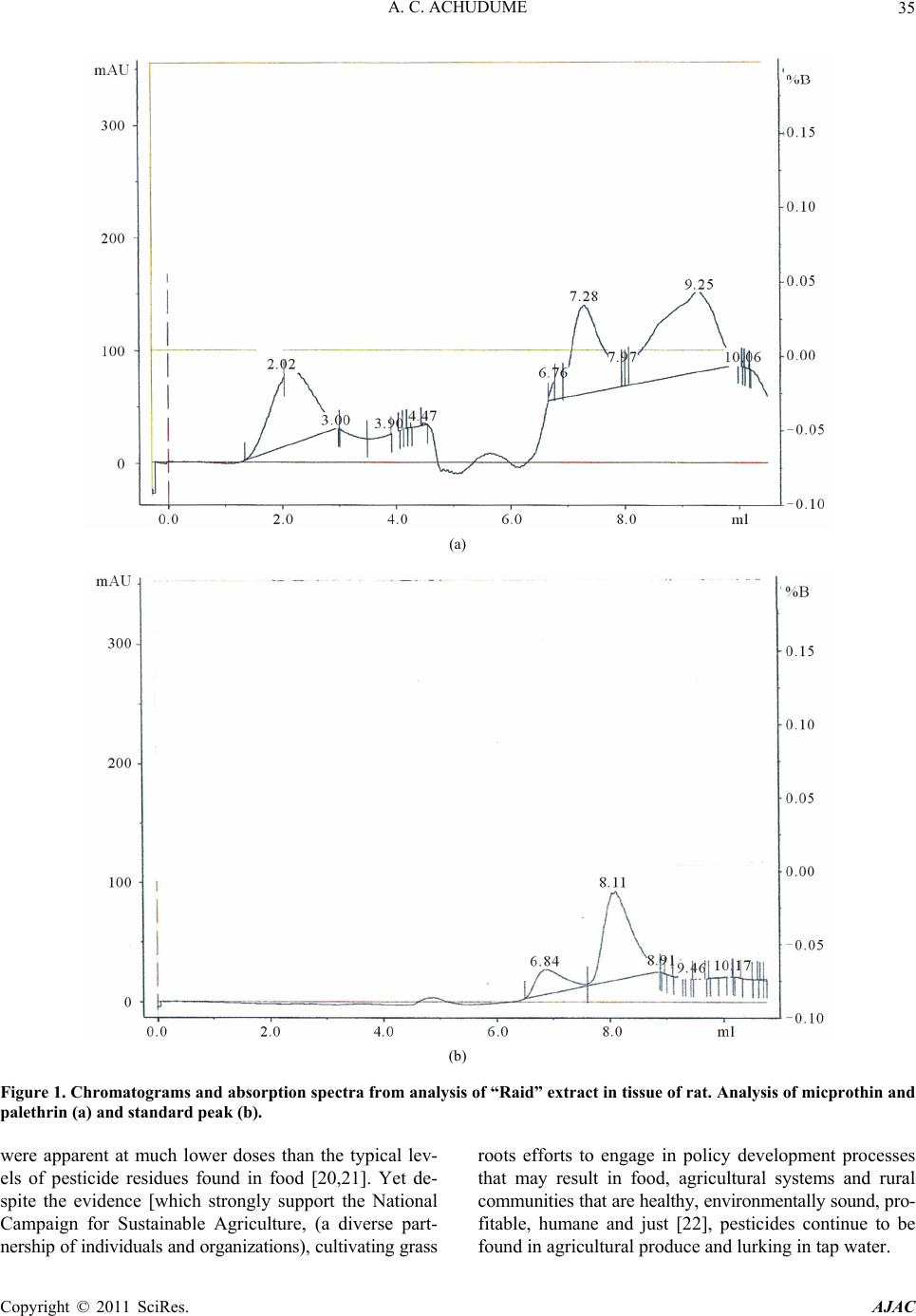 A. C. ACHUDUME35 (a) (b) Figure 1. Chromatograms and absorption spectra from analysis of “Raid” extract in tissue of rat. Analysis of micprothin and palethrin (a) and standard peak (b). were apparent at much lower doses than the typical lev- els of pesticide residues found in food [20,21]. Yet de- spite the evidence [which strongly support the National Campaign for Sustainable Agriculture, (a diverse part- nership of individuals and organizations), cultivating grass roots efforts to engage in policy development processes that may result in food, agricultural systems and rural communities that are healthy, environmentally sound, pro- fitable, humane and just [22], pesticides continue to be found in agr icultural produce and lurking in tap water. Copyright © 2011 SciRes. AJAC  A. C. ACHUDUME 36 4. References [1] D. Mondal, S. Barat and M. K. Mukhopadhyay, “Toxicity of Neem Pesticides on a Fresh Water Loach, Lepido- cephalichthys Guntea (Hamilton Buchanan) of Darjeeling in West Bengal,” Journal of Environmental Biology, Vol. 28, No. 1, 2007, pp. 119-122. [2] D. Hoffmann, E. J. Laboie and S. Hecht, “Nicotine: A Precursor for Carcinogens,” Cancer Letters, Vol. 26, No. 1, 1985, pp. 67-75. doi:10.1016/0304-3835(85)90174-0 [3] A. Paganelli, V. Gnazzo, H. Acosta, S. L. L. Pez and A. E. Carrasco, “Glyphsate-Based Herbicides Produce Terato- genic Effects on Vertebrates by Impairing Retinoic Acid Signaling,” Chemical Research in Toxicology, Vol. 23, No. 10, 2010, pp. 1586-1595. doi:10.1021/tx1001749 [4] M. Antoniou, M. E. Habid, C. V. Howard, R. Jennings, C. Leifert, R. O. Nodar, C. Robinson and J. Fagan, “Round- up and Birthdefects: Is the Public Being Kept in the Dark?” 2011. http://www.earthopensource.org/index.php/reports/17-rou ndup-and-birth-defects-is-the-public-being-kept-in-the-da rk [5] R. W. Bretveld, G. M. G. Thomas, P. T. J. Scheepers, G. Zielhuis and N. Roeleveld, “Pesticide Exposure: The Hormonal Function of the Female Reproductive System Disrupted,” Reproductive Biology and Endocrinology, Vol. 4, No. 30, 2006, pp. 1-4. [6] L. Menegaux, A. Baruche, Y. Bertrand, B. Lescoex, G. Leverger, B, Nelken, D. Sommelet, D. Hermon and J. Clavel, “Household Exposure to Pesticide and Risk of Childhood Acute Leukemia,” Occupational and Envi- ronmental Medicine, Vol. 63, No. 2, 2006, pp. 131-134. doi:10.1136/oem.2005.023036 [7] P. O. Okonkwo, C. O. Apala, H. U. Okafor, A. U. Mbah and O. Nwaiwu, “Compliance to Correct Dose of Chloro- quine in Uncomplicated Malaria Correlates with Im- provement in the Condition of Nigerian Children,” Trans- actions of the Royal Society of Tropical Medicine and Hygiene, Vol. 95, No. 3, 2001, pp. 320-324. doi:10.1016/S0035-9203(01)90252-4 [8] National Research Council, “Guide for the Care and Use of Laboratory Animals,” National Academy Press, Wash- ington DC, 1996. [9] World Medical Association (WMA), “Statement on Ani- mal Use in Biochemical Research,” Adopted by the 41st World Medical Assembly, Hong Kong, 1999 and Revised by WMA General Assembly, Pilanesberg, South Africa, 2006. [10] Anonymous, “Guidelines for the Treatment of Animals in Behavioral Research and Teaching,” Animal Behaviour, Vol. 55, No. 1, 1998, pp. 251-257. doi:10.1006/anbe.1997.0703 [11] US Food and Drug Administration, “Pesticide Analytical Manual Volume I. Method for Chlorophenoxy Acids and Pentachlorophenol,” US Department of Health and Hu- man Services, USFDA, Washington DC, 1989. [12] J. M. Siegler and M. N. Kazarinoff, “Effects of Acute and Chronic Protein Deprivation on Ornithine Decarboxylase Levels in Rat Liver and Colon,” Nutrition and Cancer, Vol. 4, No. 3, 1983, pp. 176-185. doi:10.1080/01635588209513755 [13] J. Folch, M. Lees and G. H. Sloane-Stanley, “A Simple Method for the Isolation and Purification of Total Lipides from Animal Tissues,” The Journal of Biological Chem- istry, Vol. 226, No. 1, 1957, pp. 497-509. [14] M. Alegre, C. J. Ciudad, C. Fillat and J. J. Guinovart, “Determination of Glucose-6-Phosphatase Activity Using the Glucose Dehydrogenase-Coupled Reaction,” Ana- lytical Biochemistry, Vol. 173, No. 1, 1988, pp. 185-189. doi:10.1016/0003-2697(88)90176-5 [15] M. D. Abeloff, J. O. Armitage, J. E. Niederhuber, M. B. Kastan and W. G. Mckenna, “Clinical Oncology,” 3rd Edition, Churchill Livingstone, Philadelphia, 2004. [16] O. H. Lowry, N. J. Rosenbrough, A. L. Farr and R. J Randall, “Protein Measurement with the Folin Phenol Reagent,” The Journal of Biological Chemistry, Vol. 193, No. 1, 1951, pp. 265-275. [17] A. C. Achudume, P. C. Nwoha and J. N. Ibe, “Toxicity and Bioaccumulation of Insecticide ‘Raid’ in Wistar Rats,” Environmental Toxicology, Vol. 24, No. 4, 2008, pp. 357- 361. [18] T. A. Slotkin, E. D. Levin and F. J. Seidler, “Comparative Developmental Neurotoxicity of Organophosphate Insec- ticides: Effect on Brain Development Are Separable from Systemic Toxicity,” Environ Health Perspect, Vol. 114, No. 5, 2006, pp. 41-50. [19] E. Walz, “Glyphosate Resistance Threaten Roundup He- gemony,” Nature Biotechnology, Vol. 28, No. 6, 2010, pp. 537-538. doi:10.1038/nbt0610-537 [20] J. J. Herve, “Agricultural, Public Health and Animal Health Usage,” In: J. P. Leahey, Ed., The Pyrethroid Insecticides, Taylor & Francis, London, 1985. [21] A. V. Nebeker, K. D. Dunn, W. L. Griffis and G. S. Schu- ytema, “Effects of Dieldrin in Food on Growth and Bio- accumulation in Mallard Ducklings,” Archives of Envi- ronmental Contamination and Toxicology, Vol. 26, 1994, pp. 29-32. doi:10.1007/BF00212790 [22] European Commission, “Directive 2009/128/EC of the European Parliament and of the Council of 21 October 2009 Establishing a Framework for Community Action to Achieve the Sustainable Use of Pesticides,” http://www.pesticide.gov.uk/uploadedfile/Web_Assets/P SD/Directive_2009_128_ECframework_sustanable_use_ of_pesticides.pdf 2009 Copyright © 2011 SciRes. AJAC |