

Paper Menu >>
Journal Menu >>
 Open Journal of Pediatrics, 2011, 1, 64-66 OJPed doi:10.4236/ojped.2011.14015 Published Online December 2011 (http://www.SciRP.org/journal/ojped/) Published Online December 2011 in SciRes. http://www.scirp.org/journal/OJPed Osteogenesis imperfecta and complications due to pelvic narrowing: case and review Jakob Bie Granild-Jensen1, Line Granild Bie Mertz2, Soren Rittig3, Gratien Andersen4, John Rosendahl Oestergaard5* 1Department of Paediatrics, Aarhus University Hospital, Skejby, Denmark; 2Paediatric Research Laboratory, Aarhus University Hospital, Skejby, Denmark; 3Department of Paediatric Nephrology, Aarhus University Hospital , Sk ejby, Denmark; 4Department of Radiology, Aarhus University Hospital, Skejby, Denmark; 5Center of Rare Diseases, Aarhus University Hospital, Skejby, Denmark. Email: *[email protected] Received 2 October 2011; revised 14 November 2011; accepted 26 November 2011. ABSTRACT We describe a case of pelvic narrowing in a patient with osteogenesis imperfecta (OI) type III and com- plications consisting of deep venous thrombosis, hy- dronephrosis and faeculomas. The literature is re- viewed and the management is discussed. Conclusion: Monitoring of OI type III patients for pelvic narrow- ing and complications is recommended. Keywords: Acetabular Pro trusion; Bisphosphonate; Cons- tipation; Deep Venous Thrombosis; Hydronephrosis; Pel- vis 1. INTRODUCTION Osteogenesis imperfecta (OI) is a rare genetic disorder of type 1 collagen resulting in connective tissue abnor- malities. Varying degrees of bone fragility and skeletal deformity are the main characteristics, other important features being dentinogenesis imperfecta and blue sclerae. Based on phenotype and genotype OI is divided into sub- types [1-3]. Pelvic narrowing has been described in a number of disorders of bone metabolism [4]. In OI, pelvic narrow- ing is probably a consequence of both fractures and bone remodeling. Complications due to pelvic narrowing have been described. Most frequently it is associated with constipation [5]. We report on a girl with OI type III and a triad of gas- trointestinal, vascular and urologic complications due to narrowing of the pelvis. The management and outcome is described and the literatu re is reviewed. 2. CASE REPORT The report concerns a girl born in 1997 in a family with no history of OI. OI was suspected shortly after her birth in 1997 due to congenital femur and rib fractures and blue sclerae. The diagnosis was confirmed by skin bi- opsy revealing a structural abnormality without quantita- tive deficiency of type I collagen consisten t with OI type III or IV. Exons of all abnormal peptides were sequenced, but no pathogenic mutation was identified. During the first year of life, she had more than 25 fractures. The rate of fractures was reduced by approximately 50% after repeated bisphosphonate infusions were started (pami- dronate 9 - 12 mg/kg/year from 1998 to 2007, thereafter 4.5 - 6 mg/kg/year). Furthermore, during the initial 4 years she had telescopic rods placed in the humerus, radius, femur and tibia bilaterally. During 2004-2006 sh e had few, but recurrent episodes of abdominal pain and constipation despite the use of laxatives. She had no urological complaints or urinary tract infections during her first 10 years of life. In March 2007, she was admit- ted with left-sided abdominal pain. An ultrasonography disclosed a left hydronephrosis. The pain faded after placement of a nephrostomy tube. Two months later, she was re-admitted, now with severe abdominal pain and constipation. A few weeks earlier, morphine was admin- istered due to fractures and pain. Large faecal masses were palpated in the abdomen. At this time, her left leg became red and swollen and as a thrombosis of the left iliac vein was found by ultrasonography, low molecular weight heparin administration was initiated. CT scan of the pelvic region revealed wedging of the sigmoid colon between the left superior ramus of the pubic bone and the vertebral column. She had bilateral hydronephrosis and hydro-ureter. No calculi were visible. In the sigmoid and transverse colon she had several faeculomas. During operation, it was necessary to do a resection of the sig- moid colon and perform a colostomy. After the operation 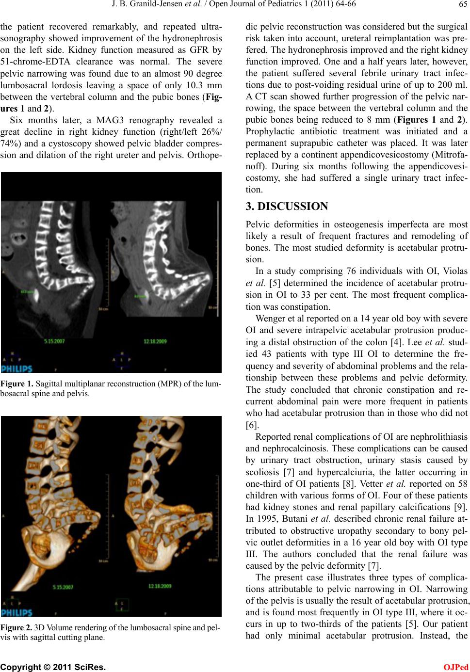 J. B. Granild-Jensen et al. / Open Journal of Pediatrics 1 (2011) 64-66 65 the patient recovered remarkably, and repeated ultra- sonography showed improvement of the hydronephrosis on the left side. Kidney function measured as GFR by 51-chrome-EDTA clearance was normal. The severe pelvic narrowing was found due to an almost 90 degree lumbosacral lordosis leaving a space of only 10.3 mm between the vertebral column and the pubic bones (Fig- ures 1 and 2). Six months later, a MAG3 renography revealed a great decline in right kidney function (right/left 26%/ 74%) and a cystoscopy showed pelvic bladder compres- sion and dilation of the right ureter and pelvis. Orthope- Figure 1. Sagittal mu lti planar reconstruction (MPR) of the lum- bosacral spine and pelvis. Figure 2. 3D Volume rendering of the lumbosacral spine and pel- vis with sagittal cutting plane. dic pelvic reconstructio n was considered but the surgical risk taken into account, ureteral reimplantation was pre- fered. The hydronephrosis improved and the right kidney function improved. One and a half years later, however, the patient suffered several febrile urinary tract infec- tions due to post-voiding residual urine of up to 200 ml. A CT scan showed further progression of the pelvic nar- rowing, the space between the vertebral column and the pubic bones being reduced to 8 mm (Figures 1 and 2). Prophylactic antibiotic treatment was initiated and a permanent suprapubic catheter was placed. It was later replaced by a continent appendicovesicostomy (Mitrofa- noff). During six months following the appendicovesi- costomy, she had suffered a single urinary tract infec- tion. 3. DISCUSSION Pelvic deformities in osteogenesis imperfecta are most likely a result of frequent fractures and remodeling of bones. The most studied deformity is acetabular protru- sion. In a study comprising 76 individuals with OI, Violas et al. [5] determined the incidence of acetabular protru- sion in OI to 33 per cent. The most frequent complica- tion was constipation. Wenger et al reported on a 14 year old boy with severe OI and severe intrapelvic acetabular protrusion produc- ing a distal obstruction of the colon [4]. Lee et al. stud- ied 43 patients with type III OI to determine the fre- quency and severity of abdominal problems and the rela- tionship between these problems and pelvic deformity. The study concluded that chronic constipation and re- current abdominal pain were more frequent in patients who had acetabular protrusion than in those who did not [6]. Reported renal complications of OI are nephrolithiasis and nephrocalcinosis. These complications can be caused by urinary tract obstruction, urinary stasis caused by scoliosis [7] and hypercalciuria, the latter occurring in one-third of OI patients [8]. Vetter et al. reported on 58 children with various forms of OI. Four of these patients had kidney stones and renal papillary calcifications [9]. In 1995, Butani et al. described chronic renal failure at- tributed to obstructive uropathy secondary to bony pel- vic outlet deformities in a 16 year old boy with OI type III. The authors concluded that the renal failure was caused by the pelvic de f ormity [7]. The present case illustrates three types of complica- tions attributable to pelvic narrowing in OI. Narrowing of the pelvis is usually the result of acetabular protrusion, and is found most frequently in OI type III, where it oc- curs in up to two-thirds of the patients [5]. Our patient had only minimal acetabular protrusion. Instead, the C opyright © 2011 SciRes. OJPed 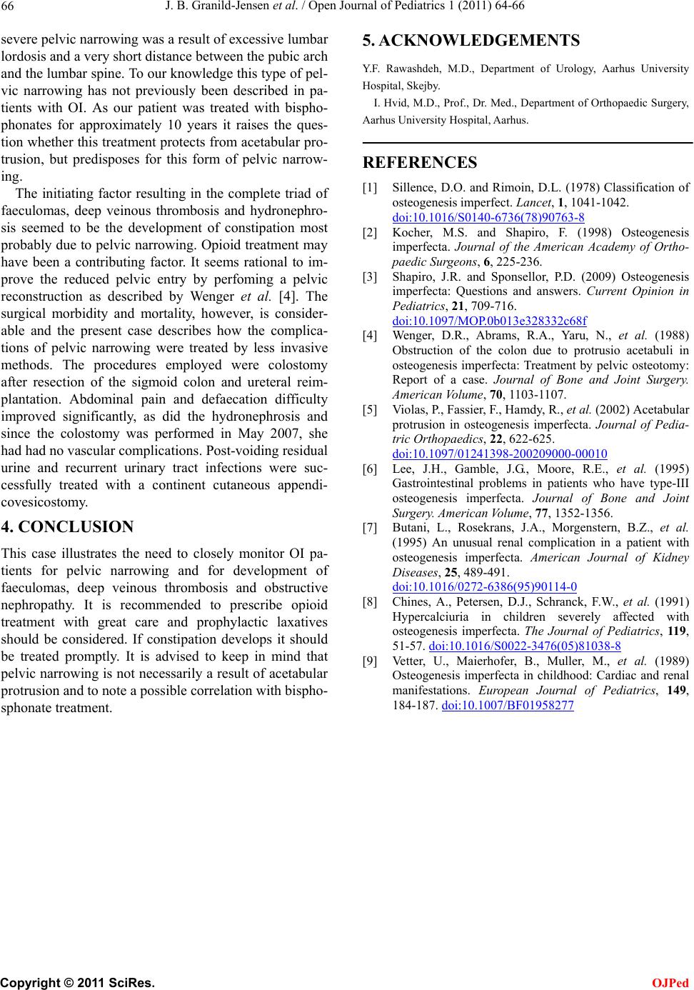 J. B. Granild-Jensen et al. / Open Journal of Pediatrics 1 (2011) 64-66 Copyright © 2011 SciRes. OJPed 66 5. ACKNOWLEDGEMENTS severe pelvic narrowing was a result of excessive lumbar lordosis and a very short distance between the pubic arch and the lumbar spine. To our knowledge this typ e of pel- vic narrowing has not previously been described in pa- tients with OI. As our patient was treated with bispho- phonates for approximately 10 years it raises the ques- tion whether this treatment protects from acetabular pro- trusion, but predisposes for this form of pelvic narrow- ing. Y.F. Rawashdeh, M.D., Department of Urology, Aarhus University Hospital, Skejby. I. Hvid, M.D., P rof., Dr. Med., Department of Orthop aedic Surgery, Aarhus University Hospital, Aarhus. REFERENCES [1] Sillence, D.O. and Rimoin, D.L. (1978) Classification of osteogenesis imperfect. Lancet, 1, 1041-1042. doi:10.1016/S0140-6736(78)90763-8 The initiating factor resulting in the complete triad of faeculomas, deep veinous thrombosis and hydronephro- sis seemed to be the development of constipation most probably due to pelvic narrowing. Opioid treatment may have been a contributing factor. It seems rational to im- prove the reduced pelvic entry by perfoming a pelvic reconstruction as described by Wenger et al. [4]. The surgical morbidity and mortality, however, is consider- able and the present case describes how the complica- tions of pelvic narrowing were treated by less invasive methods. The procedures employed were colostomy after resection of the sigmoid colon and ureteral reim- plantation. Abdominal pain and defaecation difficulty improved significantly, as did the hydronephrosis and since the colostomy was performed in May 2007, she had had no vascular complications. Post-voiding residual urine and recurrent urinary tract infections were suc- cessfully treated with a continent cutaneous appendi- covesicostomy. [2] Kocher, M.S. and Shapiro, F. (1998) Osteogenesis imperfecta. Journal of the American Academy of Ortho- paedic Surgeons, 6, 225-236. [3] Shapiro, J.R. and Sponsellor, P.D. (2009) Osteogenesis imperfecta: Questions and answers. Current Opinion in Pediatrics, 21, 709-716. doi:10.1097/MOP.0b013e328332c68f [4] Wenger, D.R., Abrams, R.A., Yaru, N., et al. (1988) Obstruction of the colon due to protrusio acetabuli in osteogenesis imperfecta: Treatment by pelvic osteotomy: Report of a case. Journal of Bone and Joint Surgery. American Volume, 70, 1103-1107. [5] Violas, P., Fassier, F., Hamdy, R., et al. (2002) Acetabular protrusion in osteogenesis imperfecta. Journal of Pedia- tric Orthopaedics, 22, 622-625. doi:10.1097/01241398-200209000-00010 [6] Lee, J.H., Gamble, J.G., Moore, R.E., et al. (1995) Gastrointestinal problems in patients who have type-III osteogenesis imperfecta. Journal of Bone and Joint Sur gery. American Volume, 77, 1352-1356. 4. CONCLUSION [7] Butani, L., Rosekrans, J.A., Morgenstern, B.Z., et al. (1995) An unusual renal complication in a patient with osteogenesis imperfecta. American Journal of Kidney Diseases, 25, 489-491. doi:10.1016/0272-6386(95)90114-0 This case illustrates the need to closely monitor OI pa- tients for pelvic narrowing and for development of faeculomas, deep veinous thrombosis and obstructive nephropathy. It is recommended to prescribe opioid treatment with great care and prophylactic laxatives should be considered. If constipation develops it should be treated promptly. It is advised to keep in mind that pelvic narrowing is not necessarily a result of acetabular protrusion and to note a possible correlation with bispho- sphonate treatment. [8] Chines, A., Petersen, D.J., Schranck, F.W., et al. (1991) Hypercalciuria in children severely affected with osteogenesis imperfecta. The Journal of Pediatrics, 119, 51-57. doi:10.1016/S0022-3476(05)81038-8 [9] Vetter, U., Maierhofer, B., Muller, M., et al. (1989) Osteogenesis imperfecta in childhood: Cardiac and renal manifestations. European Journal of Pediatrics, 149, 184-187. doi:10.1007/BF01958277 |