

Paper Menu >>
Journal Menu >>
 Open Journal of Obstetrics and Gynecology, 2011, 1, 213-216 OJOG doi:10.4236/ojog.2011.14041 Published Online December 2011 (http://www.SciRP.org/journal/ojog/). Published Online December 2011 in SciRes. http://www.scirp.org/journal/OJOG Non-Communicating inactive rudimentary horn of the uterus presenting with dysmenorrhoea—a case report of successful laparoscopic excision Rachana Dwivedi1, Keerthi Perera2, Padma Eedarapalli1 1The Royal Bournemouth and Christchurch Hospitals NHS Foundation Trust, Bournemouth, UK; 2Poole Hospital NHS Foundation Trust, Poole, UK. Email: [email protected] Received 7 September 2011; revised 17 October 2011; accepted 27 October 2011. ABSTRACT Uterine developmental abnormalities can cause both obstetric and gynaecological complications. Non-func- tioning rudimentary uterine horn is a rare cause of dysmenorrhoea which usually starts after menarche. Cases with lateral/inferior arrangement of blood su- pply to the functioning rudimentary horn and lapa- scopic removal have been described in literature. Ours is a unique case of non-functioning rudimentary horn with an unusual medial arrangement of vasculature successfully treated by laparoscopic excision. We also present a brief review of the literature. The patient presented with irretractable dysmenorrhoea despite hormonal manipulation and analgesics. Following the diagnosis using laparoscopy and hysteroscopy and MRI, the left sided non-communicating rudimentary horn with inactive endometrium was removed lapa- roscopically. Her symptoms were r esolved and this was followed by successful pregnancies. Diagnosis of ru- dimentary horn with inactive endometrium is diffi- cult. Ultrasound is unreliable. MRI, 3D CT scan and 3D ultrasound are gaining popularity. Accurate, prior identification of the type using laparoscopy and hys- teroscopy if necessary is essential. This helps in sur- gical planning. The laparoscopic approach is increa- singly being used to resect these horns due to its sa- fety and merits. Keywords: Dysmenorrhoea; Rudimentary Horn; Lapa- roscopic Excision 1. INTRODUCTION Congenital uterine anomalies result from arrested de- velopment of one or both of the Mullerian ducts and/or their defective fusion. In 80% - 90% of cases, there is no communicating channel between the two uterine cavi- ties [1]. There are many cases reported with non-commu- nicating rudimentary horn and the functioning endome- trium associated with various complications such as dys- menorrhoea, adenomyosis, ectopic pregnancy and haema- tometra [2,3]. There are also several in literature, treated with laparoscopic excision where the blood supp ly of the rudimentary horn was either lateral or inferior [4]. How- ever, to our knowledge, there are no case reports of uni- cornuate uterus and a non-communicating rudimentary horn with an inactive endometrium presenting with uni- lateral dysmenorrhoea. Moreover, none so far have de- scribed a medial arrangement of vasculature. We report a case of an 18 year old girl who presented with primary cyclic spasmodic dysmenorrhoea which was relieved after laparoscopic excision of type 3 rudimen- tary horn. The horn was non-communicating with a well developed horn on the other side. The vessels were ar- ranged medially and the endometrium was non-func- tioning. 2. CASE REPORT The nulliparous g irl was first referred to paediatrician s at the age of 14 for left loin pain which settled in 2 days. She presented a year later with similar pain which was thought to be due to renal co lic. An Ultrasound scan and an X-ray were unremarkable. Two years later she pre- sented to gynaecologists for pelvic pain and cyclical left sided dysmenorrhoea which did not respond to analge- sics. Clinical examination elicited tenderness in the left iliac fossa. Repeat ultrasounds and X-Rays were normal and the pain settled on minipill. She presented again to gynaecologists with cyclical left sided dysmenorrhoea despite minipill, tricycling com- bined pill and d epot provera. On laparoscopy, the uterine fundus appeared broad. The right tube and ovary were normal. The left tube had a small 2 cm sized swelling near the isthmic end but not continuous with the fundus 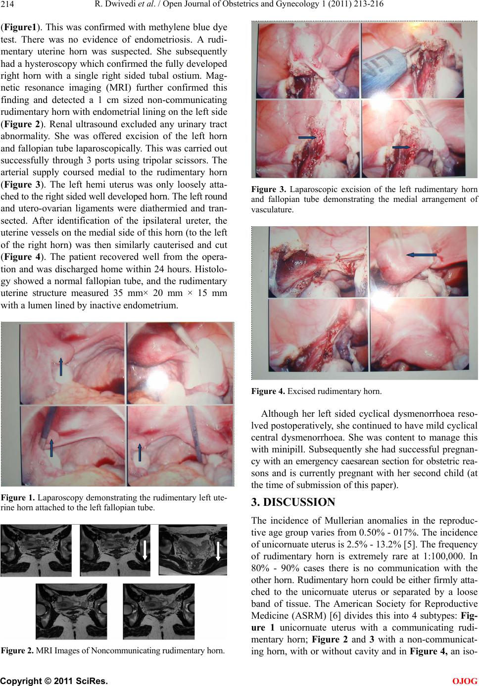 R. Dwivedi et al. / Open Journal of Obstetrics and Gynecology 1 (2011) 213-216 214 (Figure1). This was confirmed with methylene blue dye test. There was no evidence of endometriosis. A rudi- mentary uterine horn was suspected. She subsequently had a hysteroscopy which confirmed the fully developed right horn with a single right sided tubal ostium. Mag- netic resonance imaging (MRI) further confirmed this finding and detected a 1 cm sized non-communicating rudimentary horn with endometrial lining on the left side (Figure 2). Renal ultrasound excluded any urinary tract abnormality. She was offered excision of the left horn and fallopian tube laparoscopically. This was carried out successfully through 3 ports using tripolar scissors. The arterial supply coursed medial to the rudimentary horn (Figure 3). The left hemi uterus was only loosely atta- ched to the right sided well developed horn. The left round and utero-ovarian ligaments were diathermied and tran- sected. After identification of the ipsilateral ureter, the uterine vessels on the medial side of this horn (to the left of the right horn) was then similarly cauterised and cut (Figure 4). The patient recovered well from the opera- tion and was discharged home within 24 hours. Histolo- gy showed a normal fallopian tube, and the rudimentary uterine structure measured 35 mm× 20 mm × 15 mm with a lumen lined by inactive endometrium. Figure 1. Laparoscopy demonstrating the rudimentary left ute- rine horn attached to the left fallopian tube. Figure 2. MRI Images of Noncommunicating rudimentary horn. Figure 3. Laparoscopic excision of the left rudimentary horn and fallopian tube demonstrating the medial arrangement of vasculature. Figure 4. Excised rudimentary horn. Although her left sided cyclical dysmenorrhoea reso- lved postoperatively, she continued to have mild cyclical central dysmenorrhoea. She was content to manage this with minipill. Subsequently she had successful pregnan- cy with an emergency caesarean section for obstetr ic rea- sons and is currently pregnant with her second child (at the time of submission of this paper). 3. DISCUSSION The incidence of Mullerian anomalies in the reproduc- tive age group varies from 0.50% - 017%. The incidence of unicornuate uter us is 2. 5% - 13.2% [ 5] . The fre quency of rudimentary horn is extremely rare at 1:100,000. In 80% - 90% cases there is no communication with the other horn. Rudimentary horn could be either firmly atta- ched to the unicornuate uterus or separated by a loose band of tissue. The American Society for Reproductive Medicine (ASRM) [6] divides this into 4 subtypes: Fig- ure 1 unicornuate uterus with a communicating rudi- mentary horn; Figure 2 and 3 with a non-communicat- ing horn, with or without cavity and in Figure 4, an iso- C opyright © 2011 SciRes. OJOG  R. Dwivedi et al. / Open Journal of Obstetrics and Gynecology 1 (2011) 213-216 215 lated unicornuate uterus. Women with obstructive uterine anomalies usually pre- sent with chronic cyclic or continuous pelvic pain. The presentation can be delayed with increasing severity of dysmenorrhoea, with each subsequent menstrual period as a common feature. The symptoms may be severe e- nough to interfere with quality of life and may not res- pond to medication. Unlike in obstructive uterine ano- malies where symptoms are caused by the accumulation of blood in the rudimentary horn, disordered arrange- ment of uterine musculature and abnormal contraction are thought to be the cause in cases such as ours with inactive endometrium [7]. Unequal development of Mu- llerian system gives rise pain on one side of the pelvis. Torsion of the rudimentary horn has also been reported. Other complications include endometriosis and primary infertility. Anomalies of the urinary system are usually associated with uterine developmental abnormalities due to their close embryological origins. Endometriosis re- sults from retrograde menstruation from the functioning endometrial cavity through the ipsilateral fallopian tube. Obstetric complications such as rupture uterus, recurrent miscarriages, premature labour and malpresentation can also occur [8]. A high index of suspicion for Mullerian duct abnor- mality is therefore needed in teenagers with dysmenor- rhoea to prevent delays in diagnosis [9]. Ultrasonogra- phy including hysterosonography is not specific in non- functioning uterine horns as the detail of adnexa is in- sufficient. In a recent review of the literature, the sensi- tivity of ultrasonography even in cases with functioning horns was only 26%. MRI is much more accurate and detected all cases in these series and is considered to be the gold standard [10]. 3D CT scanning has been used in some cases. 3D ultrasound also may also have a future role. An early recourse to laparoscopy combined with hysteroscopy is required to confirm the diagnosis most cases. Resection not only offers symptomatic relief in cases unresponsive to medication, it also prevents the possibi- lity of pregnancy in the rudimentary horn due to trans- peritoneal migration of sperm or fertilised ovum. Accu- rate diagnosis of the anomaly is required prior to exci- sion to decide on the precise surgical approach as in so- me cases the two horns are firmly attached. This requires difficult dissection to develop a plane between the hemi uteri. If the other tube is normal, the ipsilateral fallopian tube should be removed to prevent ectopic pregnancy. If the other tube is damaged, microsurgical transposition of the ipsilateral tube may need to be considered [11]. Al- though laparotomy was used traditionally, there are seve- ral publications of the laparoscopic removal of the rudi- mentary horn [10]. This follows a similar trend in other types of gynaecological surgery. Most cases are amena- ble to laparoscopic management and this is preferred to prevent adhesions and damage to the myometrium of the well developed horn. Rudimentary horn attached to the unicornuate uterus by a band of tissue, as in our case, is relatively easy to dissect. Minimally invasive, with well established safety profile, laparoscopic approach has other advantages such as shorter hospital stay and quicker re- turn to activities [12]. Aberrant blood vessels and uro- logical anomalies increase the risk of bleeding and ina- dvertent injury. Review of MRI scans with radiologists prior to surgery allows selected cases to be referred to advanced laparoscopic surgeons. REFERENCES [1] Dadhwal, V., Mittal, S., Kumar, S. and Barua, A. (2000) Hematometra in postmenarchal adolescent girls: A report of two cases. Gynecologic and Obstetric Investigation, 50, 67-69. doi:10.1159/000010284 [2] Chakravarthi, S. and Chin, K. (2003) Rudimentary uter- ine horn: Management of a diagnostic enigma. Acta Ob- stetricia et Gynecologica Scandinavica, 82, 1153-1154. doi:10.1046/j.1600-0412.2003.00234.x [3] Goel, P., Aggarwal, A., Devi, K., Takkar, N., Saha, P.K. and Huria, A. (2005) Unicornuate with non-communi- cating rudimentary horn different clinical presentations. The Journal of Obstetrics & Ganecology of India, 55, 155-158. [4] Falcone, T., Gidwani, G., Paraiso, M., Beverly, C. and Goldberg, J. (1997) Anatomical variation in rudimentary horns of a unicornuate uterus: Implications for laparo- scopic surgery. Human Reproduction, 12, 263-265. doi:10.1093/humrep/12.2.263 [5] Green, L.K. and Harris, R.E. (1976) Uterine anomalies: Frequency of diagnosis and associated obstetric compli- cations. Obstetrics Gynecology, 47, 427-429. [6] Buttram, V.C. Jr. and Gibbons, W.E. (1979) Mullerian anomalies: A proposed classification (an analysis of 144 cases). Fertility and Sterility, 32, 40-46. [7] Jeffcoate, N. (2006) Malformation and maldevelopments of the genital tract: In principles of gynaecology. The 4th Edition, Butterworth & Co Ltd, London, 138. [8] Atmaca, R., Germen, A.T., Burak, F. and Kafkasli, A. (2005) Acute abdomen in a case with noncommunicating rudimentary horn and unicornuate uterus. Journal of the Society of Laparoendoscpic Surgeons, 9, 235-237. [9] Badawy, S.Z.A. and Montgomery, S. (2009) Severe pri- mary dysmenorrhoea due to functioning noncommunica- timg rudimentary horn. Journal of Gynecologic Surgery, 25, 23-27. doi:10.1089/gyn.2009.B-02330 [10] Spitzer, R.F., Kives, S. and Allen, L.M. (2009) Case se- ries of laparoscopically resected noncommunicating functional uterine horns. Journal of Pediatric and Ado- lescent Gynecology, 22, E23-E28. doi:10.1016/j.jpag.2007.12.001 [11] Gold Berg, J.M. and Friedman, C.I. (1988) Microsurgical fallopian tube transposition with subsequent term preg- nancy. Fertility and Sterility, 50, 661-662. C opyright © 2011 SciRes. OJOG  R. Dwivedi et al. / Open Journal of Obstetrics and Gynecology 1 (2011) 213-216 Copyright © 2011 SciRes. OJOG 216 [12] Liatsikos, S.A., Tsikouras, P., Souftas, V., Ammari, A., Prassopoulos, P., Maroulis, G. and Liberis, V. (2010) Di- agnosis and laparoscopic management of a rudimentary uterine horn in a teenage girl, presenting with hema- tometra and severe endometriosis: Our experience and review of literature. Minimally Invasive Therapy and Al- lied Technologies, 19, 241-247. doi:10.3109/13645701003644491 |