

Paper Menu >>
Journal Menu >>
 Open Journal of Obstetrics and Gynecology, 2011, 1, 191-196 doi:10.4236/ojog.2011.14037 Published Online December 2011 (http://www.SciRP.org/journal/ojog/ OJOG ). Published Online December 2011 in SciRes. http://www.scirp.org/journal/OJOG Homocysteine in pregnancies complicated by preeclampsia with and without IUGR: a comparison with normotensive pregnant women with isolated IUGR and healthy pregnant women Marzena Laskowska*, Jan Oleszczuk Chair and Department of Obstetrics and Perinatology, Medical University of Lublin, Lublin, Poland. Email: *[email protected] Received 8 October 2011; revised 18 November 2011; accepted 29 November 2011. ABSTRACT Objective: The aim of this study was to analyze ho- mocysteine levels in maternal serum in women with pregnancie s complic ated by preeclam psia and/or IUGR. Patients and methods: The study was carried out on 49 pregnant patients with normotensive pregnancies complicated by intrauterine fetal growth restriction (group IUGR), 31 patients with preeclampsia com- plicated by IUGR (group PRE-IUGR), and 35 pree- clamptic patients with appropriate-for-gestational-age weight fetuses (group PRE). The control group con- sisted of 47 healthy normotensive pregnant patients with singleton uncomplicated pregnancies and with proper intrauterine fetal growth. Results: We revea- led higher levels of maternal serum homocysteine in the group of pregnant patients with isolated fetal in- trauterine growth restriction in comparison with the control subjects. The concentrations of homocysteine were also higher in both groups of patients with pre- gnancy complicated by preeclampsia with and with- out IUGR. The highest levels of homocysteine were observed in preeclamptic women with appropriate- for-gestational-age fetal g rowth. The mean values we- re 9.004 +/– 2.820 mol/L in the IUGR group, 10.815 +/– 3.785 mol/ L in the group PRE, 9.808 +/– 2.543 mol/L in the group PRE-IUGR and 7.639 +/– 2.728 mol/L in the control group. Conclusions: Increased levels of homocysteine are involved in pathogenesis IUGR and preeclampsia and may contribute to endo- thelial cells activation or dysfunction observed in the- se pregnancy disorders. Further studies are needed to explain these aspects in order to improve the mana- gement and therapeutic strategies for pregnancies co- mplicated by IUGR and/or preeclampsia. Keywords: Homocysteine; Intrauterine Fetal Growth Restriction (IUGR); Preeclampsia 1. INTRODUCTION Intrauterine fetal growth restriction (IUGR) complicates 8% of all pregnancies and is a major cause of perinatal mortality and morbidity [1]. One of the most common maternal conditions associated with fetal growth restrict- tion is maternal hypertension and especially preeclamp- sia, which complicates 5% - 10% of all pregnancies [2]. But it is important that in many cases of pregnancies complicated by IUGR there are an idiopathic placental failure with abnormal placental development and subop- timal fetal nutrition and oxygenation without maternal hypertension or preeclampsia [3,4]. Pathophysiological processes underlying intrauterine fetal growth restriction and/or preeclampsia are compli- cated, multifactorial and unclear [5]. The impaired inva- sion of uteroplacental arteries causes changes in the pla- cental and fetal circulatory system and may be the cause of the pathological structure and function of the placenta, resulting in the inadequate fetal oxygenation and nutria- tion, and consequently leading to preeclampsia or intrau- terine fetal growth restriction.[5-7]. Endothelial dysfunction has been hypotheses to play a central role in the pathogenesis of preeclampsia [8-10]. Animal studies suggest that hyperhomocysteinemia affe- cts the blood vessel wall and causes a change in the en- dothelium and smooth muscle proliferation [11-13]. Hypehomocysteinemia is a risk factor of cardiovascu- lar diseases and vasculopathy. It may be a cause of chan- ges and lesions in endothelial cells due to vascular fibro- sis and resulting in the activation of thrombogenesis. Hyperhomocysteinemia promotes oxidation of LDL cho- lesterol molecules and alterations in the coagulation sys- tem and enhances platelet activation [11]. The aim of this study was to analyze homocysteine le-  M. Laskowska et al. / Open Journal of Obstetrics and Gynecology 1 (2011) 191-196 192 vels in maternal serum in pregnancies complicated by in- trauterine fetal growth restriction with and without pree- clampsia, and in preeclamptic pregnancies with the ap- propriate fetal growth, and in the control group of heal- thy pregnancies with normal fetal growth. The study was accepted by the local Ethics Committee (KE-0254/51/ 2010). 2. PATIENTS AND METHODS The study was carried out on 49 pregnant women with normotensive pregnancies complicated by intrauterine growth restricted fetuses (IUGR group), and 66 patients with pregnancy complicated by severe preeclampsia. In the group of preeclamptic women were 31 patients with preeclampsia complicated by intrauterine fetal growth restriction (PRE-IUGR group) and 35 preeclamptic pa- tients with appropriate-for-gestational-age fetuses (PRE group). Among preeclamptic women there were 6 patients wi- th HELLP syndrome (all in the PRE-IUGR group) and 4 patients with eclampsia (3 in the PRE-IUGR group and 1 in the PRE group). One patient from the IUGR group had fetal intrauterine death in the week 38th of gestation. The weight of her stillborn infant was 2060 g. The control group consisted of 47 healthy normoten- sive pregnant patients with singleton uncomplicated pre- gnancies without any renal, cardiac and vascular disea- ses and with normal laboratory tests and with appropria- te-for-gestational-age weight infants (CON group). Fetal biometry was based on non-invasive ultrasound method and comprised the estimation of gestational age in early gestation and diagnosis of fetal intrauterine gro- wth restriction by monitoring fetal growth later in the se- cond or third trimester of pregnancy. Intrauterine growth restricted fetuses (IUGR) were classified as such according to ultrasonographic measu- rement when the weight of the fetus was lower than ex- pected in relation to the gestational age, as determined by the standard curves characteristic of the Polish popu- lation, when the fetus was below the 5th centile for gesta- tional age and diagnosis was confirmed by the infant’s weight at birth according to the Bretelle et al. [5]. The exclusion criteria for women whose pregnancies were complicated by IUGR fetuses were the presence of a congenital malformation or chromosomal abnormality in the fetus, recent cytomegalovirus infection or drug or alcohol abuse during pregnancy. Pregnant women with multiple pregnancies were also excluded from this study. Preeclampsia was diagnosed by the increased blood pressure of >140 mmHg systolic and >90 mmHg diasto- lic in women who were normotensive before 20 weeks of gestation accompanied by proteinuria, defined as the urinary excretion of >0.3 g protein in 24 h specimen. Severe preeclampsia was defined as blood pressure >160/110 mmHg on at least 2 occasions 6h apart with proteinuria >2 g in a 24 hour urinary protein excretion, and when hypertension and proteinuria were associated with one or more of the following clinical manifestations: renal abnormalities, hematological abnormalities (throm- bocytopenia and microangiopathic hemolysis) or HELLP syndrome (hemolysis, elevated liver enzymes, low pla- telet count and right-upper quadrant pain), or neurology- cal symptoms (headache, visual disturbances and seizu- res). None of the pregnant patients with preeclampsia were affected by chronic hypertension, renal disorders and/or proteinuria before pregnancy and all were nor- motensive before 20th week of pregnancy. Preeclamptic patients were admitted to the Department of Obstetrics and Perinatology in the Medical University Hospital in Lublin because of the symptoms of the di- sease and without signs of labour. All preeclamptic wo- men were normotensive 3 months after delivery. All arterial blood pressure measurements in the con- trol group and in the group of patients with isolated fetal intrauterine growth restriction were normal and did not exceed 135/85 mmHg. None of the patients from any of these groups suffered from proteinuria. All patients in the study were non-smokers. An infor- med consent from all studied patients was obtained for peripheral blood sampling. Five milliliters of blood were collected by venipuncture from each preeclamptic pa- tient and from each woman from the control group and placed in sterile tubes. They were centrifuged for 15 min at 500 xg. The homocysteine concentrations were esti- mated using an ELISA assay according to the manufac- turer’s instructions (human sandwich ELISA kit Axis- Shield Diagnostics Ltd, UK)). In the statistical analysis results were expressed as mean +/– SD or SEM or as median values and were sta- tistically analyzed with the computer program “Statis- tica” using the Shapiro-Wilk test for the normal distribu- tion of data, and equality of variance by Levene test and, subsequently two-tailed Student’s t-tests, or (in unequal variance) the Cochran-Cox test. The Analysis of Variance—ANOVA and Kruskal-Wa- llis tests were used to test for differences among four independent groups. A statistically significant effect in ANOVA was followed up with follow-up post hoc Tu- key’s test in order to assess which group is different from which other groups. A p-value of less than 0.05 was considered to be significant. 3. RESULTS There were no statistically significant differences in gra- vidity, parity, maternal age and height in patient’s pro- files between groups. Maternal weight and BMI were C opyright © 2011 SciRes. OJOG 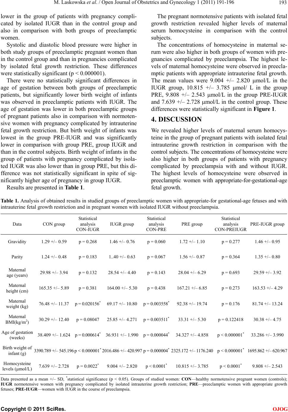 M. Laskowska et al. / Open Journal of Obstetrics and Gynecology 1 (2011) 191-196 Copyright © 2011 SciRes. 193 OJOG lower in the group of patients with pregnancy compli- cated by isolated IUGR than in the control group and also in comparison with both groups of preeclamptic women. Systolic and diastolic blood pressure were higher in both study groups of preeclamptic pregnant women than in the control group and than in pregnancies complicated by isolated fetal growth restriction. These differences were statistically significant (p < 0.000001). There were no statistically significant differences in age of gestation between both groups of preeclamptic patients, but significantly lower birth weight of infants was observed in preeclamptic patients with IUGR. The age of gestation was lower in both preeclamptic groups of pregnant patients also in comparison with normoten- sive women with pregnancy complicated by intrauterine fetal growth restriction. But birth weight of infants was lowest in the group PRE-IUGR and was significantly lower in comparison with group PRE, group IUGR and than in the control subjects. Birth weight of infants in the group of patients with pregnancy complicated by isola- ted IUGR was also lower than in group PRE, but this di- fference was not statistically significant in spite of sig- nificantly higher age of pregnancy in group IUGR. Results are presented in Table 1. The pregnant normotensive patients with isolated fetal growth restriction revealed higher levels of maternal serum homocysteine in comparison with the control subjects. The concentrations of homocysteine in maternal se- rum were also higher in both groups of women with pre- gnancies complicated by preeclampsia. The highest le- vels of maternal homocysteine were observed in preecla- mptic patients with appropriate intrauterine fetal growth. The mean values were 9.004 +/– 2.820 mol/L in the IUGR group, 10.815 +/– 3.785 mol/ L in the group PRE, 9.808 +/– 2.543 mol/L in the group PRE-IUGR and 7.639 +/– 2.728 mol/L in the control group. These differences were statistically significant in Figure 1. 4. DISCUSSION We revealed higher levels of maternal serum homocys- teine in the group of pregnant patients with isolated fetal intrauterine growth restriction in comparison with the control subjects. The concentrations of homocysteine were also higher in both groups of patients with pregnancy complicated by preeclampsia with and without IUGR. The highest levels of homocysteine were observed in preeclamptic women with appropriate-for-gestational-age fetal growth. Tab le 1 . Analysis of obtained results in studied groups of preeclamptic women with appropriate-for gestational-age fetuses and with intrauterine fetal growth restriction and in pregnant women with isolated IUGR without preeclampsia. Data CON group Statistical analysis CON-IUGR IUGR group Statistical analysis CON-PRE PRE group Statistical analysis CON-PREIUGR PRE-IUGR group Gravidity 1.29 +/– 0.59 p = 0.268 1.46 +/– 0.76 p = 0.0601.72 +/– 1.10 p = 0.277 1.46 +/– 0.95 Parity 1.24 +/– 0.48 p = 0.183 1..40 +/– 0.63 p = 0.0671.56 +/– 0.87 p = 0.364 1.35 +/– 0.80 Maternal age (years) 29.98 +/– 3.94 p = 0.132 28.54 +/– 4.40 p = 0.14328.04 +/– 6.29 p = 0.693 29.59 +/– 3.92 Maternal height (cm) 165.35 +/– 5.89 p = 0.381 164.00 +/– 5.30 p = 0.438167.21 +/– 6.85 p = 0.273 163.53 +/– 4.29 Maternal weight (kg) 76.48 +/– 11.37 p = 0.020156* 69.17 +/– 10.80 p = 0.003558*92.38 +/– 19.74 p = 0.176 81.74 +/– 13.24 Maternal BMI(kg/m2) 30.29 +/– 12.40 p = 0.08047 25.85 +/– 4.271 p = 0.003511*33.31 +/– 5.30 p = 0.122418 30.38 +/– 4.75 Age of gestation (weeks) 38.409 +/– 1.624 p = 0.000614* 36.931 +/– 1.990p = 0.000044*34.327 +/– 4.858 p < 0.000001* 33.286 +/– 3.990 Birth weight of infant (g) 3390.789 +/– 545.196p < 0.000001* 2016.486 +/– 420.997p = 0.000004*2325.172 +/– 1176.240p < 0.000001* 1695.862 +/– 620.967 Homocysteine levels (μmol/L) 7.639 +/– 2.728 p = 0.0022* 9.004 +/– 2.820 p < 0.0001*10.815 +/– 3.785 p < 0.0001* 9.808 +/– 2.543 Data presented as a mean +/– SD, *statistical significance (p < 0.05). Groups of studied women: CON—healthy normotensive pregnant women (controls); IUGR normotensive women with pregnancy complicated by isolated intrauterine growth restriction; PRE—preeclamptic women with appropriate growth fetuses; PRE-IUGR—women with IUGR in the course of preeclampsia. 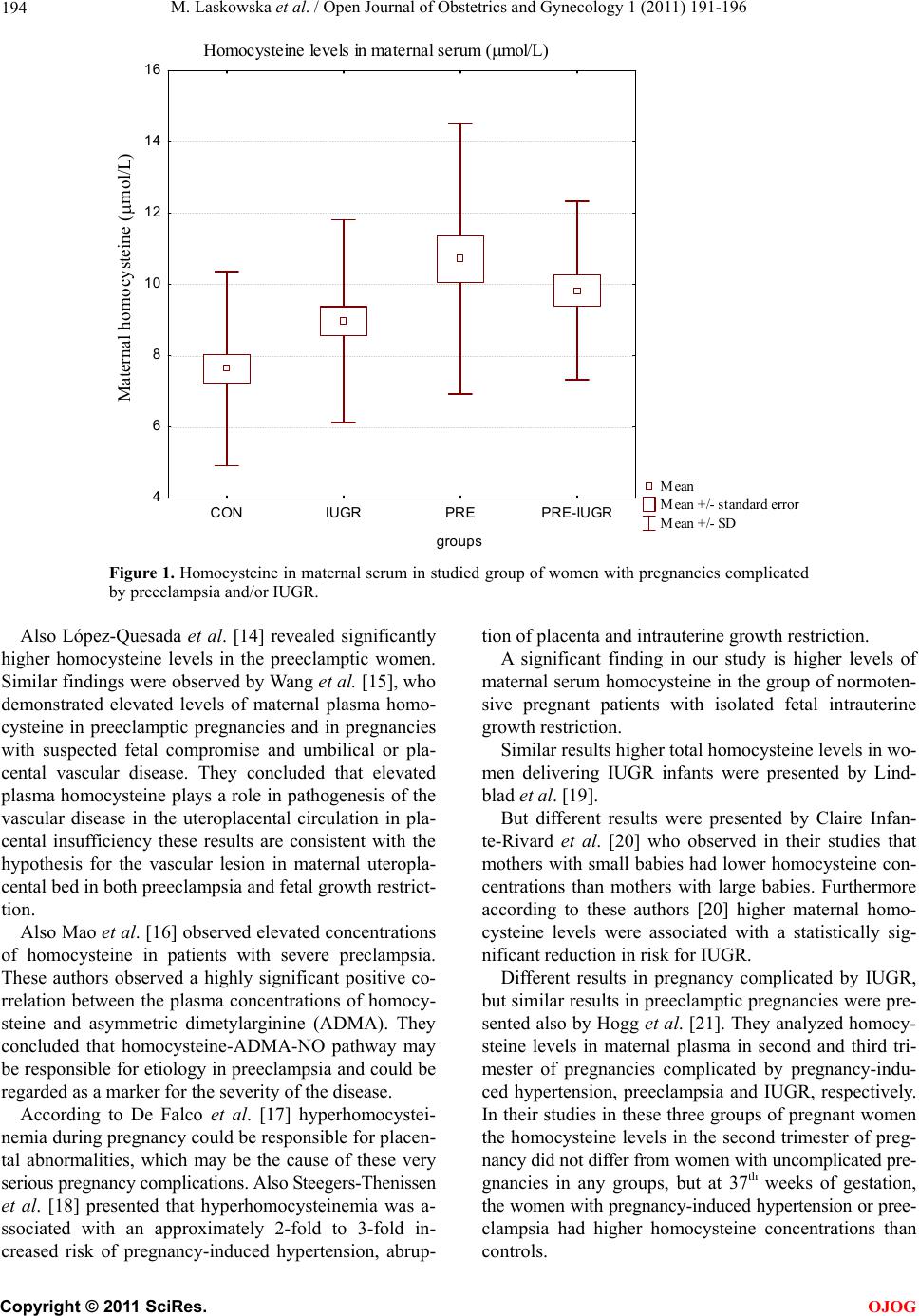 M. Laskowska et al. / Open Journal of Obstetrics and Gynecology 1 (2011) 191-196 194 Homocysteine levels in maternal serum ( mol/L) Mean Mean +/- standard error Mean +/- SD CONIUGRPRE PRE-IUGR groups 4 6 8 10 12 14 16 Maternal homocysteine (mol/L) Figure 1. Homocysteine in maternal serum in studied group of women with pregnancies complicated by preeclampsia and/or IUGR. Also López-Quesada et al. [14] revealed significantly higher homocysteine levels in the preeclamptic women. Similar findings were observed by Wang et al. [15], who demonstrated elevated levels of maternal plasma homo- cysteine in preeclamptic pregnancies and in pregnancies with suspected fetal compromise and umbilical or pla- cental vascular disease. They concluded that elevated plasma homocysteine plays a role in pathogenesis of the vascular disease in the uteroplacental circulation in pla- cental insufficiency these results are consistent with the hypothesis for the vascular lesion in maternal uteropla- cental bed in both preeclampsia and fetal growth restrict- tion. Also Mao et al. [16] observed elevated concentrations of homocysteine in patients with severe preclampsia. These authors observed a highly significant positive co- rrelation between the plasma concentrations of homocy- steine and asymmetric dimetylarginine (ADMA). They concluded that homocysteine-ADMA-NO pathway may be responsible for etiology in preeclampsia and could be regarded as a marker for the severity of the disease. According to De Falco et al. [17] hyperhomocystei- nemia during pregnancy could be responsible for placen- tal abnormalities, which may be the cause of these very serious pregnancy complications. Also Steegers-Thenissen et al. [18] presented that hyperhomocysteinemia was a- ssociated with an approximately 2-fold to 3-fold in- creased risk of pregnancy-induced hypertension, abrup- tion of placenta and intrauterine growth restriction. A significant finding in our study is higher levels of maternal serum homocysteine in the group of normoten- sive pregnant patients with isolated fetal intrauterine growth restriction. Similar results higher total homocysteine levels in wo- men delivering IUGR infants were presented by Lind- blad et al. [19]. But different results were presented by Claire Infan- te-Rivard et al. [20] who observed in their studies that mothers with small babies had lower homocysteine con- centrations than mothers with large babies. Furthermore according to these authors [20] higher maternal homo- cysteine levels were associated with a statistically sig- nificant reduction in risk for IUGR. Different results in pregnancy complicated by IUGR, but similar results in preeclamptic pregnancies were pre- sented also by Hogg et al. [21]. They analyzed homocy- steine levels in maternal plasma in second and third tri- mester of pregnancies complicated by pregnancy-indu- ced hypertension, preeclampsia and IUGR, respectively. In their studies in these three groups of pregnant women the homocysteine levels in the second trimester of preg- nancy did not differ from women with uncomplicated pre- gnancies in any groups, but at 37th weeks of gestation, the women with pregnancy-induced hypertension or pree- clampsia had higher homocysteine concentrations than controls. C opyright © 2011 SciRes. OJOG  M. Laskowska et al. / Open Journal of Obstetrics and Gynecology 1 (2011) 191-196 195 However Hogg et al. [21] did not find such differen- ces in pregnancies with IUGR. They concluded that pla- sma homocysteine concentrations in the second trimester do not predict the subsequent complications of preg- nancy-induced hypertension, preeclampsia and intraute- rine growth restriction. Contrary to our findings also D’Anna et al. [22] didn’t find any association between homocysteine levels in pre- gnancy and IUGR. Our findings about higher levels of maternal serum homocysteine in pregnancies complicated by IUGR are inconsistent with some earlier reports about the rela- tionship between higher levels of homocysteine in preg- nancy with intrauterine fetal growth restriction. This may be due to the fact that we included into a group of wo- men with pregnancy complicated by IUGR only patients with fetal weight estimated below the 5th percentile for gestational age. Furthermore we divided all pregnant patients with fetal growth restriction into two groups, one with IUGR in the course of preeclampsia and the se- cond group with isolated IUGR and normotensive preg- nancy, without any maternal or fetal condition response- ble for this pregnancy complication. The aim of this was to select the patients with the IUGR due to placental dysfunction. 5. CONCLUSIONS In conclusion, our findings seem to suggest that homo- cysteine is a mediator of endothelial activation or dys- function observed in pregnancies complicated by pree- clampsia, but also in normotensive pregnancies compli- cated by isolated IUGR. It seems that the pathomechanism underlying the de- velopment of preeclampsia and/or intrauterine fetal grow- th restriction is similar, but highest levels of homocys- teine observed in preeclamptic pregnancies may suggest that its beginning and intensity may be more enhanced in pregnancy complicated by hypertension and preeclamp- sia. Thanks to our better understanding of its physiology and pathophysiology, homocysteine may develop towa- rds a novel target for pharmacological intervention for patients with these pregnancy complications. REFERENCES [1] Mandruzzato, G., Antsaklis, A., Botet, F., Chervenak, F.A., Figueras, F., Grunebaum, A., Puerto, B., Skupski, D. and Stanojevic, M. (2008) Intrauterine restriction (IUGR). Journal of Perinatal Medicine, 36, 277-281. doi:10.1515/JPM.2008.050 [2] Maynard, S.E., Min, J.Y., Merchan, J., Lim, K.H., Li, J., Mondal, S., Libermann, T.A., Morgan, J.P., Selke, F.W., Stillman, I.E., Epstein, F.H., Sukhatme, V.P.M. and Ka- rumanchi, S.A. (2003) Excess placental soluble fms-like tyrosine kinase 1 (sFlt1) may contribute to endothelial dysfunction, hypertension, and proteinuria in preeclamp- sia. Journal of Clinical Investigation, 111 , 600-602. [3] Maršal, K. (2002) Intrauterine growth restriction. Current Opinion in Obstetrics and Gynecology, 14,127-135. doi:10.1097/00001703-200204000-00005 [4] Robinson, J.S. and Owens, J.A. (1996) Pathophysiology of intrauterine growth failure. In: Gluckman, P.D. and Heymann, M.A., Eds., Pediatrics and Perinatology: The Scientific Basis, Arnold, London, 290-297. [5] Bretelle, F., Sabatier, F., Blann, A., D’Ercole, Boutiere, B., Mutin, M., Boubli, L., Sampol, J. and Dignat-George, F. (2001) Maternal endothelial soluble cell adhesion molecules with isolated small for gestational age fetuses, comparison with preeclampsia. British Journal of Ob- stetrics Gynaecology, 108, 1277-1282. doi:10.1016/S0306-5456(01)00259-5 [6] Bamberger, A.M., Schulte, H.M., Thuneke, I., Erdmann, I., Bamberger, ChM. and Asa, S.L. (1997) Expression of the apoptosis-inducing fas ligand (FasL) in human first and third trimester placenta and choriocarcinoma cells. Journal of Clinical Endocrinology & Metabolism, 82, 3173-3175. doi:10.1210/jc.82.9.3173 [7] Hsu, ChD., hariah, H., Basherra, H. and Mor, A.G. (2001) Serum soluble fas levels in preeclampsia. Obstetrics and Gynecology, 97, 530-532. doi:10.1016/S0029-7844(00)01227-8 [8] Roberts, J.M. (1999) Objective evidence of endothelial dysfunction in preeclampsia. American Journal of Kid- ney Diseases, 33, 992-997. doi:10.1016/S0272-6386(99)70439-7 [9] Granger, J.P., Alexander, B.T., Llinas, M.T., Bennett, W.A. and Khalil, R.A. (2001) Pathophysiology of hyper- tension during preeclampsia linking placental ischemia with endothelial dysfunction. Hypertension, 38, 718-722. [10] Kassab, S., Abu-Hijleh, M.F., Al-Shaikh, H.B. and Na- galla, D.S. (2005) Hyperhomocysteinemia in pregnant rats: Effects on arterial pressure, kidneys and fetal growth. European Journal of Obstetrics & Gynecology and Reproductive, 122, 177-181. doi:10.1016/j.ejogrb.2005.02.008 [11] Aubard, Y., Darodes, N. and Cantaloube, M. (2000) Hy- perhomocysteinemia and pregnancy—review of our pre- sent understanding and therapeutic implications. Euro- pean Journal of Obstetrics & Gynecology and Reproduc- tive, 93, 57-165. [12] Onalan, R., Onalan, G., Gunenc, Z. and Karabulut, E. (2006) Combining 2nd trimester maternal serum homo- cysteine levels and uterine artery doppler for prediction of preeclampsia and isolated intrauterine growth restric- tion. Gynecology and Obstetrics Investigaton, 61, 142- 148. doi:10.1159/000090432 [13] Weir, D.G. and Scott, J.M. (1998) Homocysteine as a risk factor for cardiovascular and related disease: Nutritional implications. Nutr ition Research Reviews, 11, 311-338. doi:10.1079/NRR19980020 [14] López-Quesada, E., Also-Razo, E. and Vilaseca, M.A. (2003) Hyperhomocysteinemia during pregnancy as a risk factor of preeclampsia. Clinical Medicine, 121, 350-355. [15] Wang, J., Trudinger, B.J., Duarte, N., Wilcken, D.E. and C opyright © 2011 SciRes. OJOG  M. Laskowska et al. / Open Journal of Obstetrics and Gynecology 1 (2011) 191-196 Copyright © 2011 SciRes. 196 OJOG Wang, X.L. (2000) Elevated circulating homocysteine levels in placental vascular disease and associated pre-eclampsia. British Journal of Obstetrics and Gynae- cology, 107, 935-938. doi:10.1111/j.1471-0528.2000.tb11095.x [16] Mao, D., Che, J., Li, K., Han, S., Yue, Q., Zhu, L., Zhang, W. and Li, L. (2009) Association of homocysteine, asym- metric dimethylarginine, and nitric oxide with pree- clampsia. Archives of Gynecology and Obstetrics, 1234-1236. [17] De Falco, M., Pollio, F., Scaramelino, M., Portillo, M. and Lieto, A.D. (2000) Homocysteinemia during preg- nancy and placental disease. Clinical & Experimental Obstetrics & Gynecology, 27, 188-190. [18] Steegers-Theunissen, R.P., Van Iersel, C.A., Peer, P.G., Nelen, W.L. and Steegers, E.A. (2004) Hyperhomocys- teinemia, pregnancy complications, and the timing of in- vestigation. Obstetrics and Gynecology, 104, 336-343. doi:10.1097/01.AOG.0000129955.47943.2a [19] Lindblad, B., Zaman, S., Malik, A., Martin, H., Ekström, A.M., Amu, S., Holmgren, A. and Norman, M. (2005) Folate, vitamin B12 and homocysteine levels in South Asian women with growth-retarded fetuses. Acta Obstet- ricia et Gynecologica Scandinavica, 84, 1055-1061. [20] Infante-Rivard, C., Rivard, G.E., Gauthier, R. and Théoret, Y. (2003) Unexpected relationship between pla- sma homocysteine and intrauterine growth restriction. Clinical Chemistry, 49, 1476-1482. doi:10.1373/49.9.1476 [21] Hogg, ChD., Harirah, H., Basherra, H. and Mor, A.G. (2001) Serum soluble fas levels in preeclampsia. Obstet- rics and Gynecology, 97, 530-532. doi:10.1016/S0029-7844(00)01227-8 [22] D’Anna, R., Bariera, G., Corrido, F., Lentile, R., Granese, D. and Stella, N.C. (2004) Plasma homocysteine in early and late pregnancies complicated with preeclampsia and isolated intrauterine growth restriction. Acta Obstetricia et Gynecologica Scandinavica, 83, 155-158. |