

Paper Menu >>
Journal Menu >>
 Materials Sciences and Applicatio ns, 2011, 2, 1719-1723 doi:10.4236/msa.2011.212229 Published Online December 2011 (http://www.SciRP.org/journal/msa) Copyright © 2011 SciRes. MSA 1719 Aggregation Study of Ag-TiO2 Composites María Eugenia Noriega-Treviño1,2*, Claudia Cristina Quintero-González1, José Elpidio Morales-Sánchez1,2,3, Jesús María Guajardo-Pacheco1,2,3, Martha Eugenia Compeán-Jasso1, Facundo Ruiz4 1Science Faculty, Universidad Autónoma de San Luis Potosí, San Luis Potosí, México; 2Mathematics and Physics Department, Universidad Autónoma de San Luis Potosí, San Luis Potosí, México; 3Materials Advanced Research Center, Chihuahua, México; 4Materials Advanced Research Center, Apodaca, México. E-mail: *[email protected] Received October 21st, 2011; revised November 30th, 2011; accepted December 7th, 2011. ABSTRACT Most of the toxicity data presented in the literature are obtained in relatively simple media, like distilled water. The literature reported that nanoparticles agglomerate immediately upon being added to cell culture media and if the ag- glomerates are used directly in antimicrobial studies, the interpretation of the toxicity results tends to be complicated. Six different molar ratios Ag-TiO 2 composites were synthesized by a reduction method using two different commercial TiO2 particles as base ma terials and were used to find the agg regate size in distilled water and Mueller-Hinton Broth, and to obtain the minimum inhibitory concentrations (MIC) against E. coli and E. faecalis. To evaluate the evolution of the Ag-TiO2 particle size (z-average) three dilutions of each of the synthesized composites 100 µg/ml, 250 µg/mL and 500 µg/ml were realized in deionized water an d Mueller Hinton broth. It was found that Ag-TiO2 composites increased in size after being diluted in Mueller-Hinton Broth, but once they grew in size, they remained constant for 24 minutes, and after this time, did not affect the MIC for the microorganisms invo lved. Keywords: Ag-TiO2, Composites, Nanoparticles, Particles, Aggregate Size, MIC 1. Introduction The most common method of producing silver nanopar- ticles is a chemical reduction of silver salt dissolved in water with a reducing compound [1,2]. Silver nanomate- rials exhibit broad-spectrum biocidal activity toward bacteria, fungi, viruses and algae [3]. Several factors have been reported to influence silver nanoparticle toxicity like particle size, shape, pH, ionic strength and the presence of divalent cations and mac- romolecules [4-8]. The stability of silver nanoparticles also influences toxicity since the formation of aggregates tends to decrease biocidal activity [9,10]. Most of the toxicity data presented in the literature are obtained in relatively simple media like distilled water. It was found that TiO2, when irradiated with UV radia- tion, acted as an antimicrobial [11]. It had been reported that photocatalytic and antimicrobial properties of TiO2 can be improved by growing particles of a noble metal like Ag, Au or Cu over its surface or inside a matrix [12-14]. Composites of silver coatings over titanium di- oxide nanoparticles are used in products to produce anti- bacterial activity [15]. The literature reported that nanoparticles agglomerate immediately upon being added to cell culture media and if the agglomerates are used directly in antimicrobial studies, the interpretation of the toxicity results tends to be complicated. Agglomerates of nanoparticles have been shown to exert lower antibacterial effects as compared to well dispersed nanoparticles [16]. It was reported that the presence of proteins within the nanoparticle solution can stabilize the silver nanoparticles against aggregation [16- 19]. It was the purpose of this study to evaluate the aggre- gate size of Ag-TiO2 composites in deionized water and Mueller Hinton Broth, and analyze the bactericidal activ- ity of the composites using two bacterial strains. 2. Materials and Methods 2.1. Materials Two commercial TiO2 particles were used as a base ma- terials, Degussa P25 and DuPont™ Ti-Pure R-902, AgNO3 (Sigma Aldrich, ACS reagent) was used as a precursor, NaBH4 (Sigma Aldrich, ACS reagent) were used as a reducing agent and NH4OH (30% w/w aqueous solution, Sigma Aldrich, ACS reagent), were used to ad- just de pH. 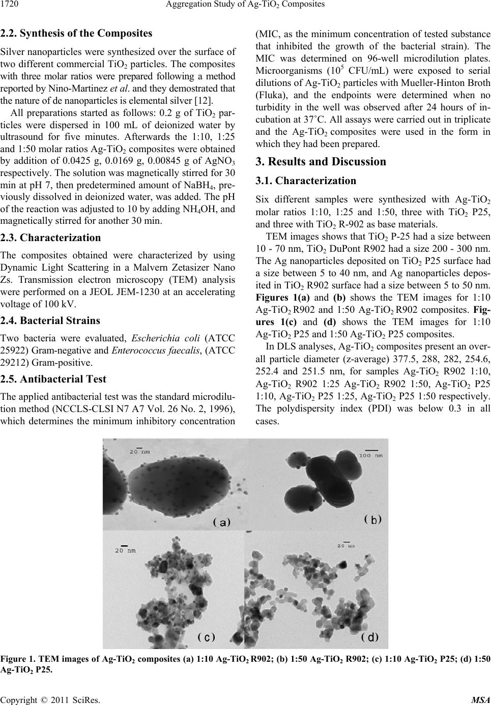 Aggregation Study of Ag-TiO Composites 1720 2 2.2. Synthesis of the Composites Silver nanoparticles were synthesized over the surface of two different commercial TiO2 particles. The composites with three molar ratios were prepared following a method reported by Nino-Martinez et al. and they demostrated that the nature of de nanoparticles is elemental silver [12]. All preparations started as follows: 0.2 g of TiO2 par- ticles were dispersed in 100 mL of deionized water by ultrasound for five minutes. Afterwards the 1:10, 1:25 and 1:50 molar ratios Ag-TiO2 composites were obtained by addition of 0.0425 g, 0.0169 g, 0.00845 g of AgNO3 respectively. The solution was magnetically stirred for 30 min at pH 7, then predetermined amount of NaBH4, pre- viously dissolved in deionized water, was added. The pH of the reaction was adjusted to 10 by adding NH4OH, and magnetically stirred for another 30 min. 2.3. Characterization The composites obtained were characterized by using Dynamic Light Scattering in a Malvern Zetasizer Nano Zs. Transmission electron microscopy (TEM) analysis were performed on a JEOL JEM-1230 at an accelerating voltage of 100 kV. 2.4. Bacterial Strains Two bacteria were evaluated, Escherichia coli (ATCC 25922) Gram-negative and Enterococcus faecalis, (ATCC 29212) Gram-positive. 2.5. Antibacterial Test The applied antibacterial test was the standard microdilu- tion method (NCCLS-CLSI N7 A7 Vol. 26 No. 2, 1996), which determines the minimum inhibitory concentration (MIC, as the minimum concentration of tested substance that inhibited the growth of the bacterial strain). The MIC was determined on 96-well microdilution plates. Microorganisms (105 CFU/mL) were exposed to serial dilutions of Ag-TiO2 particles with Mueller-Hinton Broth (Fluka), and the endpoints were determined when no turbidity in the well was observed after 24 hours of in- cubation at 37˚C. All assays were carried out in triplicate and the Ag-TiO2 composites were used in the form in which they had been prepared. 3. Results and Discussion 3.1. Characterization Six different samples were synthesized with Ag-TiO2 molar ratios 1:10, 1:25 and 1:50, three with TiO2 P25, and three with TiO2 R-902 as base materials. TEM images shows that TiO2 P-25 had a size between 10 - 70 nm, TiO2 DuPont R902 had a size 200 - 300 nm. The Ag nanoparticles deposited on TiO2 P25 surface had a size between 5 to 40 nm, and Ag nanoparticles depos- ited in TiO2 R902 surface had a size between 5 to 50 nm. Figures 1(a) and (b) shows the TEM images for 1:10 Ag-TiO2 R902 and 1:50 Ag-TiO2 R902 composites. Fig- ures 1(c) and (d) shows the TEM images for 1:10 Ag-TiO2 P25 and 1:50 Ag-TiO2 P25 composites. In DLS analyses, Ag-TiO2 composites present an over- all particle diameter (z-average) 377.5, 288, 282, 254.6, 252.4 and 251.5 nm, for samples Ag-TiO2 R902 1:10, Ag-TiO2 R902 1:25 Ag-TiO2 R902 1:50, Ag-TiO2 P25 1:10, Ag-TiO2 P25 1:25, Ag-TiO2 P25 1:50 respectively. The polydispersity index (PDI) was below 0.3 in all cases. ( ) ( ) ( ) ( ) Figure 1. TEM images of Ag-TiO2 composites (a) 1:10 Ag-TiO2 R902; (b) 1:50 Ag-TiO2 R902; (c) 1:10 Ag-TiO2 P25; (d) 1:50 Ag-TiO2 P25. Copyright © 2011 SciRes. MSA  Aggregation Study of Ag-TiO Composites1721 2 The zeta potential of the same samples were –58.8 ± 6.92, –47.8 ± 6.03, –46.78 ± 5.9, –37.7.8 ± 4.8, –49.8 ± 5.89 and –58.8 ± 6.92. Figures 2(a) and (b) show the DLS results for 1:10 Ag-TiO2 R902 and 1:50 Ag-TiO2 R902 composites. Figures 2(c) and (d) show the DLS results for 1:10 Ag-TiO2 P25 and 1:50 Ag-TiO2 P25 composites. Acording to the DVLO Theory, the stability of particle dispersions depends on the balance between attractive and repulsive forces between the particles. With electro- static stabilization, the zeta potential of the particles pro- vides a repulsive force. In practice if the zeta potential of the particles is higher than 30 mV or lower than −30 mV the dispersion is stable [19]. To evaluate the evolution of the Ag-TiO2 particle size (z-average) there were realized three dilutions of each of the six synthesized composites 100 µg/ml, 250 µg/mL and 500 µg/ml in deionized water and Mueller Hinton broth. It is interesting to note that the particles size re- mained stable in deionized water for about 24 minutes, 235 - 290 nm for Ag-TiO2 R902 1:10 (Figure 3(a)), 240 - 290 for Ag-TiO2 R902 1:25, 257 - 285 nm for Ag-TiO2 R902 1:50, 170 - 190 nm for Ag-TiO2 P25 1:10, 160 - 200 nm for Ag-TiO2 P25 1:25 and 190 - 210 nm f Ag-TiO2 P25 1:50. In contrast the particles became larger in Mueller Hinton Broth but once they grew in size, they remained constant for about 24 minutes, 280 - 360 nm for Ag-TiO2 R 9021:10 (Figure 3(b)), 260 - 360 nm for Ag-TiO2 R 902 1:25, 260 - 350 nm for Ag-TiO2 R 902 1:50,190 - 215 nm for Ag-TiO2 P25 1:10, 180 - 220 nm for Ag-TiO2 P25 1:25 and 210 - 240 nm for TiO2 P25 1:50. The size of de Ag-TiO2 composites was influenced by the TiO2 base materials in both media. Steric stabilization is used for nanoparticle dispersion stabilization, where a stabilizer is added to the dispersion and it is adsorbed onto the particle surface, preventing them from coming close to one another [16-19]. It is be- lieved that nanoparticles are covered by proteins imme- diately upon contact with a cell culture media and physiological environment, resulting in a protein ar- rangement also referred to as protein corona on the parti- cle surface [16] and this protein corona is exchanging with other nearby proteins [20]. Mueller-Hinton Broth is a complex system containing a lot of different proteins. Therefore, it is possible that Ag-TiO2 composite adsorb proteins from Mueller-Hinton Broth, as indicated by an increase in the particle size (z-average), which could make them become more stable. The identification of the protein corona composition was not the focus of this work. 3.2. Antibacterial Results Minimum inhibitory concentration values were obtained (Table 1). The six different samples have antibacterial activity. The TiO2 particles present no antibacterial activ- ity. The test was performed on dark and it is reported that TiO2 particles in dark condition present no antibacterial activity [12,15] which is consistent with our results. The six composites show antibacterial activity without light. We evaluated the antibacterial activity after 24 minutes of exposure of the Ag-TiO2 composites in Mueller-Hin- ton Broth, and this did not affect the MIC for the micro- organisms involved. According to Lynch and coworkers [20] if the protein corona exchanges with other proteins in the medium faster than the time it takes for the particle to attach to the bacteria surface, then the particle-bacteria interactions will not be greatly affected by the presence of the corona. Therefore the MIC of the composite would not be affected. We found the best results in Ag-TiO2 1:10 composite. Figure 2. DLS results (a) 1:10 Ag-TiO2 R902; (b) 1:50 Ag-TiO2 R902; (c) 1:10 Ag-TiO2 P25; (d) 1:50 Ag-TiO2 P25. Copyright © 2011 SciRes. MSA  Aggregation Study of Ag-TiO Composites 1722 2 (a) (b) Figure 3. Temporal evolution of the Ag-TiO2 DuPont 1:10 composite size (z-average): (a) deionized water; (b) Mueller Hinton Broth. Table 1. Minimu n inhibitory concentr ation of Ag-TiO2 nano- particles against E. coli and E. faecalis. Material MIC (µg/mL) E. coli (Gram-negative) E. faecalis (Gram-positive) Ag-TiO2 1:10 (P25) 500 ± 0 500 ± 0a 500 ± 0 500 ± 0a Ag-TiO2 1:10 (R902) 500 ± 0 500 ± 0a 375 ± 144.3 375 ± 144.3a Ag-TiO2 1:25 (P25) 500 ± 0 500 ± 0a 1000 ± 0 1000 ± 0a Ag-TiO2 1:25 (R902) 500 ± 0 500 ± 0a 1000 ± 0 1000 ± 0a Ag-TiO2 1:50 (P25) 1000 ± 0 1000 ± 0a 1000 ± 0 1000 ± 0a Ag-TiO2 1:50 (R902) 1000± 0 1000 ± 0a 1000 ± 0 1000 ± 0a TiO2 (P25) TiO2 (R902) >2000b >2000b >2000b >2000b 4. Conclusions Six different molar ratios Ag-TiO2 composites were pre- pared and characterized. We found that Ag-TiO2 com- posites increased in size with respect to deionized water when be diluted in Muller-Hinton Broth, but once they grew in size, they remained constant for 24 minutes, and did not affect the MIC for the microorganisms involved. The Ag-TiO2 1:10 composites showed promising results as an antibacterial agent against E. faecalis Gram-posi- tive and E. coli. Gram-negative bacteria. 5. Acknowledgements M. E. Noriega-Treviño, J. E. Morales-Sánchez and J. M. Guajardo-Pacheco would like to thank CONACYT for grant of scholarships. REFERENCES [1] G. A. Martinez-Castanon, N. Nino-Martinez, J. P. Loyola- Rodriguez, N. Patino-Marin, J. R. Martinez-Mendoza and F. Ruiz, “Synthesis of Silver Particles with Different Sizes and Morphologies,” Materials Letters, Vol. 63, No. 15, 2009, pp. 1266-1268. doi:10.1016/j.matlet.2009.02.061 [2] A. Panacek, L. Kvitek, R. Prucek, M. Kolar, R. Vecerova, N. Pizurova, V. Sharma, T. Nevecna and R. Zboril, “Sil- ver Colloid Nanoparticles: Synthesis, Characterization, and Their Antibacterial Activity,” Journal of Physical Chemistry B, Vol. 110, No. 33, 2006, pp. 16248-16253. doi:10.1021/jp063826h [3] C. Marambio-Jones and E. M. V. Hoek, “A Review of the Antibacterial Effects of Silver Nanomaterials and Poten- Copyright © 2011 SciRes. MSA  Aggregation Study of Ag-TiO Composites1723 2 tial Implications for Human Health and the Environ- ment,” Journal of Nanoparticle Research, Vol. 12, No. 5, 2010, pp. 1531-1551. doi:10.1007/s11051-010-9900-y [4] O. Choi and Z. Hu, “Size Dependent and Reactive Oxy- gen Species Related Nanosilver Toxicity to Nitrifying Bacteria,” Environmental Science & Technology, Vol. 42, No. 12, 2008, pp. 4583-4588. doi:10.1021/es703238h [5] G. A. Martinez-Castanon, N. Nino-Martinez, F. Martinez- Gutierrez, J. R. Martinez-Mendoza and F. Ruiz, “Synthe- sis and Antibacterial Activity of Silver Nanoparticles with Different Sizes,” Journal of Nanoparticle Research, Vol. 10, No. 8, 2008, pp. 1343-1348. doi:10.1007/s11051-008-9428-6 [6] A. Smetana, K. Klabunde, G. Marchin and C. Sorensen, “Biocidal Activity of Nanocrystalline Silver Powders and Particles,” Langmuir, Vol. 24, No. 14, 2008, pp. 7457- 7464. doi:10.1021/la800091y [7] S. Pal, Y. Tak and J. Song, “Does the Antibacterial Ac- tivity of Silver Nanoparticles Depend on the Shape of the Nanoparticle? A Study of the Gram-Negative Bacterium Escherichia coli,” Applied and Environmental Microbi- ology, Vol. 73, No. 6, 2007, pp. 1712-1720. doi:10.1128/AEM.02218-06 [8] J. R. Morones, J. L. Elechiguerra, A. Camacho, K. Holt, J. B. Kouri, J. T. Ramirez and M. J. Yacaman, “The Bacte- ricidal Effect of Silver Nanoparticles,” Nanotechnology, Vol. 16, No. 10, 2005, pp. 2346-2353. doi:10.1088/0957-4484/16/10/059 [9] L. Kvitek, A. Panacek, J. Soukupova, M. Kolar, R. Ve- cerova, R. Prucek, M. Holecova and R. Zboril, “Effect of Surfactants and Polymers on Stability and Antibacterial Activity of Silver Nanoparticles (NPs),” Journal of Phy- sical Chem is try C, Vol. 112, No. 15, 2008, pp. 5825-5834. doi:10.1021/jp711616v [10] S. Shrivastava, T. Bera, A. Roy, G. Singh, P. Ramachandra- rao and D. Dash, “Characterization of Enhanced Antibac- terial Effects of Novel Silver Nanoparticles,” Nanotech- nology, Vol. 18, No. 22, 2007, pp. 225103-225111. doi:10.1088/0957-4484/18/22/225103 [11] K. Page, R. G. Palgrave, I. P. Parkin, M. Wilson, S. L. P. Savin and A. V. Chadwick, “Titania and Silver-Titania Composite Films on Glass-Potent Antimicrobial Coat- ings,” Journal of Materials Chemistry, Vol. 17, No. 35, 2007, pp. 95-104. doi:10.1039/b611740f [12] N. Nino-Martinez, G. A. Martinez-Castanon, A. Aragon- Pina, F. Martinez-Gutierrez, J. R. Martinez-Mendoza and F. Ruiz, “Characterization of Silver Nanoparticles Synthe- sized on Titanium Dioxide Fine Particles,” Nanotechnol- ogy, Vol. 19, No. 6, 2008, Article ID: 065711. doi:10.1088/0957-4484/19/6/065711 [13] G. Fu, P. S. Vary and C. Lin, “Anatase TiO 2 Nanocom- posites for Antimicrobial Coatings,” Journal of Physical Chemistry B, Vol. 109, No. 18, 2005, pp. 8889-8898. doi:10.1021/jp0502196 [14] J. Thiel, L. Pakstis, S. Buzby, M. Raffi, C. Ni, D. J. Pochan and S. I. Shah, “Antibacterial Properties of Silver- Doped Titania,” Small, Vol. 3, No. 5, 2007, pp. 799-803. doi:10.1002/smll.200600481 [15] M. Yeo and M. Kang, “Effects of Nanometer Sized Silver Materials on Biological Toxicity during Zebrafish Em- bryogenesis,” Bulletin of the Korean Chemical Society, Vol. 29, No. 6, 2008, pp. 1179-1184. doi:10.5012/bkcs.2008.29.6.1179 [16] P. Bihari, M. Vippola, S. Schultes, M. Praetner, A. Khan- doga, C. A. Reichel, C. Coester T. Tuomi, M. Rehberg and F. Krombach, “Optimized Dispersion of Nanoparticles for Biological in Vitro and in Vivo Studies,” Particle and Fi- bre Toxicology, Vol. 5, No. 14, 2008, pp. 1-14. [17] R. MacCuspie, “Colloidal Stability of Silver Nanoparti- cles in Biologically Relevant Conditions,” Journal of Nanoparticle Research, Vol. 13, No. 7, 2011, pp. 2893- 2908. doi:10.1007/s11051-010-0178-x [18] S. Kitller, G. Greulich, J. S. Gebauer, J. Diendorf, L. Treuel, L. Ruiz, J. M. G. Calbet, M. Vallet-Regi, R. Zell- ner, M. Köller and M. Epple, “The Infuence of Proteins on the Dispersability and Cell-Biollogical Activity of Silver Nanoparticles,” Journal of Materials Chemistry, Vol. 20, No. 3, 2010, pp. 512-518. doi:10.1039/b914875b [19] Z. Ji, X. Jin, S. George, T. Xia, H. Meng, X. Wang, E. Suarez, H. Zhang, E. M. H. Hoek, H. Godwin, A. E. Nel and J. I. Zink, “Dispersion and Stability Optimization of TiO2. Nanoparticles in Cell Culture Media,” Environ- mental Science & Technology, Vol. 44, No. 19, 2010, pp. 7309-7314. doi:10.1021/es100417s [20] I. Lynch, A. Salvati and K. A. Dawson, “Protein-Nano- particle Interactions. What Does the Cell See?” Nature Nanotechnology, Vol. 4, No. 9, 2009, pp. 546-547. doi:10.1038/nnano.2009.248 Copyright © 2011 SciRes. MSA |