

Paper Menu >>
Journal Menu >>
 Advances in Infectious Diseases, 2011, 1, 27-28 doi:10.4236/aid.2011.12004 Published Online December 2011 (http://www.SciRP.org/journal/aid) Copyright © 2011 SciRes. AID 27 Bacteremi a with Cuta neo us Nodules, Due to Pseudomonas Aerugi nosa Ali Akbar Heydari1, Maryam Mojtabavi2 1Imam Reza Ho spi tal, Mashhad Universi ty of Medical Scien ces, Mashhad, Iran; 2Mashhad Uni versit y of Medical S cien ces, Mashhad, Iran. Email: [email protected] Received November 1st, 2011; r evised D ecember 2nd , 201 1; accepted December 10th, 2011. ABSTRACT Pseudomonas aeruginosa bacteremia rarely occurs in non-immunocompromised adults and can be difficult to be treated. We report a case of 35-year-old woman who presented with respiratory distress, fever and skin lesions. There was pleural effusion in chest radiograph, and CT scan of thorax showed necrotizing pneumonia and loculated empyema. Among the cultures of blood, empyema, sputum and the skin nodules aspirates, all were positive for Pseudomonas ae- ruginosa. The patient was treated with ciprofloxacin, amikacin and then pleural decortications. Laboratory tests failed to reveal any immunological deficits. After a period of 21 days of antibiotic therapy, the patient was discharged from hospital with a good condition. Keywords: Pseudomonas, Aeruginosa, Bacteremia, Cutaneous Nodules. 1. Introduction Pseudomonas aeruginosa (PA) is an opportunistic patho- gen which rarely causes a disease in healthy persons, ei- ther children or adults [1-3]. These infections often occur in patients with underlying conditions. Our patient pre- sented with Pseudomonas aeruginosa bacteremia, empy- ma, lung abscess and the rare form of metastatic lesion (skin nodul es). 2. Case Presentation A 35 -years-old female was ad mitted to the Infectio us Dis - eases department, for respiratory distress. The illnes s had begun 18 days earlier with abdominal pain in association with pleuretic-type flank pain, which were accompanied by fever and rigors. Following aggravation of the ab- dominal pain, laparotomy was performed with the prob- able diagnosis of appendicitis, but there were no abnor- mal findings in the abdomen. Her fever was continued despite antimicrobial therapy and her condition was wo r- sened progressively. She was ill and her extremities were cyanotic and there were multiple subcutaneous scalded nodules (Fig ur e 1) on he r limbs. There was no history of previous disease including human immunodeficienc y vi rus infec t ion. High resolution co mputed tomo graphy o f the chest was performed, and it showed an abscess in apical-posterior Figure 1. Subcut aneous nodule i n a patie nt w ith pseudomo- nas aerog i nos a bacteremia. segment of right lower lobe, with louculated pleural ef- fusion. Among the cultures of blood, empeyma, sputum, and the skin nodules aspirates, all were positive for pseu- domonas aeruginosa. 3. Discussion The onl y poi nt di ffe re nti ati ng t his e nti ty fro m othe r ca use s of gram negative sepsis is the skin lesion known as ec- thyma gangrenosum (EG). Infection with the bacteria should be suspected in the pati ents who presented with EG but this lesio n is no t pathognomonic. 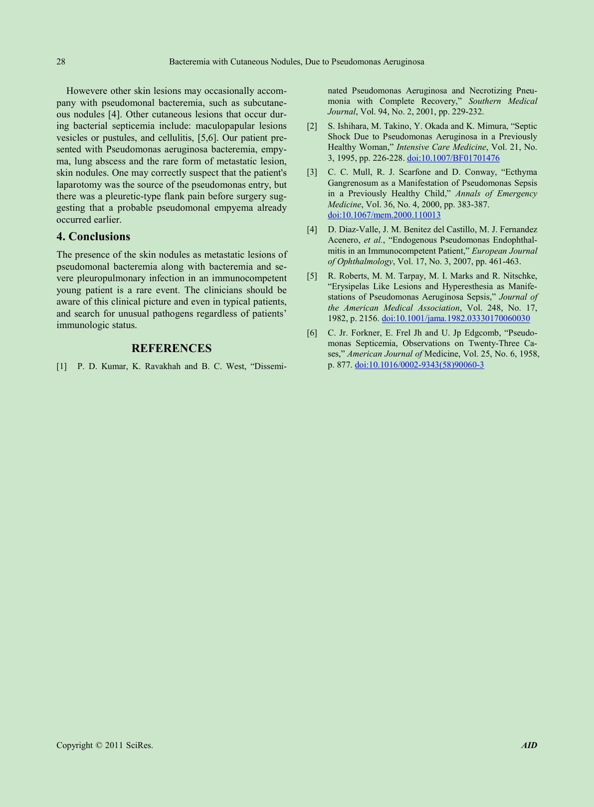 Bacteremia with Cutaneous Nodules, Due to Pseudomonas Aeruginosa Copyright © 2011 SciRes. AID 28 Howevere other skin lesions may occasionally accom- pany with pseudomonal bacteremia, such as subcutane- ous nodules [4]. Other cutaneous lesions that occur dur- ing bacterial septicemia include: maculopapular lesions vesicles or pustules, and cellulitis, [5,6]. Our patient pre- sented with Pseudomonas aeruginosa bacteremia, empy- ma, lung abscess and the rare form of metastatic lesion, skin no dule s. O ne ma y cor re ctl y suspe ct that the patient's laparotomy was the source of the pseudomonas entry, but there was a pleuretic-type flank pain before surgery sug- gesting that a probable pseudomonal empyema already occurred earlier. 4. Conclusions The presence of the skin nodules as metastatic lesions o f pseudomonal bacteremia along with bacteremia and se- vere pleuropulmonary infection in an immunocompetent young patient is a rare event. The clinicians should be aware of this clinical picture and even in typical patients, and search for unusual pathogens regardless of patients’ immu nol o gic s tatus. REFERENCES [1] P. D. Kumar, K. Ravakhah and B. C. West, “Dissemi- nated Pseudomonas Aeruginosa and Necrotizing Pneu- monia with Complete Recovery, ” Southern Medical Journal, Vol . 94, No. 2, 20 01 , pp. 229-232. [2] S. Ish ih ara, M . Takin o, Y. Okad a and K. Mimura, “Sep ti c Shock Due to Pseudomonas Aeruginosa in a Previously Healthy Woman,” Intensive Care Medicine, Vol. 21, No. 3, 1995, pp. 226-228. doi:10.1007/BF01701476 [3] C. C. Mull, R. J. Scarfone and D. Conway, “Ecthyma Gangrenosum as a Manifestation of Pseudomonas Sepsis in a Previously Healthy Child,” Annals of Emergency Medicine, Vol. 36, No. 4, 2000, pp. 383-387. doi:10.1067/mem.2000.110013 [4] D. Diaz-V all e, J. M . Benitez del Castillo, M. J. Fernandez Acenero, et al., “Endogenous Pseudomonas Endophthal- mitis in an Immunocompetent Patient,” European Journal of O phthalmology , Vol. 17, No. 3, 2007, pp. 461-463. [5] R. Roberts, M. M. Tarpay, M. I. Marks and R. Nitschke, “Erysipelas Like Lesions and Hyperesthesia as Manife- stations of Pseudomonas Aeruginosa Sepsis,” Journal of the American Medical Association, Vol. 248, No. 17, 1982, p. 2156. doi:10.1001/jama.1982.03330170060030 [6] C. Jr. Forkner, E. Frel Jh and U. Jp Edgcomb, “Pseudo- monas Septicemia, Obser vat ions on Twen ty-Three Ca- ses,” Amer ican Journal of Medicine, Vol. 25, No. 6, 1958, p. 877. doi:1 0.1016 /0002-9343(58)90060-3 |