

Paper Menu >>
Journal Menu >>
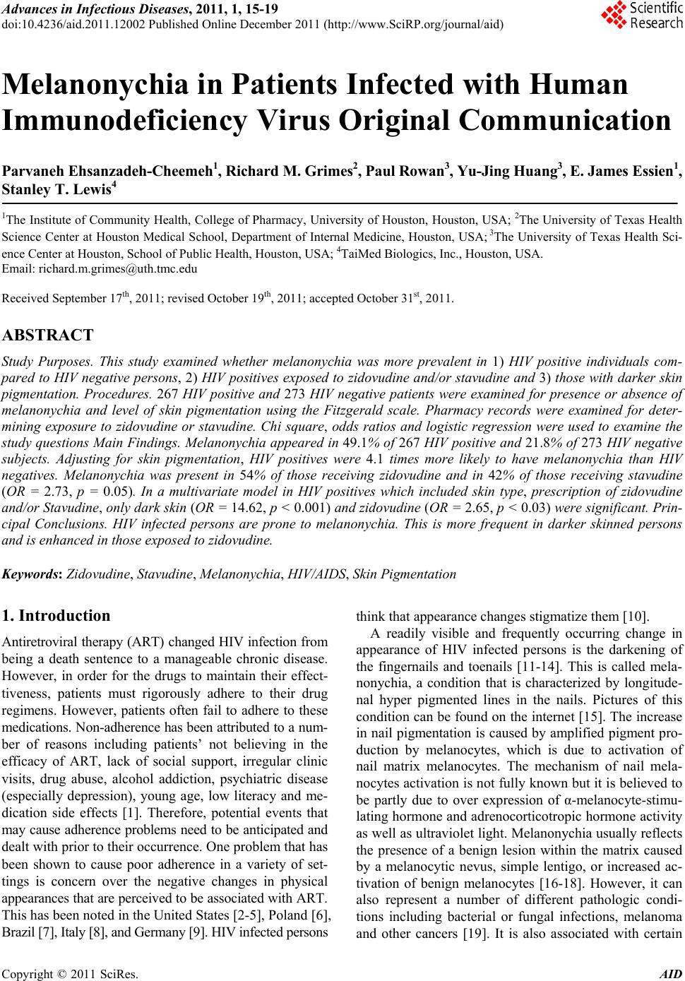 Advances in Infectious Diseases, 2011, 1, 15-19 doi:10.4236/aid.2011.12002 Published Online December 2011 (http://www.SciRP.org/journal/aid) Copyright © 2011 SciRes. AID 15 Melanonychia in Patients Infected with Human Immunodeficiency Virus Original Communication Parvaneh Ehsanzadeh-Cheemeh1, Richard M. Grimes2, Paul Rowan3, Yu-Jing Huang3, E. James Essien1, Stanley T. Lewis4 1The Institute of Community Health, College of Pharmacy, University of Houston, Houston, USA; 2The University of Texas Health Science Center at Houston Medical School, Department of Internal Medicine, Houston, USA; 3The University of Texas Health Sci- ence Center at Houston, School of Public Health, Houston, USA; 4TaiMed Biologics, Inc., Houston, USA. Email: [email protected] Received September 17th, 2011; revised October 19th, 2011; accepted October 31st, 2011. ABSTRACT Study Purposes. This study examined whether melanonychia was more prevalent in 1) HIV positive individuals com- pared to HIV negative persons, 2) HIV positives exposed to zidovudine and/or stavudine and 3) those with darker skin pigmentation. Procedures. 267 HIV positive and 273 HIV negative patients were examined for presence or absence of melanonychia and level of skin pigmentation using the Fitzgerald scale. Pharmacy records were examined for deter- mining exposure to zidovudine or stavudine. Chi square, odds ratios and logistic regression were used to examine the study questions Main Findings. Melanonychia appeared in 49.1% of 267 HIV positive an d 21.8% of 273 HIV negative subjects. Adjusting for skin pigmentation, HIV positives were 4.1 times more likely to have melanonychia than HIV negatives. Melanonychia was present in 54% of those receiving zidovudine and in 42% of those receiving stavudine (OR = 2.73, p = 0.05). In a multivariate model in HIV positives which included skin type, prescription of zidovudine and/or Stavudine, only dark skin (OR = 14.62, p < 0.001) an d zidovudine (OR = 2.65, p < 0.03) were significant. Prin- cipal Conclusions. HIV infected persons are prone to melanonychia. This is more frequent in darker skinned persons and is enhanced in those exposed to zidovudine. Keywords: Zidovudine, St avudine, Melanonychia, HIV/AIDS, Skin Pigmentation 1. Introduction Antiretroviral therapy (ART) changed HIV infection from being a death sentence to a manageable chronic disease. However, in order for the drugs to maintain their effect- tiveness, patients must rigorously adhere to their drug regimens. However, patients often fail to adhere to these medications. Non-adherence has been attributed to a num- ber of reasons including patients’ not believing in the efficacy of ART, lack of social support, irregular clinic visits, drug abuse, alcohol addiction, psychiatric disease (especially depression), young age, low literacy and me- dication side effects [1]. Therefore, potential events that may cause adherence problems need to be anticipated and dealt with prior to their occurrence. One problem that has been shown to cause poor adherence in a variety of set- tings is concern over the negative changes in physical appearances that are perceived to be associated with ART. This has been noted in the United States [2-5], Poland [6], Brazil [7], Italy [8], and Germany [9]. HIV infected persons think that appearance changes stigmatize them [10]. A readily visible and frequently occurring change in appearance of HIV infected persons is the darkening of the fingernails and toenails [11-14]. This is called mela- nonychia, a condition that is characterized by longitude- nal hyper pigmented lines in the nails. Pictures of this condition can be found on the internet [15]. The increase in nail pigmentation is caused by amplified pigment pro- duction by melanocytes, which is due to activation of nail matrix melanocytes. The mechanism of nail mela- nocytes activation is not fully known but it is believed to be partly due to over expression of α-melanocyte-stimu- lating hormone and adrenocorticotropic hormone activity as well as ultraviolet light. Melanonychia usually reflects the presence of a benign lesion within the matrix caused by a melanocytic nevus, simple lentigo, or increased ac- tivation of benign melanocytes [16-18]. However, it can also represent a number of different pathologic condi- tions including bacterial or fungal infections, melanoma and other cancers [19]. It is also associated with certain 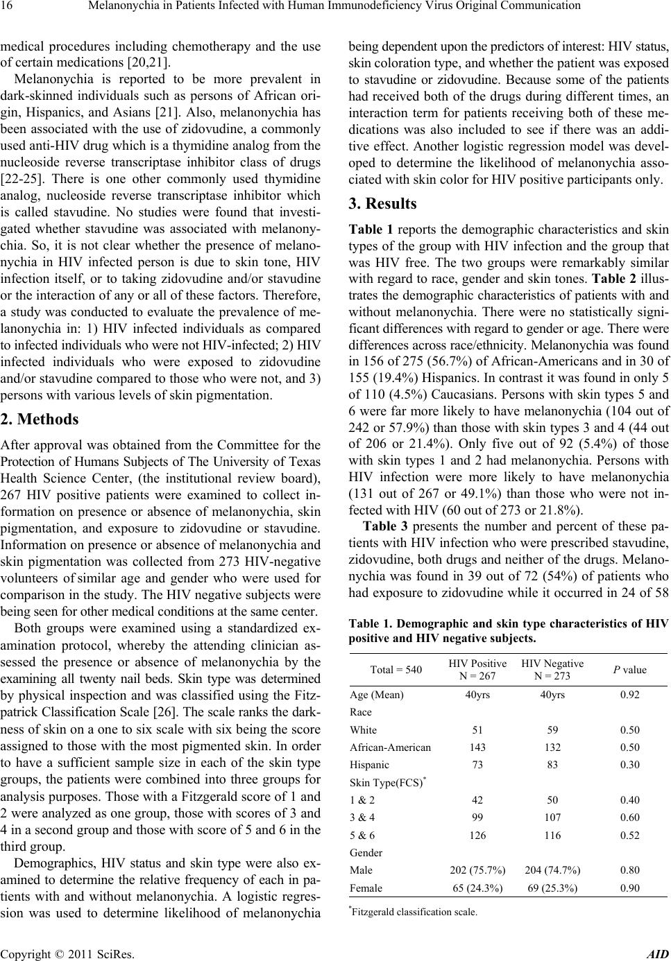 Melanonychia in Patients Infected with Human Immunodeficiency Virus Original Communication 16 medical procedures including chemotherapy and the use of certain medications [20,21]. Melanonychia is reported to be more prevalent in dark-skinned individuals such as persons of African ori- gin, Hispanics, and Asians [21]. Also, melanonychia has been associated with the use of zidovudine, a commonly used anti-HIV drug which is a thymidine analog from the nucleoside reverse transcriptase inhibitor class of drugs [22-25]. There is one other commonly used thymidine analog, nucleoside reverse transcriptase inhibitor which is called stavudine. No studies were found that investi- gated whether stavudine was associated with melanony- chia. So, it is not clear whether the presence of melano- nychia in HIV infected person is due to skin tone, HIV infection itself, or to taking zidovudine and/or stavudine or the interaction of any or all of these factors. Therefore, a study was conducted to evaluate the prevalence of me- lanonychia in: 1) HIV infected individuals as compared to infected individuals who were not HIV-infected; 2) HIV infected individuals who were exposed to zidovudine and/or stavudine compared to those who were not, and 3) persons with various levels of skin pigmentation. 2. Methods After approval was obtained from the Committee for the Protection of Humans Subjects of The University of Texas Health Science Center, (the institutional review board), 267 HIV positive patients were examined to collect in- formation on presence or absence of melanonychia, skin pigmentation, and exposure to zidovudine or stavudine. Information on presence or absence of melanonychia and skin pigmentation was collected from 273 HIV-negative volunteers of similar age and gender who were used for comparison in the study. The HIV negative subjects were being seen for other medical conditions at the same center. Both groups were examined using a standardized ex- amination protocol, whereby the attending clinician as- sessed the presence or absence of melanonychia by the examining all twenty nail beds. Skin type was determined by physical inspection and was classified using the Fitz- patrick Classification Scale [26]. The scale ranks the dark- ness of skin on a one to six scale with six being the score assigned to those with the most pigmented skin. In order to have a sufficient sample size in each of the skin type groups, the patients were combined into three groups for analysis purposes. Those with a Fitzgerald score of 1 and 2 were analyzed as one group, those with scores of 3 and 4 in a second group and those with score of 5 and 6 in the third group. Demographics, HIV status and skin type were also ex- amined to determine the relative frequency of each in pa- tients with and without melanonychia. A logistic regres- sion was used to determine likelihood of melanonychia being dependent upon the predictors of interest: HIV status, skin coloration type, and whether the patient was exposed to stavudine or zidovudine. Because some of the patients had received both of the drugs during different times, an interaction term for patients receiving both of these me- dications was also included to see if there was an addi- tive effect. Another logistic regression model was devel- oped to determine the likelihood of melanonychia asso- ciated with skin color for HIV positive participants only. 3. Results Table 1 reports the demographic characteristics and skin types of the group with HIV infection and the group that was HIV free. The two groups were remarkably similar with regard to race, gender and skin tones. Table 2 illus- trates the demographic characteristics of patients with and without melanonychia. There were no statistically signi- ficant differences with regard to gender or age. There were differences across race/ethnicity. Melanonychia was found in 156 of 275 (56.7%) of African-Americans and in 30 of 155 (19.4%) Hispanics. In contrast it was found in only 5 of 110 (4.5%) Caucasians. Persons with skin types 5 and 6 were far more likely to have melanonychia (104 out of 242 or 57.9%) than those with skin types 3 and 4 (44 out of 206 or 21.4%). Only five out of 92 (5.4%) of those with skin types 1 and 2 had melanonychia. Persons with HIV infection were more likely to have melanonychia (131 out of 267 or 49.1%) than those who were not in- fected with HIV (60 out of 273 or 21.8%). Table 3 presents the number and percent of these pa- tients with HIV infection who were prescribed stavudine, zidovudine, both drugs and neither of the drugs. Melano- nychia was found in 39 out of 72 (54%) of patients who had exposure to zidovudine while it occurred in 24 of 58 Table 1. Demographic and skin type characteristics of HIV positive and HIV negative subjects. Total = 540 HIV Positive N = 267 HIV Negative N = 273 P value Age (Mean) 40yrs 40yrs 0.92 Race White 51 59 0.50 African-American 143 132 0.50 Hispanic 73 83 0.30 Skin Type(FCS)* 1 & 2 42 50 0.40 3 & 4 99 107 0.60 5 & 6 126 116 0.52 Gender Male 202 (75.7%) 204 (74.7%) 0.80 Female 65 (24.3%)69 (25.3%) 0.90 *Fitzgerald classification scale. Copyright © 2011 SciRes. AID 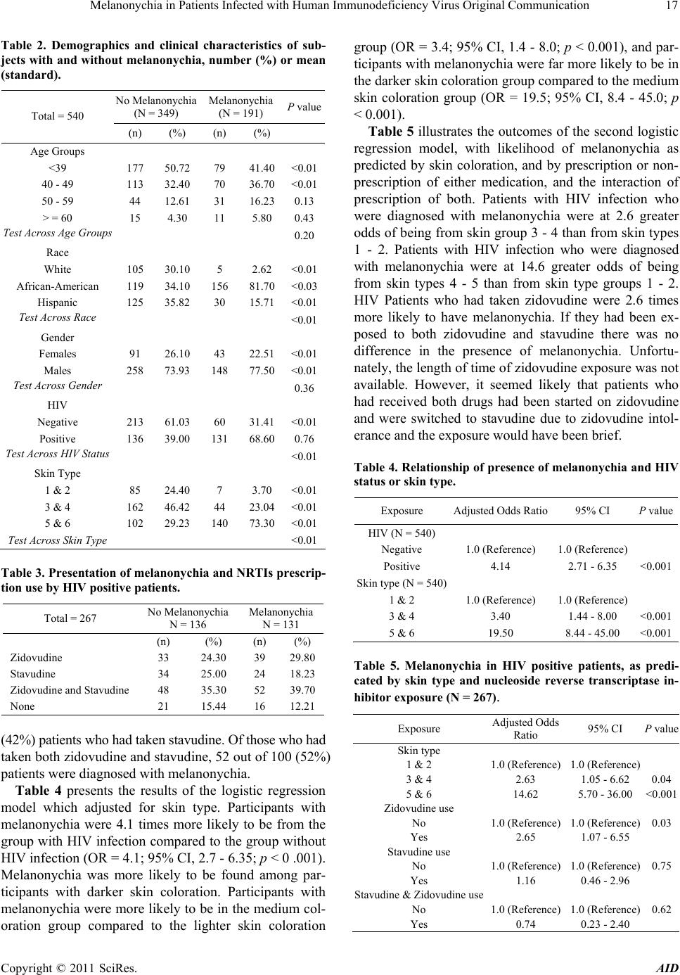 Melanonychia in Patients Infected with Human Immunodeficiency Virus Original Communication17 Table 2. Demographics and clinical characteristics of sub- jects with and without melanonychia, number (%) or mean (standard). No Melanonychia (N = 349) Melanonychia (N = 191) P value Total = 540 (n) (%) (n) (%) Age Groups <39 177 50.72 79 41.40<0.01 40 - 49 113 32.40 70 36.70<0.01 50 - 59 44 12.61 31 16.230.13 > = 60 15 4.30 11 5.80 0.43 Test Across Age Groups 0.20 Race White 105 30.10 5 2.62 <0.01 African-American 119 34.10 156 81.70<0.03 Hispanic 125 35.82 30 15.71<0.01 Test Across Race <0.01 Gender Females 91 26.10 43 22.51<0.01 Males 258 73.93 148 77.50<0.01 Test Across Gender 0.36 HIV Negative 213 61.03 60 31.41<0.01 Positive 136 39.00 131 68.600.76 Test Across HIV Status <0.01 Skin Type 1 & 2 85 24.40 7 3.70 <0.01 3 & 4 162 46.42 44 23.04<0.01 5 & 6 102 29.23 140 73.30<0.01 Test Across Skin Type <0.01 Table 3. Presentation of melanonychia and NRTIs prescrip- tion use by HIV positive patients. Total = 267 No Melanonychia N = 136 Melanonychia N = 131 (n) (%) (n) (%) Zidovudine 33 24.30 39 29.80 Stavudine 34 25.00 24 18.23 Zidovudine and Stavudine 48 35.30 52 39.70 None 21 15.44 16 12.21 (42%) patients who had taken stavudine. Of those who had taken both zidovudine and stavudine, 52 out of 100 (52%) patients were diagnosed with melanonychia. Table 4 presents the results of the logistic regression model which adjusted for skin type. Participants with melanonychia were 4.1 times more likely to be from the group with HIV infection compared to the group without HIV infection (OR = 4.1; 95% CI, 2.7 - 6.35; p < 0 .001). Melanonychia was more likely to be found among par- ticipants with darker skin coloration. Participants with melanonychia were more likely to be in the medium col- oration group compared to the lighter skin coloration group (OR = 3.4; 95% CI, 1.4 - 8.0; p < 0.001), and par- ticipants with melanonychia were far more likely to be in the darker skin coloration group compared to the medium skin coloration group (OR = 19.5; 95% CI, 8.4 - 45.0; p < 0.001). Table 5 illustrates the outcomes of the second logistic regression model, with likelihood of melanonychia as predicted by skin coloration, and by prescription or non- prescription of either medication, and the interaction of prescription of both. Patients with HIV infection who were diagnosed with melanonychia were at 2.6 greater odds of being from skin group 3 - 4 than from skin types 1 - 2. Patients with HIV infection who were diagnosed with melanonychia were at 14.6 greater odds of being from skin types 4 - 5 than from skin type groups 1 - 2. HIV Patients who had taken zidovudine were 2.6 times more likely to have melanonychia. If they had been ex- posed to both zidovudine and stavudine there was no difference in the presence of melanonychia. Unfortu- nately, the length of time of zidovudine exposure was not available. However, it seemed likely that patients who had received both drugs had been started on zidovudine and were switched to stavudine due to zidovudine intol- erance and the exposure would have been brief. Table 4. Relationship of presence of melanonychia and HIV status or skin type. Exposure Adjusted Odds Ratio 95% CI P value HIV (N = 540) Negative 1.0 (Reference) 1.0 (Reference) Positive 4.14 2.71 - 6.35 <0.001 Skin type (N = 540) 1 & 2 1.0 (Reference) 1.0 (Reference) 3 & 4 3.40 1.44 - 8.00 <0.001 5 & 6 19.50 8.44 - 45.00 <0.001 Table 5. Melanonychia in HIV positive patients, as predi- cated by skin type and nucleoside reverse transcriptase in- hibitor exposure (N = 267). Exposure Adjusted Odds Ratio 95% CI P value Skin type 1 & 2 1.0 (Reference) 1.0 (Reference) 3 & 4 2.63 1.05 - 6.62 0.04 5 & 6 14.62 5.70 - 36.00 <0.001 Zidovudine use No 1.0 (Reference) 1.0 (Reference)0.03 Yes 2.65 1.07 - 6.55 Stavudine use No 1.0 (Reference) 1.0 (Reference)0.75 Yes 1.16 0.46 - 2.96 Stavudine & Zidovudine use No 1.0 (Reference) 1.0 (Reference)0.62 Yes 0.74 0.23 - 2.40 Copyright © 2011 SciRes. AID  Melanonychia in Patients Infected with Human Immunodeficiency Virus Original Communication 18 4. Discussion This study demonstrates that melanonychia is highly pre- valent in HIV infected individuals with pigmented skin. Given the much higher rate in HIV infected persons than in the uninfected, it is quite possible that it will occur after infection. As a result patients may connect it to their HIV infection or to their taking antiretroviral medications. Patients may be concerned that it is a serious medical condition and/or they may think that it is cosmetically displeasing. As a result patients may question the clini- cian as to its origin and cause. Reassurance as to its be- nign nature may help the patient to cope with their me- dical concerns. However, patients may discover that me- lanonychia has been associated with zidovudine use on the internet and/or through information provided by non- governmental organizations. If patients believe that zi- dovudine or other HIV medications are causing a disfig- urement of his or her nails, they may stop taking that medication with negative consequences to their overall well being and a reduction of their future treatment op- tions. Physicians should be alert to this possibility in pa- tients who raise questions about their melanonychia and should reassure patients that it has not been linked to any medication except zidovudine. If the patient is taking zi- dovudine there are many other medications that can be substituted for zidovudine (including stavudine). Because patients may link melanonychia to antiretrovi- ral therapy (even if they are not taking zidovudine) clini- cians may wish to forewarn their patients that nail changes might may occur and that it is not a reason to stop taking their antiretrovirals. In forewarning or reas- suring them, clinicians should emphasize that the condi- tion is common in those who not HIV infected (21.8% of those who were not infected in our study) and so its ap- pearance should not be seen as something that will reveal their HIV status nor is it a reasons to stop taking their an- tiretrovirals. If a patient who is taking zidovudine has the condition and is concerned about it and insists on chang- ing medications they should be made aware that it will take a long time for the lesions to disappear. Clinicians should also recognize that while melanony- chia is almost always a benign condition that is idiopa- thic in origin, it is occasionally linked to more serious con- ditions, particularly melanoma. Tosti et al. [20] offered several clinical clues as to whether melanonychia requires additional investigation for the presence of melanoma. These were 1) discoloration bands in the nails that are wider than 3mm and have irregular borders; 2) extension of the band into the proximal and/or lateral nail fold (Hutchinson’s sign); 3) a lesion with a triangular shape; 4) nails that are split or have fissures; and 5) if the pigmen- tation is not homogenous. Given the extremely high prevalence of melanonychia in these HIV positive patients (>49%) clinicians might consider its presence as a potential clue to HIV positivity in previously untested patients. Alone or together with other signs or patient history, it might induce a discus- sion of the need to rule out HIV infection. It is seldom that a clinician has such a visible sign that might be in- dicative of undiagnosed HIV infection. 5. Acknowledgements This study was supported by the Baylor-UT Houston Center for AIDS Research (CFAR), a program funded by the US National Institutes of Health (NIH) (AI036211). REFERENCES [1] A. K. Patel and K. K. Patel, “Future Implications: Com- pliance and Failure with Antiretroviral Treatment,” Jour- nal of Post-Graduate Medicine, Vol. 52, No. 3, 2006, pp. 197-200. [2] T. Hawkinsm, “Appearance-Related Side Effects of HIV-1 Treatment,” Aids Patient Care and STDs, Vol. 20, No. 1, 2006, pp. 6-18. doi:10.1089/apc.2006.20.6 [3] R. Power, H. L. Tate, S. M. McGill and C. Taylor, “A Qualitative Study of the Psychosocial Implications of Li- podystrophy Syndrome on HIV Positive Individuals,” Sexually Transmitted Infections, Vol. 4, No. 2, 2003, pp. 137-141. doi:10.1136/sti.79.2.137 [4] I. B. Corless, K. M. Kirksey, J. Kemppainen, et al., “Li- podystrophy-Associated Symptoms and Medication Ad- herence in HIV-AIDS,” AIDS Patient Care and STDs, Vol. 19, No. 9, 2005, pp. 577-586. doi:10.1089/apc.2005.19.577 [5] N. R. Reynolds, J. L. Neidig, A. W. Wu, A. L. Gifford and W. C. Holmes, “Balancing Disfigurement and Fear of Disease Progression: Patient Perceptions of HIV Body Fat Redistribution,” AIDS Care, Vol. 18, No. 7, 2006, pp. 663-673. doi:10.1080/09540120500287051 [6] D. Rogowska-Szadkowska, S. Chlabicz, M. A. Oltar- zewska and J. Sawicka-Powierza, “Which Factors Hinder the Decision of Polish HIV-Positive Patients to Take Up Antiretroviral Therapy?” AIDS Care, Vol. 21, No. 3, 2009, pp. 280-283. doi:10.1080/09540120802241871 [7] C. P. Santos, Y. X. Felipe, P. E. Braga, D. Ramos, R. O. Lima and A. C. Segurado, “Self-Perception of Body Changes in Persons Living with HIV/AIDS: Prevalence and Associated Factors,” AIDS, Vol. 19, 2005, pp. S14-21. doi:10.1097/01.aids.0000191485.92285.c7 [8] A. Ammassari, A. Antinor, A. Cozzi-Lepri, et al., “Rela- tionship between HAART Adherence and Adipose Tissue Alterations,” Journal of Acquired Immune Deficiency Syndromes and Human Retrovirology, Vol. 31, 2002, pp. S140-144. [9] M. Oette, P. Juretzko, A. Kroidl, et al., “Lipodystrophy Syndrome and Self-Assessment of Well-Being and Phy- sical Appearance in HIV-Positive Patients,” AIDS Patient Copyright © 2011 SciRes. AID  Melanonychia in Patients Infected with Human Immunodeficiency Virus Original Communication Copyright © 2011 SciRes. AID 19 Care and STDs, Vol. 16, No. 9, 2002, pp. 413-417. doi:10.1089/108729102760330254 [10] B. E. Berger, C. E. Ferrans and F. R. Lashley, “Measur- ing Stigma in People with HIV: Psychometric Assess- ment of the HIV Stigma Scale,” Research in Nursing & Health, Vol. 24, No. 6, 2001, p. 518. doi:10.1002/nur.10011 [11] B. Cribier, M. Mena, D. Rey, M. Partisani, V. Fabien, J. Lang, et al., “Nail Changes in Patients Infected with Hu- man Immunodeficiency Virus: A Prospective Controlled Study,” Archive of Dermatology, Vol. 134, No. 10, 1998, pp. 1216-1220. doi:10.1001/archderm.134.10.1216 [12] B. Fisher and L. Warner, “Cutaneous Manifestations of the Acquired Immunodeficiency Syndrome,” International Journal of Dermatology, Vol. 26, No. 6, 1987, pp. 615- 630. doi:10.1111/j.1365-4362.1987.tb02267.x [13] A. P. Panwalker, “Nail Pigmentation in the Acquired Immunodeficiency Syndrome,” Annals of Internal Medi- cine, Vol. 107, No. 6, 1987, pp. 943-944. [14] P. Chandrasekar, “Nail Discoloration and Human Immu- nodeficiency Virus Infection,” American Journal of Medicine, Vol. 86, No. 4, 1989, pp. 506-507. doi:10.1016/0002-9343(89)90365-3 [15] PicSearch. Pictures of Longitudinal Melanonychia, 2011. http://www.picsearch.com/pictures/Health/Cutaneous%20 conditions/Longitudinal%20melanonychia.html [16] K. J. Smith, H. G. Skelton, J. Yeager, R. Ledsky, W. McCarthy, D. Baxter, et al., “Cutaneous Findings in HIV-1-Positive Patients: A 42-Month Prospective Study, Military Medical Consortium for the Advancement of Retroviral Research,” Journal of the American Academy of Dermatology, Vol. 31, No. 5, 1994, pp. 746-754. doi:10.1016/S0190-9622(94)70236-5 [17] D. A. Glaser and K. Remlinger, “Blue Nails and Acquired Immunodeficiency Syndrome: Not Always Associated with Azidothymidine Use,” Cutis, Vol. 57, No. 4, 1996, pp. 243-244. [18] B. M. Piraccini, M. Iorizzo, A. Antonucci and A. Tosti, “Drug-I Induced Nail Abnormalities,” Expert Opinion on Drug Safety, No. 1, 2004, pp. 57-65. doi:10.1517/14740338.3.1.57 [19] E. Haneke and R. Baran, “Longitudinal Melanonychia,” Dermatologic Surgery, Vol. 27, No. 6, 2001, pp. 580-584. doi:10.1111/j.1524-4725.2001.01916.x [20] R. Baran and P. Kechijian, “Longitudinal Melanonychia (Melanonychia Striata): Diagnosis and Management,” Journal of the American Academy of Dermatology, Vol. 21, No. 6, 1989, pp. 1165-1175. doi:10.1016/S0190-9622(89)70324-8 [21] A. Tosti, Piraccini, M. Bianca and D. C. De Farias, “Dealing with Melanonychia, Seminars in Cutaneous,” Medicine and Surgery, Vol. 28, No. 1, 2009, pp. 49-54. [22] A. Tosti, G. Gaddoni, P. A. Fanti, A. D’Antuono and F. Albertini, “Longitudinal Melanonychia Induced by 3’- Azidodeoxythymidine: Report of 9 Cases,” Derma- tologica, Vol. 180, No. 4, 1990, pp. 217-220. doi:10.1159/000248033 [23] R. G. Greenberg and T. G. Berger, “Nail and Mucocuta- neous Hyperpigmentation with Azidothymidine Ther- apy,” Journal of the American Academy of Dermatology, Vol. 22, No. 2, 1990, pp. 327-330. doi:10.1016/0190-9622(90)70039-K [24] G. Rahav and S. Maayan, “Nail Pigmentation Associated with Zidovudine: A Review and Report of a Case,” Scan- dinavian Journal of Infectious Diseases, Vol. 24: No. 5, 1992, pp. 557-561. doi:10.3109/00365549209054640 [25] J. Sahai, B. Conway, D. Cameron, G. Garber, “Zi- dovudine-Associated Hypertrichosis and Nail Pigmenta- tion in an HIV-Infected Patient,” AIDS, Vol. 5, No. 1, 1991, pp. 1395-1396. doi:10.1097/00002030-199111000-00025 [26] H. Brannon, Fitzpatrick Classification Scale, 2008. http://dermatology.about.com/od/cosmeticprocedure/a/fitz patrick.htm. |