

Paper Menu >>
Journal Menu >>
 Open Journal of Pediatrics, 2011, 1, 75-78 doi:10.4236/ojped.2011.14018 Published Online December 2011 (http://www.SciRP.org/journal/ojped/ OJPed ) Published Online December 2011 in SciRes. http://www.scirp.org/journal/OJPed A rare clinical presentation of pulmonary hypoplasia Figen Gülen1, Sakine Kar1, Levent Midyat1*, Esen Demir1, Süphan Özyurt2, Arif Ruhi Özyürek3, Remziye Tanaç1, Naim Ceylan4 1Department of Pediatric Pulmonology-Allerg y, Faculty of Medicine, Ege University, Izmir, Turkey; 2Department of Pediatric Pulmonology-Allergy, Social Security Tepecik Children’s Hospital, Izmir, Turkey; 3Department of Pediatric Cardiology, Faculty of Medicine, Ege Universi ty, Izmir, Turkey; 4Department of Radiology, Faculty of Medicine, Ege University, Izmir, Turkey. Email: *[email protected] Received 7 October 2011; revised 18 November 2011; accepted 24 November 2011. ABSTRACT Pulmonary hypoplasia is a rare congenital anomaly, which is frequently associated with other congenital anomalies. Clinical symptoms vary depending on the other system anomalies and severity of pulmonary hypoplasia. Although it is usually diagnosed in in- fancy and childhood, some cases do not show any symptoms until the adolescent ages. In adolescents and adults with unilateral hypolucent lung, although it is seen rarely, pulmonary hypoplasia should always be kept in mind. In this article, we present a case with pulmonary hypoplasia who remained asympto- matic until puberty. Keywords: Lung; Unilateral; Hypoplasia; Adolescent 1. INTRODUCTION Unilateral pulmonary hypoplasia is a rare congenital anomaly. Although its exact prevalence is not well known, it is estimated to be seen in about 1 - 2 out of every 12.000 births. There is a decrease in number or size of the airways, vessels and alveoli; however, gross morphology of the lung is essentially unremarkable [1]. Hypoplastic region of the lung becomes small, fibrotic and unfunctional as a consequence of developmental delay in the alveolar tissue. Anomalies may be seen in several systems, especially in the cardiovascular, gastro- intestinal, urinary and musculoskeletal systems [2-6]. Depending on the severity of hypoplasia, the patients may either be asymptomatic or may present with severe respiratory distress, and the disease usually shows its symptoms in the neonatal, infancy or childhood period [1]. In this article, a case report of a patient with this anomaly is presented who remained asymptomatic until puberty, which is a rare clinical presentation of pulmo- nary hypoplasia. 2. CASE REPORT 14-years-old male patient was admitted to our clinic with complaints of acute respiratory distress, cough, and chest pain for one day and with presumptive diagnoses of for- eign body aspiratio n or endobronchial mass, which were considered upon finding of total volume loss in the left lung on posteroanterior chest radiograph. It was noted that medical and family history of the patient was unre- markable and he was completely asymptomatic for res- piratory system and other systems until his admission to the hospital. On physical examination, reduced respira- tory sounds on the left hemi-thorax, matity on percus- sion, systolic murmur of 2/6 intensity on the pulmonary focus was found in the patient as well as scoliosis and right medial strabismus that was noted to be congenital. Volume loss in the left lung, compensatory increase in aeration of the right lung, mediastin al sh ift to the left and elevation on the left diaphragm was found on pos- teroanterior chest radiograph of the patient whose rou- tine hemogram, biochemical results and PPD (purified protein derivative) test were normal (Figure 1). On bronchoscopic examination of the patient, no en- dobronchial lesion was found, and concentric narrowing of the left main bronchus and the left lower bronchus was observed. His respiratory function test showed re- strictive and reversible obstructive p attern. The left main pulmonary artery and the left lung was hypoplastic on thoracic high-resolution computed tomography (CT) examination. Hypoplasia was found on the left main pulmonary artery also on thoracic CT and magnetic resonance (MR) angiographic examinations of the pa- tient whose GI (gastrointestinal) barium X-ray which was taken to investigate congen ital diaphragmatic hernia was normal (Figure 2). Cardiac catheterization of the patient revealed that the left main pulmonary artery was hypoplastic and he had no additional cardiac anomaly. It was decided to give the patient symptomatic treatment 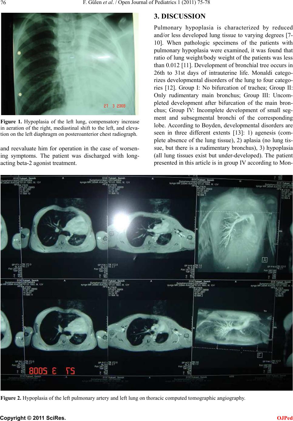 F. Gülen et al. / Open Journal of Pediatrics 1 (2011) 75-78 76 Figure 1. Hypoplasia of the left lung, compensatory increase in aeration of the right, mediastinal shift to the left, and eleva- tion on the left diaphragm on posteroanterior chest radiograph. and reevaluate him for operation in the case of worsen- ing symptoms. The patient was discharged with long- acting beta-2 agon ist treatmen t. 3. DISCUSSION Pulmonary hypoplasia is characterized by reduced and/or less developed lung tissue to varying degrees [7- 10]. When pathologic specimens of the patients with pulmonary hypoplasia were examined, it was found that ratio of lung weight/body weight of the patients was less than 0.012 [11]. Development of bronchial tree occurs in 26th to 31st days of intrauterine life. Monaldi catego- rizes developmental disorders of the lung to four catego- ries [12]. Group I: No bifurcation of trachea; Group II: Only rudimentary main bronchus; Group III: Uncom- pleted development after bifurcation of the main bron- chus; Group IV: Incomplete development of small seg- ment and subsegmental bronchi of the corresponding lobe. According to Boyden, developmental disorders are seen in three different extents [13]: 1) agenesis (com- plete absence of the lung tissue), 2) aplasia (no lung tis- sue, but there is a rudimentary bronchus), 3) hypoplasia (all lung tissues exist but under-developed). The patient presented in this article is in g roup IV according to Mon- F igure 2. Hypoplasia of the left pulmonary artery and left lung on thorac ic comp uted tomographic angiography. C opyright © 2011 SciRes. OJPed  F. Gülen et al. / Open Journal of Pediatrics 1 (2011) 75-78 77 aldi classification and is of third degree according to Boyden. There are primary (idiopathic) and secondary causes in the etiology of the pulmonary hypoplasia. It usually occurs secondary to the intrauterine diseases impairing normal lung development. Deformities of the chest wall, pleural effusion associated with hydrops fetalis, cystic adenomatoid malformation, and diaphragmatic hernia physically limits the development of the lung. Further- more, conditions leading to oligohydramnios such as early rupture of the membranes or fetal renal insuffi- ciency also impair development of the lungs [1]. Hy- poplasia may be bilateral in thoracic dystrophies and oligohydramnios. In such cases, development of the air- ways and arterial truncation is inhibited and th u s, surface area required for alveolo-capillary gas exchange be- comes limited [1,7]. Isolated pulmonary hypoplasia is rare; it is generally accompanied by anomalies in other systems, especially in the cardiovascular, gastrointestinal, musculoskeletal and urogenital systems. Severity of the clinical symp- toms and timing of their occurrence is related to the de- gree of hypoplasia [14]. It usually presents with respira- tory distress or pulmonary hypertension in the newborn period. Respiratory distress in the first five minutes fol- lowing birth is typical for these patients, and they usu- ally require mechanical ventilation. Apgar scores are usually found to be lower, although babies with com- pletely normal Apgar scores may also be seen [11, 15-19]. However, as in our patient, patients who remain asymptomatic until pub erty have also be en reported [7,9 , 10]. Total atelectasia, pleural effusion, pneumonia, dia- phragmatic elevation, diaphragmatic hernia, pulmonary aplasia, and pneumonectomy should be considered in differential diagnosis of these patients. Postero-anterior chest radiograph, thoracic CT, CT angiography, MR an- giography and bronchoscopy may be used for diagnosis [14]. Some pathologists suggested that radial alveolar counting (RAC) might be used for confirming the diag- nosis of hypoplasia [20]. In this method, number of the alveoli taken from a region between the respiratory bronchiole and the closest connective tissue is compared to a normal control sample in the same laboratory. This rate was found to be reduced in two published studies [11,15,21-24], although another study did not support this assumption [18]. Life-span of the patients depends on severity of anomalies of other systems and degree of the pulmonary hypoplasia [1]. Mortality has been reported to be high (71% - 95%) especially in those cases manifesting in the newborn period [11,25]. Membrane rupture earlier than 25th week of the gestation, presence of severe oligohy- dramnios (amniotic fluid index < 4) for more than 2 weeks, and preterm labor have been reported to be con- ditions increasing mortality [25]. Some prenatal inter- ventions may be attempted to increase fetal lung capac- ity or to provide lung development, depending on the underlying etiologic reason. Tocolitics for early mem- brane rupture, antibiotics, steroids, use of fibrin glue to correct the lesion transcervically, and consecutive use of amnio-infusions are methods used currently in several centers and of which benefits have been reported in some references [25]. Additionally, other methods in the postnatal period include mechanical ventilation, provid- ing ECMO (extracorporeal membrane oxygenation) and surfactant support, and early initiation of dialysis to support renal function. No method with proven efficacy has been found although some surgical techniques are being tried [25]. Unilateral hypoplasia is a congenital ano maly causing a wide spectrum of clinical manifestations from mild, non-specific findings to severe respiratory symptoms. Our case was presented with nonspecific findings, who remained asymptomatic until puberty, which is a rare clinical presentation of pulmonary hypoplasia. In pa- tients with unilateral hypolucen t lung, although it is seen rarely, pulmonary hypoplasia should always be kept in mind. REFERENCES [1] Kant, S. (2007) Unilateral pulmonary hypoplasia—A case report. Lung India, 24, 69-71. doi:10.4103/0970-2113.44215 [2] Hislop, A., Hey, E. and Reid, L. (1979) The lungs in congenital bilateral renal agenesis and dysplasia. Ar- chives of Disease in Childhood, 54, 32-38. doi:10.1136/adc.54.1.32 [3] Areechon, W. and Reid, L. (1963) Hypoplasia of lung with congenital diaphragmatic hernia. British Medical Journal, 1, 230-233. doi:10.1136/bmj.1.5325.230 [4] Johnson, R.J. and Haworth, S.G. (1982) Pulmonary vas- cular and alveolar development in tetralogy of fallot: A recommendation for early correction. Thorax, 37, 893- 901. doi:10.1136/thx.37.12.893 [5] Cooney, T.P. and Thurlbeck, W.M. (1985) Lung growth and development in anencephly and hydranencephaly. The American Review of Respiratory Disease, 13 2, 596- 601. [6] Cooney, T.P. and Thurlbeck, W.M. (1982) Pulmonary hypoplasia in Down syndrome. New England Journal of Medicine, 307, 1170-1173. doi:10.1056/NEJM198211043071902 [7] Deniz, Ö., Tozkoparan, E., Çiftçi, F., Özkan, M., et al. (2004) Sol pulmoner aplazi (agenezi) (olgu sunumu). Gülhane Tıp Dergisi, 46, 56-58. [8] Fraser, R.S., et al. (Eds.) (1994) Pulmonary abnormalities of developmental origin. Synopsis of diseases of the ch est , Se con d Edi ti on, W. B. Saunders Company, P hil del - phia, 256-286. C opyright © 2011 SciRes. OJPed  F. Gülen et al. / Open Journal of Pediatrics 1 (2011) 75-78 78 [9] Naclerio, E.A. (1959) Congenital diseases, agenesis of lung. Bronchopulmonary Diseases, 35, 309-313. [10] Roque, A.S., Burton, E.M., Boedy, R.F., Falls, G., et al. (1997) Unilateral pulmonary agenesis without mediasti- nal displacement. Southern Medical Journal, 90, 335- 337. [11] Odd, D.E., Batin, M.R., Halam, L. and Knight, D.B. (2003) Primary pulmonary hypoplasia: A case report and review of the literature. Journal of Paediatrics and Child Health, 39, 467-469. doi:10.1046/j.1440-1754.2003.00191.x [12] Monaldi, V. (1960) Malformative bronchopulmonary diseases caused by anatomical defects. Minerva Medica, 51, 3474-3478. [13] Boyden, E.A. (1955) Developmental anomalies of the lungs. American Journal of Surgery, 89, 79-89. doi:10.1016/0002-9610(55)90510-9 [14] Haddad, G.G. and Fontan, J.J.P. (2000) Development of the respiratory system. Nelson Textbook of Pediatrics, 16th Edition, 1235-1237. [15] Swischuk, L.E., Richardson, C.J., Nichols, M.M. and Ingman, M.J. (1979) Primary pulmonary hypoplasia in the neonate. Journal of Pediatric, 95, 573-577. doi:10.1016/S0022-3476(79)80773-8 [16] Sunam, G. and Ceran, S. (2009) Pulmonary artery agen- esis and kung hypoplasia. European Journal of General Medicine, 6, 265-267. [17] Mendelsohn, G. and Hutchins, G.M. (1971) Primary pulmonary hypoplasia. Report of a case with polyhy- dramnios. American Journal of Diseases of Children, 131, 1220-1223. [18] Frey, B., Fleishauer, A. and Gersbach, M. (1994) Familial isolated pulmonary hypoplasia: A case report, suggesting autosomal recessive inheritance. European Journal of Pediatrics, 153, 460-463. doi:10.1007/BF01983413 [19] Hamel, B.C.J. (1995) Familial primary pulmonary hy- poplasia. European Journal of Pediatrics, 154, 336. doi:10.1007/BF01957378 [20] Askenazi, S.S. and Perlman, M. (1979) Pulmonary hy- poplasia: Lung weight and radial alveolar count as crite- ria of diagnosis. Archives of Disease in Childhood, 54, 614-618. doi:10.1136/adc.54.8.614 [21] Langer, R. and Kaufmann, H.J. (1986) Primary (isolated) bilateral pulmonary hypoplasia: A comparative study of radiologic findings and autopsy results. Pediatric Radi- ology, 16, 175-179. doi:10.1007/BF02456280 [22] Laudy, J.A., Tibboel, D., Robben, S.G., de Krijger, R.R., et al. (2002) Prenatal prediction of pulmonary hypoplasia. Clinical, biometric, and doppler velocity correlates. Pe- diatrics, 109, 250-258. doi:10.1542/peds.109.2.250 [23] Chang, N., Hertzle r, J.H., Gregg, R.H., Lofti, M.W., et al. (1968) Congenital stenosis of the right mainstem bron- chus. A case report. Pediatrics, 41, 739-742. [24] Goldstein, J.D. and Reid, L.M. (1980) Pulmonary hy- poplasia resulting from phrenic nerve agenesis and dia- phragmatic amyoplasia. Journal of Pediatric, 97, 282- 287. doi:10.1016/S0022-3476(80)80496-3 [25] Chin, T. (2009) Pulmonary hypoplasia. Cited March 2009. http://www.emedicine.com/ped/TOPIC2627.HTM C opyright © 2011 SciRes. OJPed |