

Paper Menu >>
Journal Menu >>
 Open Journal of Pediatrics, 2011, 1, 67-71 doi:10.4236/ojped.2011.14016 Published Online December 2011 (http://www.SciRP.org/journal/ojped/ OJPed ) Published Online December 2011 in SciRes. http://www.scirp.org/journal/OJPed An atypical presentation of Kikuchi-Fujimoto disease mimicking systemic lupus erythematosus: case report and literature review Diane Belder-Preston1*, Catherine-Maude Pound1,2, Roman Jurencak1,3 1Department of Pediatrics, Children’s Hospital of Eastern Ontario, Ottawa, Canada; 2Division of Pediatric Medicine, Children’s Hospital of Eastern Ontario, Ottawa, Canada; 3Division of Rheumatology, Children’s Hospital of Eastern Ontario, Ottawa, Canada. Email: *[email protected] Received 1 October 2011; revised 5 November 2011; accepted 18 November 2011. ABSTRACT Purpose: To report a case of atypical Kikuchi-Fuji- moto disease (KFD) that illustrates several overlap- ping features with systemic lupus erythematosus (SLE). Methods: A case is reported followed by a re- view of the current literature. Case Report: A 16- year- old boy with an unusual manifestation of Kikuchi- Fujimoto disease (KFD) is described. The patient presented with fever, weight loss and severe abdomi- nal pain, due to extensive necrotizing retroperitoneal and mesenteric lymphadenopathy. During the course of his illness, he developed several symptoms sugges- tive of systemic lupus erythematosus (SLE): a peri- cardial effusion, cotton wool spots on the retina and antibodies against nuclear antigens (ANA), Smith (Sm) and ribonucleoprotein (RNP) antigens. However, no additional features of SLE were found. The pa- tient subsequently fully recovered within two months, without initiation of immunosuppressive therapy. His autoantibodies became negative five months after initial presentation and he remains well at his 23 month follow up visit. Discussion: We hypothesize that the autoantibodies developed by our patient were secondary to self-antigen induced autoimmunity related to his extensive tissue necrosis. Despite ini- tially having clinical features suggestive of SLE, our patient’s full and spontaneous recovery strongly supports the diagnosis of KFD. This illustrates the need for careful diagnosis, in order to avoid unneces- sary and potentially toxic treatment with immuno- suppressive agents. Keywords: Kikuchi-Fujimoto’s Disease; Retroperitoneal Lymphadenitis; Systemic Lupus Erythematosus (SLE); Antinuclear Antibodies (ANA) 1. INTRODUCTION Kikuchi disease, also called Kikuchi-Fujimoto Disease (KFD) and Histiocytic Necrotizing Lymphadenopathy, is a rare, benign, self-limited disease of unknown etiology. KFD was first reported in 1972 by Kikuchi [1] as well as independently, by Fujimoto et al. [2]. Typical features of the disease include subacute regional lymphadenopathy predominantly involv ing cerv ical lymph nodes as well as fevers, often accompanied by leukopenia, high erythro- cyte sedimentation rate (ESR), and anemia [3]. Exci- sional biopsy of the affected lymph node shows non- specific histopathologic features of cortical and paracor- tical necrosis. No specific diagnostic imaging or labora- tory tests are available and KFD remains a diagnosis of exclusion. Clinicians must rule out other causes of ne- crotizing lymphadenopathy, such as malignancy, sys- temic connective tissue disorders, and infectious lym- phadenitis, before diagnosing KFD. These other diagno- ses have different therapeutic and prognostic implica- tions. This can result in costly and invasive investiga- tions, potentially causing significant distress to the pa- tient and family. We describe the case of a young man who presented to a pediatric tertiary care centre with atypical features of Kikuchi-Fujimoto Disease. 2. CASE REPORT A 16-year-old, previously healthy, Caucasian boy pre- sented to a pediatric tertiary care centre with a three- week history of intermittent abdominal pain, vomiting, anorexia and a seven-kilogram weight loss. He also had a two-week history of intermittent fevers, up to 40 de- grees Celsius, associated with a faint diffuse erythema- tous rash involving the face and upper torso. Personal and family histories were noncontributory. Physical examination on admission revealed a pale, 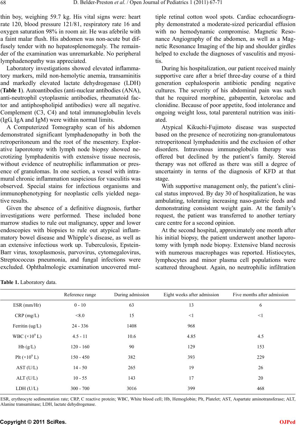 D. Belder-Preston et al. / Open Journal of Pediatrics 1 (2011) 67-71 68 thin boy, weighing 59.7 kg. His vital signs were: heart rate 120, blood pressure 121/81, respiratory rate 16 and oxygen saturation 98% in room air. He was afebrile with a faint malar flush. His abdomen was non-acute but dif- fusely tender with no hepatosplenomegaly. The remain- der of the examination was unremarkable. No peripheral lymphadenopathy was appreciated. Laboratory investigations showed elevated inflamma- tory markers, mild non-hemolytic anemia, transaminitis and markedly elevated lactate dehydrogenase (LDH) (Table 1). Autoantibodies (anti-nuclear antibodies (ANA), anti-neutrophil cytoplasmic antibodies, rheumatoid fac- tor and antiphospholipid antibodies) were all negative. Complement (C3, C4) and total immunoglobulin levels (IgG, IgA and IgM) were within normal limits. A Computerized Tomography scan of his abdomen demonstrated significant lymphadenopathy in both the retroperitoneum and the root of the mesentery. Explor- ative laporotomy with lymph node biopsy showed ne- crotizing lymphadenitis with extensive tissue necrosis, without evidence of neutrophilic inflammation or pres- ence of granulomas. In one section, a vessel with intra- mural chronic inflammation suspicious for vasculitis was observed. Special stains for infectious organisms and immunophenotyping for neoplastic cells yielded nega- tive results. Given the absence of a definitive diagnosis, further investigations were performed. These included bone marrow studies to rule out malignancy, upper and lower endoscopies with biopsies to rule out atypical inflam- matory bowel disease and Whipple’s disease, as well as an extensive infectious work up. Tuberculosis, Epstein- Barr virus, toxoplasmosis, parvovirus, cytomegalovirus, Streptococcus pneumonia, and fungal infections were excluded. Ophthalmologic examination uncovered mul- tiple retinal cotton wool spots. Cardiac echocardiogra- phy demonstrated a moderate-sized pericardial effusion with no hemodynamic compromise. Magnetic Reso- nance Angiography of the abdomen, as well as a Mag- netic Resonance Imaging of the hip and shoulder girdles helped to exclude the diagnoses of vascu litis and myosi- tis. During his hospitalization, our patient received mainly supportive care after a brief three-day course of a third generation cephalosporin antibiotic pending negative cultures. The severity of his abdominal pain was such that he required morphine, gabapentin, ketorolac and clonidine. Because of poor ap petite, food intoleran ce and ongoing weight loss, total parenteral nutrition was initi- ated. Atypical Kikuchi-Fujimoto disease was suspected based on the presence of necrotizing non-granulomatous retroperitoneal lymphadenitis and the exclusion of other disorders. Intravenous immunoglobulin therapy was offered but declined by the patient’s family. Steroid therapy was not offered as there was still a degree of uncertainty in terms of the diagnosis of KFD at that stage. With supportive management only, the patient’s clini- cal status improved. By day 30 of hospitalization, he was ambulating, tolerating increasing naso-gastric feeds and demonstrating consistent weight gain. At the family’s request, the patient was transferred to another tertiary care centre for a second opinion. At the second hospital, approximately one month after his initial biopsy, the patient underwent another laporo- tomy with lymph node biopsy. Extensive bland necrosis with numerous macrophages was reported. Histiocytes, lymphocytes and minor plasma cell populations were scattered throughout. Again, no neutrophilic infiltration Table 1. Laboratory data. Reference range During admission Eight weeks after admission Five months after admission ESR (mm/Hr) 0 - 10 63 13 6 CRP (mg/L) <8.0 15 < 1 <1 Ferritin (ug/L) 24 - 336 1408 968 WBC (×109 L) 4.5 - 11 10.6 4.85 4.5 Hb (g/L) 120 - 160 90 129 153 Plt (×109 L) 150 - 450 382 393 229 AST (U/L ) 14 - 50 265 19 26 ALT (U/L) 10 - 55 143 17 20 LDH (U/L) 300 - 700 3016 399 468 ESR, erythrocyte sedimentation rate; CRP, C reactive protein; WBC, White blood cell; Hb, Hemoglobin; Plt, Platelet; AST, Aspartate aminotransferase; ALT, lanine transaminase; LDH, lactate dehydrogenase. A C opyright © 2011 SciRes. OJPed 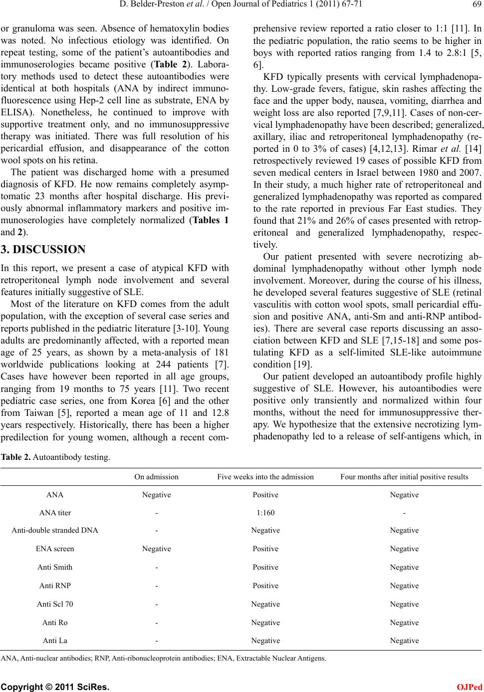 D. Belder-Preston et al. / Open Journal of Pediatrics 1 (2011) 67-71 69 or granuloma was seen. Absence of hematoxylin bodies was noted. No infectious etiology was identified. On repeat testing, some of the patient’s autoantibodies and immunoserologies became positive (Table 2). Labora- tory methods used to detect these autoantibodies were identical at both hospitals (ANA by indirect immuno- fluorescence using Hep-2 cell line as substrate, ENA by ELISA). Nonetheless, he continued to improve with supportive treatment only, and no immunosuppressive therapy was initiated. There was full resolution of his pericardial effusion, and disappearance of the cotton wool spots on his retina. The patient was discharged home with a presumed diagnosis of KFD. He now remains completely asymp- tomatic 23 months after hospital discharge. His previ- ously abnormal inflammatory markers and positive im- munoserologies have completely normalized (Tables 1 and 2). 3. DISCUSSION In this report, we present a case of atypical KFD with retroperitoneal lymph node involvement and several features initially suggestive of SLE. Most of the literature on KFD comes from the adult population, with the exception of several case series and reports published in the pediatric literature [3-10]. Young adults are predominantly affected, with a reported mean age of 25 years, as shown by a meta-analysis of 181 worldwide publications looking at 244 patients [7]. Cases have however been reported in all age groups, ranging from 19 months to 75 years [11]. Two recent pediatric case series, one from Korea [6] and the other from Taiwan [5], reported a mean age of 11 and 12.8 years respectively. Historically, there has been a higher predilection for young women, although a recent com- prehensive review reported a ratio closer to 1:1 [11]. In the pediatric population, the ratio seems to be higher in boys with reported ratios ranging from 1.4 to 2.8:1 [5, 6]. KFD typically presents with cervical lymphadenopa- thy. Low-grade fevers, fatigue, skin rashes affecting the face and the upper body, nausea, vomiting, diarrhea and weight loss are also reported [7,9,11]. Cases of non-cer- vical lymphadenopathy have been described; generalized, axillary, iliac and retroperitoneal lymphadenopathy (re- ported in 0 to 3% of cases) [4,12,13]. Rimar et al. [14] retrospectively reviewed 19 cases of possible KFD from seven medical centers in Israel between 1980 and 2007. In their study, a much higher rate of retroperitoneal and generalized lymphadenopathy was reported as compared to the rate reported in previous Far East studies. They found that 21% and 26% of cases presented with retro p- eritoneal and generalized lymphadenopathy, respec- tively. Our patient presented with severe necrotizing ab- dominal lymphadenopathy without other lymph node involvement. Moreover, during the course of his illness, he developed several features suggestive of SLE (retinal vasculitis with cotton wool spots, small pericardial effu- sion and positive ANA, anti-Sm and anti-RNP antibod- ies). There are several case reports discussing an asso- ciation between KFD and SLE [7,15-18] and some pos- tulating KFD as a self-limited SLE-like autoimmune condition [19]. Our patient developed an autoantibody profile highly suggestive of SLE. However, his autoantibodies were positive only transiently and normalized within four months, without the need for immunosuppressive ther- apy. We hypothesize that the extensive necrotizing lym- phadenopathy led to a release of self-antigens which, in Table 2. Autoantibody t esting. On admission Five weeks into the admission Four months after initial positive r e s u l t s ANA Negative Positive Negative ANA titer - 1:160 - Anti-double stranded DNA - Negative Negative ENA screen Negative Positive Negative Anti Smith - Positive Negative Anti RNP - Positive Negative Anti Scl 70 - Negative Negative Anti Ro - Negative Negative Anti La - Negative Negative A NA, Anti-nuclear antibodies; RNP, Anti-ribonucleoprotein antibodies; ENA, Extractable Nuclear Antigens. C opyright © 2011 SciRes. OJPed  D. Belder-Preston et al. / Open Journal of Pediatrics 1 (2011) 67-71 70 the hyperinflammatory state, enabled mounting of a pathologic immune response with production of autoan- tibodies. Upon removal of the antigenic stimulation, the pathologic production of autoantibodies ceased, leading to their disappearance from the circulation. This hy- pothesis is supported by the fact that, 23 months after initial presentation, the patient remains off any therapy and healthy. Cotton wool spots, which were ob ser v ed in our patien t, often indicate a serious systemic disease. They represent acute, focal, inner retinal ischemia and may occur in any disease that compromises arteriolar circulation to the inner retinal layers which includes SLE and systemic vasculitides where ischemia is caused by thrombogenic effects from antigen-antibody complexes [20]. However, similar to our patient, isolated, self-limited cotton wool spots in patients with no identifiable systemic disease have also been reported [21]. The presence of pericardial effusion in our patient could indicate SLE as pericarditis is the most common cardiac manifestation of this disease. However, transient asymptomatic pericardial effusions are often seen in instances of significant systemic inflammation, espe- cially in the presence of accompanying hypoalbumine- mia [22]. Our patient did have hypoalbuminemia (data not shown). The pericardial effusion in our patient was asymptomatic with spontaneous resolution. Pericarditis accompanying KFD has not been previously described. Histologically, SLE and KFD can be very challenging to differentiate and at times impossible. In KFD, histo- pathological findings include varying degrees of necrosis, histiocytic proliferation with activated T lymphocytes, small lymphocytes and plasma cells without granuloma- tous inflammation with absent neutrophils and eosino- phils. Adjacent vessels may be thrombosed. In contrast to KFD, SLE lymphadenitis, demonstrates hematoxylin bodies (aggregates of degenerated nuclear debris) and Azzopardi phenomenon (degenerated nuclear material aggregated in the walls of blood vessels). In SLE, abun- dant plasma cells, prominent reactive follicular hyper- plasia, sparse cytoxic T cells and capsular and pericap- sular inflammation are seen [23,24]. In our patient’s bi- opsy specimens, the presence of extensive necrosis without neutrophilic inflammation or granulomas and the absence of hematoxylin bodies support the diagnosis of KFD rather than SLE or systemic vasculitis. In conclusion, this report discusses a case of atypical KFD and illustrates several overlapping features of KFD and SLE. Our patient presented with retroperitoneal lymphadenopathy, an unusual manifestation of KFD. He demonstrated a transient elevation of autoantibodies typically associated with SLE, as well as retinal vascu- litis and pericardial effusion, not previously described in patients with KFD. This illustrates the need for careful differential diagnosis, as KFD is self-limited and, con- trary to SLE, does not require long-term immunosup- pressive therapy; ra ther, often only supportive ther apy is needed. REFERENCES [1] Kikuchi, M. (1972) Lymphadenitis showing focal reticu- lum cell hyperplasia with nuclear debris and phagocytes. Acta Haematologica Japanese, 35, 379-380. [2] Fujimoto, Y., Kozima, Y. and Yamaguchi, K. (1972) Cer- vical subacute necrotizing lymphadenitis. Naika, 30, 920- 927. [3] Han HJ, Lim G-Y, Yeo D-M., et al. (2009) Kikuchi’s disease in children: Clinical manifestations and imaging features. Journal of Korean Medical Science, 24, 1105- 1109. doi:10.3346/jkms.2009.24.6.1105 [4] Saini, A.P., Jamil, S. and Dulai, M. (2010) Kikuchi’s disease causing persistent fever and iliac lymphadenopa- thy in an 8-year-old boy. Clinical Pediatrics (Philadel- phia), 49, 601-604. doi:10.1177/0009922809350497 [5] Lin, H.C., Su, C.Y. and Huang, S.C. (2005) Kikuchi’s disease in Asian children. Pediatrics, 11 5, e92-e96. [6] Lee, K.Y., Yeon, Y.H. and Lee, B.C. (2004) Kikuchi- Fujimoto disease with prolonged fever in children. Pedi- atrics, 11 4, e752-e756. doi:10.1542/peds.2004-0485 [7] Kucukardali, Y., Solmazgul, E., Kunter, E., et al. (2007) Kikuchi-fujimoto disease: Analysis of 244 cases. Clinical Rheumatology, 26, 50-54. doi:10.1007/s10067-006-0230-5 [8] Wang, T.J., Yang, Y.H., Lin, Y.T. and Chiang, B.L. (2004) Kikuchi-Fujimoto disease in children: Clinical features and disease course. Journal of Microbiology, Immunol- ogy and Infection, 37, 219-224. [9] Chuang, C.H., Yan, D.C., Chiu, C.H., et al. (2005) Clini- cal and laboratory manifestations of Kikuchi’s disease in children and differences between patients with and with- out prolonged fever. Pediatric Infectious Disease Journal, 24, 551-554. doi:10.1097/01.inf.0000167246.24500.21 [10] Payne, J.H., Evans, M. and Gerrard, M.P. (2003) Kiku- chi-Fujimoto disease: A rare but important cause of lym- phadenopathy. Acta Paedia t rica, 92, 261-264. doi:10.1111/j.1651-2227.2003.tb00539.x [11] Bosch, X., Guilabert, A., Miquel, R. and Campo, E. (2004) Enigmatic Kikuchi-Fujimoto disease: A compre- hensive review. American Journal of Clinical Pathology, 122, 141-152. doi:10.1309/YF081L4TKYWVYVPQ [12] Rudniki, C., Kessler, E., Zarfati, M., et al. (1988) Kiku- chi’s necrotizing lymphadenitis: A cause of fever of un- known origin and splenomegaly. Act a Ha e mat ol ogi c a, 79, 99-102. doi:10.1159/000205730 [13] Kita, Y., Kikuchi, M., Nakae, T., et al. (1997) A case of Kikuchi’s disease with abdominal manifestations. Sur- gery, 122, 962-963. doi:10.1016/S0039-6060(97)90338-X [14] Rimar, D., Zisman, D., Schendler, Y. , et al. (2010) Kiku- chi-Fujimoto disease in israel-more than a pain in the neck. Seminars in Arthritis and Rheumatism, 39, 515-520. doi:10.1016/j.semarthrit.2009.03.003 C opyright © 2011 SciRes. OJPed  D. Belder-Preston et al. / Open Journal of Pediatrics 1 (2011) 67-71 71 [15] Martinez-Vazquez, C., Hughes, G., Bordon, J., et al. (1997) Histiocytic necrotizing lymphadenitis, Kikuchi- Fujimoto’s disease, associated with systemic lupus ery- themotosus. QJM: An International Journal of Medicine, 90, 531-533. doi:10.1093/qjmed/90.8.531 [16] Fernandez, S.H. (2000) Kikuchi’s lymphadenitis (ne- crotizing lymphadenitis) and systemic lupus erythema- tosus: A case report. Malaysian Journal of Pathology, 22, 25-29. [17] Goldblatt, F., Andrew, J., Russell, A. and Isenberg, D. (2008) Association of Kikuchi-Fujimoto’s disease with SLE. Rheumatology (Oxford), 47, 553-554. doi:10.1093/rheumatology/ken008 [18] Tsaousis, G., Kolevris, N., Vaidakis, E., et al. (1999) Kikuchi’s disease and systemic lupus erythematosus: Case report and review. European Journal of Internal Medicine, 10, 53-55. doi:10.1016/S0953-6205(99)00010-2 [19] Nieman, R.B. (1990) Kikuchi’s disease. Lancet, 335, 1563. doi:10.1016/0140-6736(90)91382-K [20] Arevalo, J.F., Lowder, C.Y. and Muci-Mendoza, R. (2002) Ocular manifestations of systemic lupus erythematosus. Current Opinion in Ophthalmology, 13, 404-410. doi:10.1097/00055735-200212000-00011 [21] Remky, A. and Arend, O. (2000) Single isolated cotton- wool spots. Ophthalmologica, 214, 143-148. doi:10.1159/000027485 [22] Sugiura, T., Kumon, Y., Kataoka, H., et al. (2008) As- ymptomatic pericardial effusion in patients with rheu- matoid arthritis. Cardiology, 110, 87-91. doi:10.1159/000110485 [23] Hu, S., Kuo, T.T. and Hong, H.S. (2003) Lupus lympha- denitis simulating Kikuchi’s lymphadenitis in patients with systemic lupus erythematosus: A clinicopathological analysis of six cases and review of the literature. Pathol- ogy International, 53, 221-226. doi:10.1046/j.1320-5463.2003.01458.x [24] Hutchinson, C.B. and Wang, E. (2010) Kikuchi-Fujimoto disease. Archives of Pathology & Laboratory Medicine, 134, 289-293. ABBREVIATIONS Kikuchi-Fujimoto disease (KFD), Systemic lupus erythematosus (SLE), Antinuclear antibodies (ANA), lactate dehydrogenase (LDH), erythrocyte sedimentation rate (ESR), smith (Sm), ribonucleoprotein (RNP), C reactive protein (CRP), White blood cell (WBC), Hemoglobin (Hb), Platelet (Plt), Aspartate aminotransferase (AST), Alanine transaminase (ALT). C opyright © 2011 SciRes. OJPed |