

Paper Menu >>
Journal Menu >>
 Open Journal of Pediatrics, 2011, 1, 87-89 doi:10.4236/ojped.2011.14020 Published Online December 2011 (http://www.SciRP.org/journal/ojped/ OJPed ) Published Online December 2011 in SciRes. http://www.scirp.org/journal/OJPed Cardiac tamponade due to group a streptococcal pericarditis in a 10-month-old boy and a review of the literature Matthew C. Schwartz1*, Matthew J. Gillespie1, Paul Stephens Jr.1, Brian Fisher2 1Division of Cardiology, The Children’s Hospital of Philadelphia, Philadelph ia, USA; 2Division of Infectious Disease and Center for Clinical Epidemiology and Biostatistics, The Children’s Hospital of Philadelphia, Philadelphia, USA. Email: *[email protected] Received 18 September 2011; revised 27 October 2011; accepted 8 November 2011. ABSTRACT Group A streptococcus (GAS) is a rare cause of pu- rulent pericarditis in pediatric patients as only 7 cases have been reported. We present a 10-month-old boy who developed cardiac tamponade from GAS and was successfully treated with subxiphoid tube drainage and 4 weeks of antibiotics at the Children’s Hospital of Philadelphia. Keywords: Streptococcus; Pericarditis; Pericardiocente- sis; Tamponade 1. INTRODUCTION Group A streptococcus (GAS) is a rare cause of bacterial pericarditis in children with only 7 pediatric cases pre- viously reported [1-5]. We describe a 10-month-old who developed cardiac tamponade due to GAS pericarditis and was successfully treated. 2. CASE REPORT A 10-month-old boy presented to the Children’s Hospital of Philadelphia (CHOP) Emergency Department (ED) with 9 days of fever accompanied by a transient papular rash, but no other localizing symptoms. He underwent outpatient evaluation on day 6 and was diagnosed with roseola. The patient’s fever continued prompting his presentation. His birth and past medical histories were unremark- able. He lived in Philadelphia and had no exposures or travel. Several days into the child’s illness, his father was diagnosed with GAS pharyngitis by rapid strepto- coccal antigen test and was treated. In the ED, he had a rectal temperature of 38.2˚C. The remainder of his vital signs and physical exam were normal except for mild bilateral anterior cervical lym- phadenopathy. Initial laboratory testing showed a white blood cell count of 20,500/uL (63% neutrophils, 26% lymphocytes), a hemoglobin of 10.2 g/dL, a platelet count of 57,400/u L, an erythrocyte sedimentation rate of 75 mm/hr, a C-reactive protein of 6.2 mg/dL, an albumin of 3.7 g/dL, an aspartate aminotransferase of 109 U/L, and an alanine aminotransferase <6 U/L. Urinalysis and chest radiograph were normal. Urine and blood cultures were sent as was a respiratory virus polymerase chain reaction (PCR) panel on respiratory secretions. The patient was admitted for evaluation. On hospital day 1, the respiratory virus PCR panel returned negative for adenovirus, respiratory syncytial virus, influenza A and B, parainfluenza 1 - 3, metapneumovirus, and rhino- virus. Serum was sent for adenovirus PCR, human her- pes virus-6 (HHV6) PCR , cytomegalovi r us ( C M V) PCR, and Ebstein Barr virus (EBV) serology. To consider oc- cult osteomyelitis, a whole body magnetic resonance imaging scan was obtained and was negative. In light of possible Kawasaki Disease, an echocardiogram was performed, revealing a small, 4 mm posterior pericardial effusion, normal biventricular systolic function, and mild, diffuse ectasia of the right coronary artery (proximal right coronary artery z-score 2.62). Due to fever, elevated inflammatory markers, an ele- vated aspartate transferase, and ectasia of the right coro- nary artery, incomplete Kawasaki Disease was suspected. On hospital day 2, the patient was treated with intrave- nous immunoglobulin and aspirin was started. From hospital day 3 through 5, the patient’s fever continued and his inflammatory markers increased. Serial blood cultures remained negative and urine culture at the time of admission was negative. The serum adenovirus PCR, CMV PCR, and EBV serologies returned negative. The patient’s HHV6 serum PCR returned as mildly positive (1981 copies/mL). The Infectious Disease Service was consulted and felt HHV6 not likely responsible for the illness. By hospital day 6, the patient remained febrile and had increased work of breathing. Chest x-ray showed an 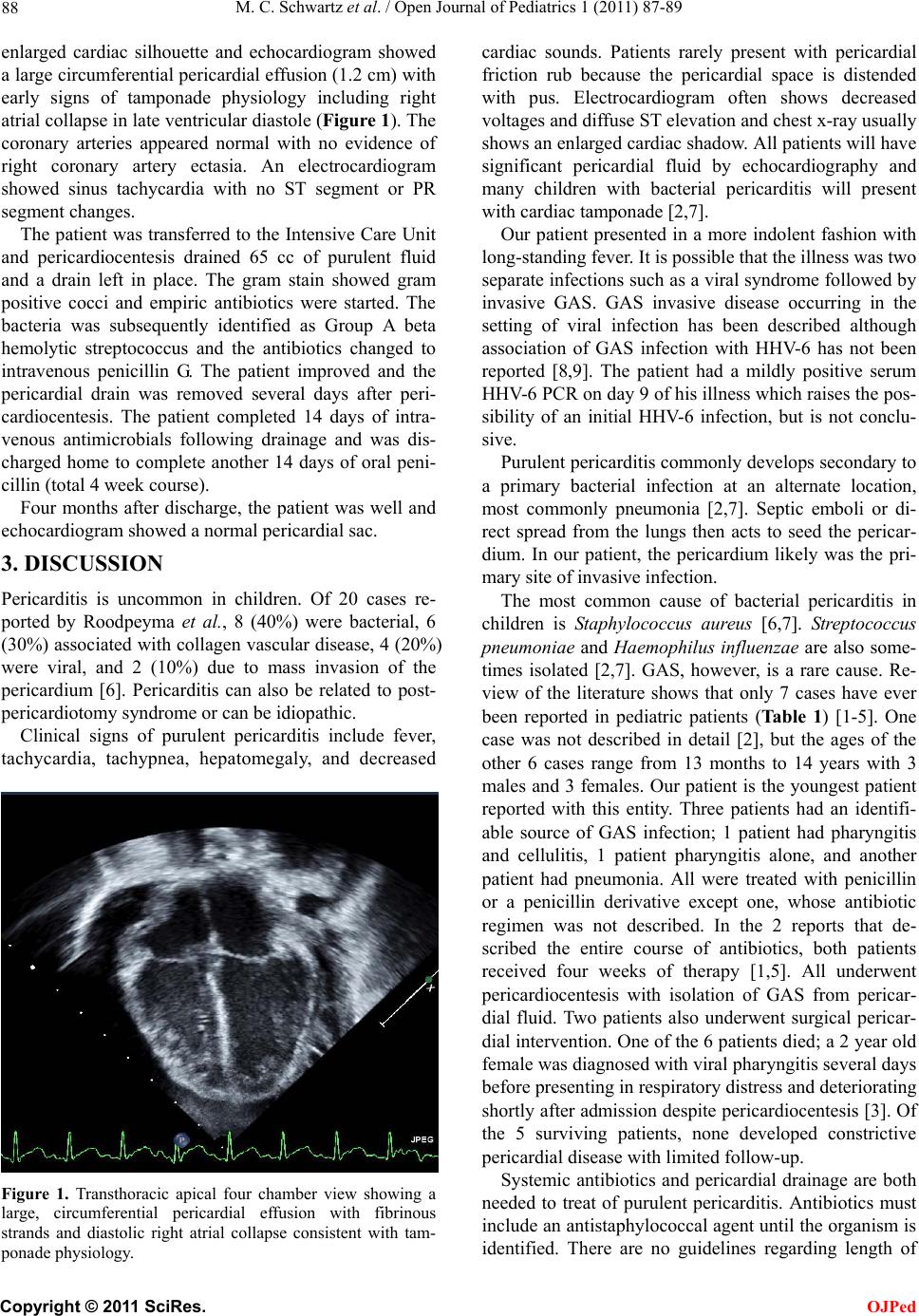 M. C. Schwartz et al. / Open Journal of Pediatrics 1 (2011) 87-89 88 enlarged cardiac silhouette and echocardiogram showed a large circumferential pericardial effusion (1.2 cm) with early signs of tamponade physiology including right atrial collapse in late ventricular diastole (Figure 1). The coronary arteries appeared normal with no evidence of right coronary artery ectasia. An electrocardiogram showed sinus tachycardia with no ST segment or PR segment changes. The patient was transferred to the Intensive Care Unit and pericardiocentesis drained 65 cc of purulent fluid and a drain left in place. The gram stain showed gram positive cocci and empiric antibiotics were started. The bacteria was subsequently identified as Group A beta hemolytic streptococcus and the antibiotics changed to intravenous penicillin G. The patient improved and the pericardial drain was removed several days after peri- cardiocentesis. The patient completed 14 days of intra- venous antimicrobials following drainage and was dis- charged home to complete another 14 days of oral peni- cillin (total 4 week course). Four months after discharge, the patient was well and echocardiogram showed a normal pericardial sac. 3. DISCUSSION Pericarditis is uncommon in children. Of 20 cases re- ported by Roodpeyma et al., 8 (40%) were bacterial, 6 (30%) associated with collagen vascular disease, 4 (20%) were viral, and 2 (10%) due to mass invasion of the pericardium [6]. Pericarditis can also be related to post- pericardiotomy syndrome or can be idiopathic. Clinical signs of purulent pericarditis include fever, tachycardia, tachypnea, hepatomegaly, and decreased Figure 1. Transthoracic apical four chamber view showing a large, circumferential pericardial effusion with fibrinous strands and diastolic right atrial collapse consistent with tam- ponade physiology. cardiac sounds. Patients rarely present with pericardial friction rub because the pericardial space is distended with pus. Electrocardiogram often shows decreased voltages and diffuse ST elevation and chest x-ray usually shows an enlarged cardiac shadow. All patients will have significant pericardial fluid by echocardiography and many children with bacterial pericarditis will present with cardiac tamponade [2,7]. Our patient presented in a more indolent fashion with long-standing fever. It is possible that the illness was two separate infections such as a viral syndro me followed by invasive GAS. GAS invasive disease occurring in the setting of viral infection has been described although association of GAS infection with HHV-6 has not been reported [8,9]. The patient had a mildly positive serum HHV-6 PCR on day 9 of his illness which raises the pos- sibility of an initial HHV-6 infection, but is not conclu- sive. Purulent pericarditis co mmonly develops seco ndary to a primary bacterial infection at an alternate location, most commonly pneumonia [2,7]. Septic emboli or di- rect spread from the lungs then acts to seed the pericar- dium. In our patient, the pericardium likely was the pri- mary site of invasive infection. The most common cause of bacterial pericarditis in children is Staphylococcus aureus [6,7]. Streptococcus pneumonia e and Haemophilus influenzae are also some- times isolated [2,7]. GAS, however, is a rare cause. Re- view of the literature shows that only 7 cases have ever been reported in pediatric patients (Ta b l e 1) [1-5]. One case was not described in detail [2], but the ages of the other 6 cases range from 13 months to 14 years with 3 males and 3 females. Our patient is the youngest patient reported with this entity. Three patients had an identifi- able source of GAS infection; 1 patient had pharyngitis and cellulitis, 1 patient pharyngitis alone, and another patient had pneumonia. All were treated with penicillin or a penicillin derivative except one, whose antibiotic regimen was not described. In the 2 reports that de- scribed the entire course of antibiotics, both patients received four weeks of therapy [1,5]. All underwent pericardiocentesis with isolation of GAS from pericar- dial fluid. Two patients also underwent surgical pericar- dial intervention. One of the 6 patients died; a 2 year old female was diagnosed with viral pharyngitis several days before presenting in respiratory distress and deteriorating shortly after admission despite pericardiocentesis [3]. Of the 5 surviving patients, none developed constrictive pericardial disease with limited follow-up. Systemic antibiotics and pericardial drainage are both needed to treat of purulent pericarditis. Antibiotics must include an antistaphylococcal agent until the organism is identified. There are no guidelines regarding length of C opyright © 2011 SciRes. OJPed 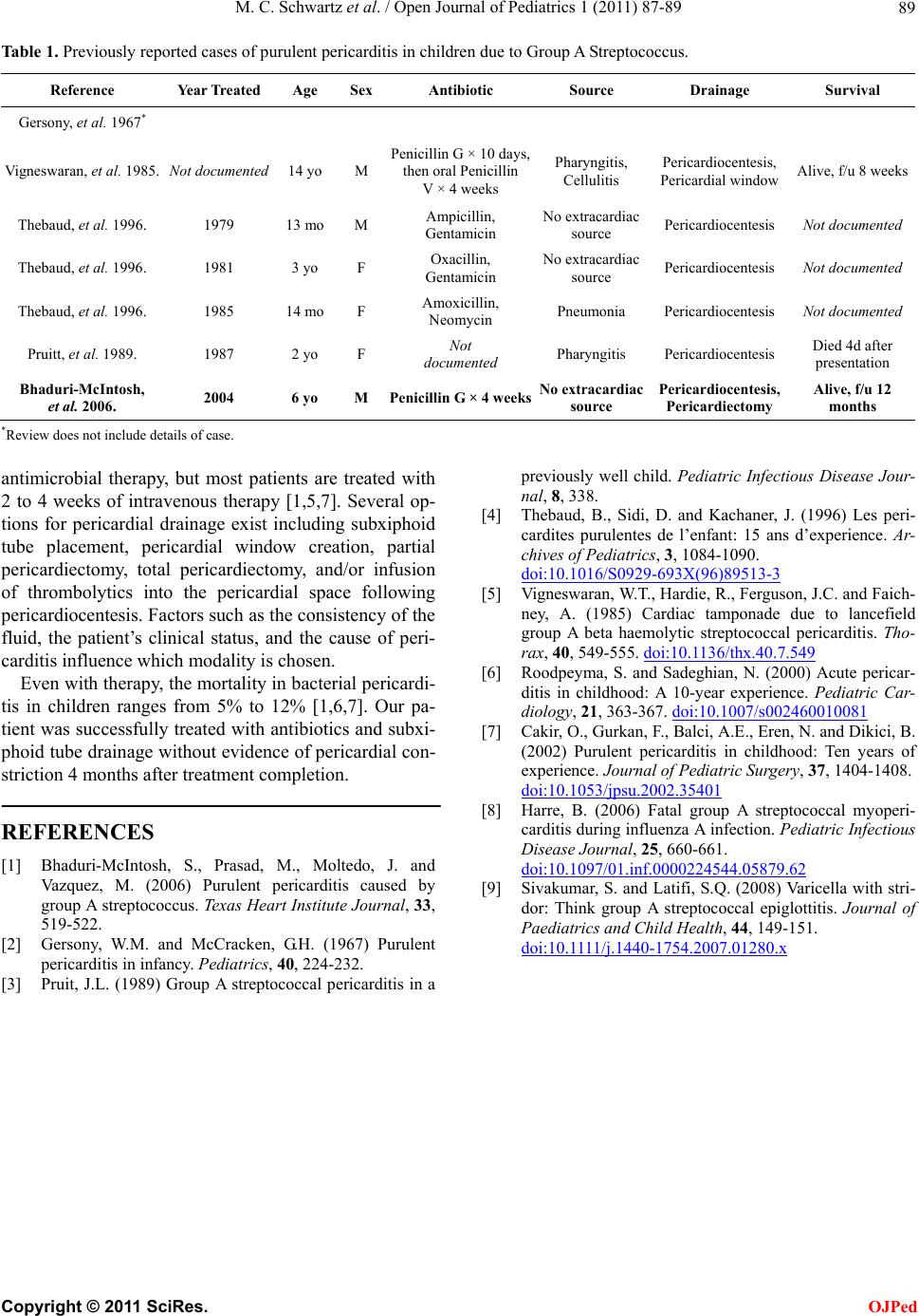 M. C. Schwartz et al. / Open Journal of Pediatrics 1 (2011) 87-89 Copyright © 2011 SciRes. 89 Table 1. Previously reported cases of purulent pericarditis in children due to Group A Streptococcus. Reference Year Treated Age SexAntibiotic Source Drainage Survival Gersony, et al. 1967* Vigneswaran, et al. 1985. Not documented 14 yo M Penicillin G ×10 days, then oral Penicillin V × 4 weeks Pharyngitis, Cellulitis Pericardiocentesis, Pericardial window Alive, f/u 8 weeks Thebaud, et al. 1996. 1979 13 mo M Ampicillin, Gentamicin No extracardiac source Pericardiocentesis Not documented Thebaud, et al. 1996. 1981 3 yo F Oxacillin, Gentamicin No extracardiac source Pericardiocentesis Not documented Thebaud, et al. 1996. 1985 14 mo F Amoxicillin, Neomycin Pneumonia Pericardiocentesis Not documented Pruitt, et al. 1989. 1987 2 yo F Not documented Pharyngitis Pericardiocentesis Died 4d after presentation Bhaduri-McIntosh, et al. 2006. 2004 6 yo M Penicillin G × 4 weeksNo extracardiac source Pericardiocentesis, Pericardiectomy Alive, f/u 12 months *Review does not incl ud e details of case. antimicrobial therapy, but most patients are treated with 2 to 4 weeks of intravenous therapy [1,5,7]. Several op- tions for pericardial drainage exist including subxiphoid tube placement, pericardial window creation, partial pericardiectomy, total pericardiectomy, and/or infusion of thrombolytics into the pericardial space following pericardiocentesis. Factors such as the consistency of the fluid, the patient’s clinical status, and the cause of peri- carditis influence which modality is chosen. OJPed Even with therapy, the mortality in bacterial pericardi- tis in children ranges from 5% to 12% [1,6,7]. Our pa- tient was successfully treated with antibiotics and subxi- phoid tube drainage without evidence of pericardial con- striction 4 months after treatment completion. REFERENCES [1] Bhaduri-McIntosh, S., Prasad, M., Moltedo, J. and Vazquez, M. (2006) Purulent pericarditis caused by group A streptococcus. Texas Heart Institute Journal, 33, 519-522. [2] Gersony, W.M. and McCracken, G.H. (1967) Purulent pericarditis in infancy. Pediatrics, 40, 224-232. [3] Pruit, J.L. (1989) Group A streptococcal pericarditis in a previously well child. Pediatric Infectious Disease Jour- nal, 8, 338. [4] Thebaud, B., Sidi, D. and Kachaner, J. (1996) Les peri- cardites purulentes de l’enfant: 15 ans d’experience. Ar- chives of Pediatrics, 3, 1084-1090. doi:10.1016/S0929-693X(96)89513-3 [5] Vigneswaran, W.T., Hardie, R., Ferguson, J.C. and Faich- ney, A. (1985) Cardiac tamponade due to lancefield group A beta haemolytic streptococcal pericarditis. Tho- rax, 40, 549-555. doi:10.1136/thx.40.7.549 [6] Roodpeyma, S. and Sadeghian, N. (2000) Acute pericar- ditis in childhood: A 10-year experience. Pediatric Car- diology, 21, 363-367. doi:10.1007/s002460010081 [7] Cakir, O., Gurkan, F., Balci, A.E., Eren, N. and Dikici, B. (2002) Purulent pericarditis in childhood: Ten years of experience. Journal of Pediatric Surgery, 37, 1404-1408. doi:10.1053/jpsu.2002.35401 [8] Harre, B. (2006) Fatal group A streptococcal myoperi- carditis during influenza A infection. Pediatric Infectious Disease Journal, 25, 660-661. doi:10.1097/01.inf.0000224544.05879.62 [9] Sivakumar, S. and Latifi, S.Q. (2008) Varicella with stri- dor: Think group A streptococcal epiglottitis. Journal of Paediatrics and Child Health, 44, 149-151. d oi: 10.1111/j.1440-1754.2007.01280.x |