

Paper Menu >>
Journal Menu >>
 Materials Sciences and Applications, 2010, 1, 1-7 doi:10.4236/msa.2010.11001 Published Online April 2010 (http://www.SciRP.org/journal/msa) Copyright © 2010 SciRes. MSA 1 Synthesis of Crystalline Ag Nanoparticles (AgNPs) from Microorganisms Seema Sharma1, Naheed Ahmad2, Anuradha Prakash3, Vidya Nand Singh4, Ashok Kumar Ghosh3, Bodh Raj Mehta4 1Department of Physics, A.N College, Patna, India; 2University Department of Botany, Patna University, Patna, India; 3Department of Water Management, A.N College, Patna, India; 4Thin Film Laboratory, Department of Physics, Indian Institute of Technology, New Delhi, India. Email: [email protected] Received January 26th, 2010; revised February 5th, 2010; accepted February 9th, 2010. ABSTRACT Bacteria obtained from the isolates of the biodiversity of riverine coast of Ganga identified as Bacillus Koriensis, when challenged with silver nitrate solution accumulated silver nanoparticles on the surface of its cell wall. These nanoparti- cles showed an absorption peak at 438 nm in UV-visible spectrum corresponding to the plasmon resonance of AgNPs. The transmission electron micrographs of nanoparticles in aqueous solution showed the production of reasonably monodisperse AgNPs (average particle size: 9.92 ± 1.311 nm) by the bacteria. X-ray diffraction spectrum of the nanoparticles confirmed the formation of metallic silver. Keywords: Crystal Structure, Nanostructures, Metal, AgNPs 1. Introduction Nanostructured materials are being viewed as the future material and for various diverse applications in areas such as biomedical science, optics, mechanics, magnetics catalysts, biosensors and energy science [1-3]. However most synthesis techniques employed involve huge inputs in terms of capital and energy. In the era of growing awareness about increasing pollution and global warming, developing new methods of synthesis of nanomaterials with green technologies is a challenge. Biodiversity is the best resource for biotechnological innovations as organic materials of nanometer dimensions form the basis of life. This diversity of life especially microorganisms is being used as ecofriendly nanofactories for bioproduction and synthesis of different compounds of nanometer size [4-6]. The concept of exploiting locally available species with potential to synthesize extracellular nanoparticles is a novel concept for bioprospecting. It provides valuable leads for new product development and new applications of biological species that have not been studied earlier. The metal microbe interaction has made the material scientist aware of the immense potential of the microorg- anisms as ecofriendly nanofactories [6-8]. Biological sy- nthesis of such nanomaterials has gained significant in- terest due to the use of mild experimental conditions of temperature, pH and pressure. If harnessed to their full potential, biological synthesis could present extra advan- tages over chemical methods such as higher productivity and lower cost. One new dimension in the metal-microbial interaction has emerged in the last few years, is that the syn- thesis of metallic nanoparticles have been reported from bac- teria, yeast, fungi, and other biological sources [9-16]. Bacteria are ubiquitous and constantly exposed to str- essful situations; hence they develop diverse ways to res- ist and survive. When exposed to metals or other toxic substances beyond a certain level they develop many me- chanisms such as efflux, alteration in the solubility and toxicity by change in redox state of the metalion, extrac- ellular complexation or precipitation of metals and the lack of specific metal transport systems [17-19]. The exact mechanism leading to the formation of reduction to silver ions and formation of nanoparticles is not fully understood. However, it is known that the tolerance of bacterial cells and formation of mineral particles is very much dependant on the composition of the growth envi- ronment. Various workers have exploited various microorgan- isms for nanosynthesis. Bacteria as for instance Bacillus subtilis 168 was reported to reduce Au+ to nanoscale di- mensions [20,21].Other bacterial strains like Pseudomo- nas stutzeri Ag 259 were found to be silver resistant and were able to produce nanosize silver [22].Other workers have used the fungi like Fusarium oxysporum and Veri- 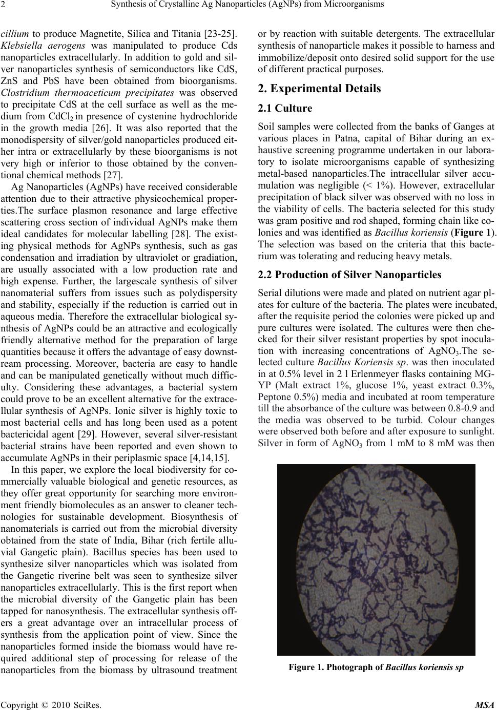 Synthesis of Crystalline Ag Nanoparticles (AgNPs) from Microorganisms 2 cillium to produce Magnetite, Silica and Titania [23-25]. Klebsiella aerogens was manipulated to produce Cds nanoparticles extracellularly. In addition to gold and sil- ver nanoparticles synthesis of semiconductors like CdS, ZnS and PbS have been obtained from bioorganisms. Clostridium thermoaceticum precipitates was observed to precipitate CdS at the cell surface as well as the me- dium from CdCl2 in presence of cystenine hydrochloride in the growth media [26]. It was also reported that the monodispersity of silver/gold nanoparticles produced eit- her intra or extracellularly by these bioorganisms is not very high or inferior to those obtained by the conven- tional chemical methods [27]. Ag Nanoparticles (AgNPs) have received considerable attention due to their attractive physicochemical proper- ties.The surface plasmon resonance and large effective scattering cross section of individual AgNPs make them ideal candidates for molecular labelling [28]. The exist- ing physical methods for AgNPs synthesis, such as gas condensation and irradiation by ultraviolet or gradiation, are usually associated with a low production rate and high expense. Further, the largescale synthesis of silver nanomaterial suffers from issues such as polydispersity and stability, especially if the reduction is carried out in aqueous media. Therefore the extracellular biological sy- nthesis of AgNPs could be an attractive and ecologically friendly alternative method for the preparation of large quantities because it offers the advantage of easy downst- ream processing. Moreover, bacteria are easy to handle and can be manipulated genetically without much diffic- ulty. Considering these advantages, a bacterial system could prove to be an excellent alternative for the extrace- llular synthesis of AgNPs. Ionic silver is highly toxic to most bacterial cells and has long been used as a potent bactericidal agent [29]. However, several silver-resistant bacterial strains have been reported and even shown to accumulate AgNPs in their periplasmic space [4,14,15]. In this paper, we explore the local biodiversity for co- mmercially valuable biological and genetic resources, as they offer great opportunity for searching more environ- ment friendly biomolecules as an answer to cleaner tech- nologies for sustainable development. Biosynthesis of nanomaterials is carried out from the microbial diversity obtained from the state of India, Bihar (rich fertile allu- vial Gangetic plain). Bacillus species has been used to synthesize silver nanoparticles which was isolated from the Gangetic riverine belt was seen to synthesize silver nanoparticles extracellularly. This is the first report when the microbial diversity of the Gangetic plain has been tapped for nanosynthesis. The extracellular synthesis off- ers a great advantage over an intracellular process of synthesis from the application point of view. Since the nanoparticles formed inside the biomass would have re- quired additional step of processing for release of the nanoparticles from the biomass by ultrasound treatment or by reaction with suitable detergents. The extracellular synthesis of nanoparticle makes it possible to harness and immobilize/deposit onto desired solid support for the use of different practical purposes. 2. Experimental Details 2.1 Culture Soil samples were collected from the banks of Ganges at various places in Patna, capital of Bihar during an ex- haustive screening programme undertaken in our labora- tory to isolate microorganisms capable of synthesizing metal-based nanoparticles.The intracellular silver accu- mulation was negligible (< 1%). However, extracellular precipitation of black silver was observed with no loss in the viability of cells. The bacteria selected for this study was gram positive and rod shaped, forming chain like co- lonies and was identified as Bacillus koriensis (Figure 1). The selection was based on the criteria that this bacte- rium was tolerating and reducing heavy metals. 2.2 Production of Silver Nanoparticles Serial dilutions were made and plated on nutrient agar pl- ates for culture of the bacteria. The plates were incubated, after the requisite period the colonies were picked up and pure cultures were isolated. The cultures were then che- cked for their silver resistant properties by spot inocula- tion with increasing concentrations of AgNO3.The se- lected culture Bacillus Koriensis sp. was then inoculated in at 0.5% level in 2 l Erlenmeyer flasks containing MG- YP (Malt extract 1%, glucose 1%, yeast extract 0.3%, Peptone 0.5%) media and incubated at room temperature till the absorbance of the culture was between 0.8-0.9 and the media was observed to be turbid. Colour changes were observed both before and after exposure to sunlight. Silver in form of AgNO3 from 1 mM to 8 mM was then Figure 1. Photograph of Bacillus koriensis sp Copyright © 2010 SciRes. MSA  Synthesis of Crystalline Ag Nanoparticles (AgNPs) from Microorganisms3 added and left for further incubation.The culture super- natant that was obtained at different time intervals after the addition of aqueous AgNO3 solution was centrifuged at 3000 rpm for 5 min prior to harvesting bacterial cells; the supernatant was filtered through a 0.22-mm filter, and centrifuged again at 8000 rpm for 15 min to precipi- tate AgNPs. While the Bacillus Koriensis sp. incubated with deionized water (positive control) retained its origi- nal colour, the silver nitrate treated organism turned dark red after 24 h due to the deposition of silver nanoparti- cles.This colour is primarily due to the surface plasmon resonance of deposited silver nanoparticles. In case of negative control (silver nitrate solution alone), no change in colour was observed even after 10 days. 2.3 Characterization of AgNPs 2.3.1 UV–Visible Spectral Analysis The optical absorbance of silver nano particles suspended in distilled water was recorded on UV VIS spectrophoto- meter (Systronics 2202 double beam model) from wave- lengths 200-800 nm. The formation and quality of com- pounds were checked by XRD technique. 2.3.2 X-Ray Diffraction Analysis The X-ray diffraction (XRD) pattern measurements of drop-coated films AgNPs on glass substrate were recor- ded in a wide range of Bragg angles 2 at a scanning rate of 20min-1, carried out on a Philips PW 1830 instrument that was operated at a voltage of 40 kV and a current of 30 mA with Cu Kα radiation (1.5405Å). 2.3.3 TEM and Electron Diffraction Analysis High Resolution Transmission Electron Microscopy (HR- TEM) was performed by TECHNAI G20-STWIN (200 KV) machine with a line resolution 2.32 (in angstrom). These images were taken by drop coating AgNPs on a carbon- coated copper grid. Energy Dispersive Absorption Spec- troscopy photograph of AgNPs were carried out by the HRTEM equipment as mentioned above. 3. Results and Discussions Figure 2(a) shows the characteristic Surface Plasmon Resonance (SPR) peak for AgNPs [30] obtained from Baccilus koriensis, which was absent in all appropriate simultaneously run controls. The AgNP production was not detected even in a closely related genus of the same family of E. coli, which indicates that the phenomenon could be specific to silver-resistant Baccilus koriensis. Production optimization of AgNPs was performed with respect to concentrations of Ag+ ions, which ranged from 1 to 8 mM. It clearly showed an increase in the intensity of the SPR up to 4 mM concentration, and an intensity decrease at higher concentrations. The results clearly indicated that 44 mM concentration of Ag+ ions was most appropriate for the synthesis of AgNPs from Bac- cilus koriensis. While no absorption band was observed 0 1 2 3 4 250 450 650 850 Wav e le ng th(n m) Absorbanc e Absorbance 4 3 2 1 0 250 450 650 850 Wavelength (nm) (a) -0.5 0.5 1.5 2.5 3.5 200 300 400 500600 700 800 Wavelength (nm) 15 mins 30mins 8h 10day 1month (b) Figure 2. UV/Vis spectra recorded from the culture super- natant that shows the production of AgNPs (a) after 24 h of reaction; (b) stability on storage of AgNPs in both controls (positive and negative), a characteristic surface Plasmon absorption band at 438 nm was ob- served at 6 h that attained the maximum intensity after 24 h. After 24 h of incubation, no change in intensity at 438 nm was observed indicating complete reduction of silver ions. The Plasmon bands are broad with an absorption tail in the longer wavelengths, which could be in princi- ple due to the size distribution of the particles [31]. Since the varying intensity of the plasmon resonance depends on the cluster size, the number of particles cannot be re- lated linearly to the absorbance intensities [31]. Nanoparticles find various applications in electronics, optoelectronics etc. in some of these applications it is a requirement that the particles are stable in certain organic solvents. Hence, the stability of the culture in organic solvents such as ethanol, acetone and diethyl ether was determined. It was observed that the Ag solution was soluble in ethanol and no shift in absorbance maxima was observed when the absorbance measurements of the silver nanoparticles suspended in ethanol were taken. However, the particles did not disperse uniformly in acetone and diethyl ether and precipitated. The stability of the synthesized silver nanoparticles was studied by Copyright © 2010 SciRes. MSA  Synthesis of Crystalline Ag Nanoparticles (AgNPs) from Microorganisms 4 measuring its intensity over a period of 1 month in room temperature. A narrow peak at ∼280 nm can be attributed to the presence of contaminating proteins in the sample. No significant change in the intensity was observed which proved its stability over a period of 1 month indi- cating that the coating polymer is not allowing the ag- glomeration of the particles (Figure 2(b)). This may be described by the fact that bacterial cells to protect itself from the toxic environment of the silver takes advantage of the detoxification mechanism by precipitation of silver to elemental silver, following the interaction of metal with chemical reactive group located at the bacterial sur- face or its translocation into the cell. These sites nulcle- ate deposition of metal chemical precipitate, thus leading to formation of more metals as chemical precipitate and lead to the formation of mineralized crystalline particles. Such a response results in detoxification of Ag+ because AgO is less toxic. The silver cation (Ag+) is a highly re- active chemical structure which binds strongly to elec- tron donor groups containing sulfur, oxygen, or nitrogen and these bindings with biomolecules like protein could restrict the size of the particle [32]. Whatever the mecha- nism, the production of silver based crystalline particles seems to be connected with the ability of bacterial cell to survive in an environment that would be highly toxic to other bacteria. The bacteria were found to be reducing the silver extr- acellularly. The biosynthesis and reduction of metal was indicated by change in colour. The colour changed from golden yellow which turned to tea brown on addition of AgNO3 after 24 hours (Figure 3).There was perceptible reduction in time of biosynthesis when the bacteria in log phase with AgNO3 was exposed to sunlight. X-ray diffraction (XRD) pattern (Figure 4) shows in- tense Bragg’s reflections that can be indexed on the basis of the fcc structure of silver [33]. The XRD pattern thus obtained clearly shows [111], [200], [220], and [311] planes, and exhibit that the synthesized AgNPs by the Bacillus koriensis were crystalline in nature. The diffrac- tion peaks were found to be broad around their bases ind- icating that the silver particles are in nanosizes. The peak broadening at half maximum intensity of the X-ray dif- fraction lines is due to a reduction in crystallite size, flat- tening and micro-strains within the diffracting domains. Scherrer’s equation for broadening resulting from a small crystalline size, the mean, effective or apparent di- mension of the crystalline composing the powder is Phkl = k / 1/2 cos where and have their usual meaning, is the breadth of the pure diffraction profile in radians on 2 scale and k is a constant approximately equal to unity and related both to the crystalline shape and to the way in which is defined. The best possible value of k has been estimated as 0.89. The particle sizes of all the samples in our study (a) (b) Figure 3. Photographs showing change in colour after add- ing AgNO3. (a) after 6hrs; (b) after 24hrs Figure 4. X-ray diffractogram of silver nanoparticles: (a) angular and background corrected raw data with structure factors of Ag represented by vertical bars have been estimated by using the above Scherrer’s equa- tion and was found to be ~10nm for the strongest peak. Transmission electron microscopy (TEM) images of AgNPs that were synthesized by Bacillus koriensis, indi- cated that the nanoparticles were in the size range of ~10 Copyright © 2010 SciRes. MSA 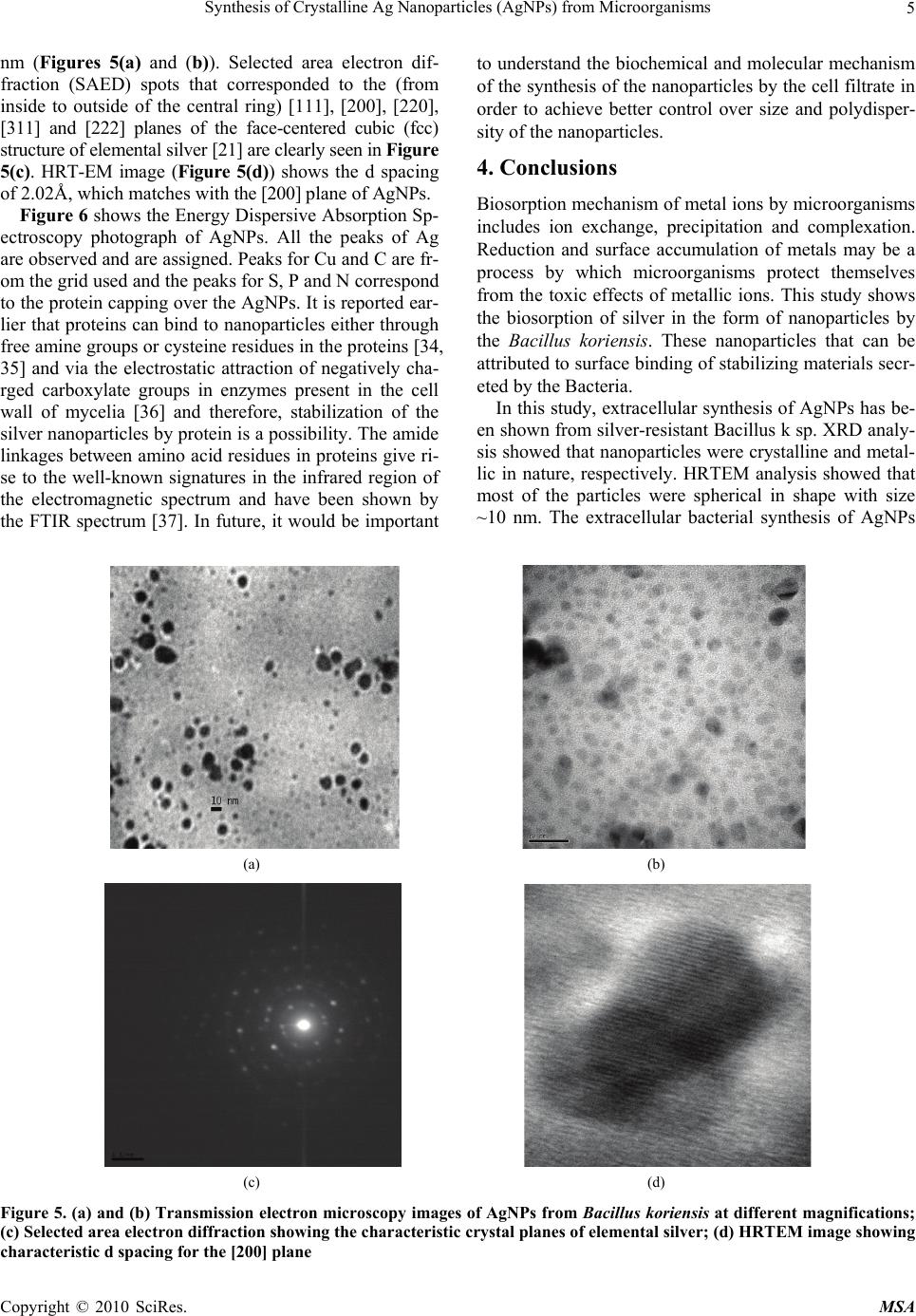 Synthesis of Crystalline Ag Nanoparticles (AgNPs) from Microorganisms Copyright © 2010 SciRes. MSA 5 nm (Figures 5(a) and (b)). Selected area electron dif- fraction (SAED) spots that corresponded to the (from inside to outside of the central ring) [111], [200], [220], [311] and [222] planes of the face-centered cubic (fcc) structure of elemental silver [21] are clearly seen in Figure 5(c). HRT-EM image (Figure 5(d)) shows the d spacing of 2.02Å, which matches with the [200] plane of AgNPs. to understand the biochemical and molecular mechanism of the synthesis of the nanoparticles by the cell filtrate in order to achieve better control over size and polydisper- sity of the nanoparticles. 4. Conclusions Biosorption mechanism of metal ions by microorganisms includes ion exchange, precipitation and complexation. Reduction and surface accumulation of metals may be a process by which microorganisms protect themselves from the toxic effects of metallic ions. This study shows the biosorption of silver in the form of nanoparticles by the Bacillus koriensis. These nanoparticles that can be attributed to surface binding of stabilizing materials secr- eted by the Bacteria. Figure 6 shows the Energy Dispersive Absorption Sp- ectroscopy photograph of AgNPs. All the peaks of Ag are observed and are assigned. Peaks for Cu and C are fr- om the grid used and the peaks for S, P and N correspond to the protein capping over the AgNPs. It is reported ear- lier that proteins can bind to nanoparticles either through free amine groups or cysteine residues in the proteins [34, 35] and via the electrostatic attraction of negatively cha- rged carboxylate groups in enzymes present in the cell wall of mycelia [36] and therefore, stabilization of the silver nanoparticles by protein is a possibility. The amide linkages between amino acid residues in proteins give ri- se to the well-known signatures in the infrared region of the electromagnetic spectrum and have been shown by the FTIR spectrum [37]. In future, it would be important In this study, extracellular synthesis of AgNPs has be- en shown from silver-resistant Bacillus k sp. XRD analy- sis showed that nanoparticles were crystalline and metal- lic in nature, respectively. HRTEM analysis showed that most of the particles were spherical in shape with size ~10 nm. The extracellular bacterial synthesis of AgNPs (a) (b) (c) (d) Figure 5. (a) and (b) Transmission electron microscopy images of AgNPs from Bacillus koriensis at different magnifications; (c) Selected area electron diffraction showing the characteristic crystal planes of elemental silver; (d) HRTEM image showing characteristic d spacing for the [200] plane 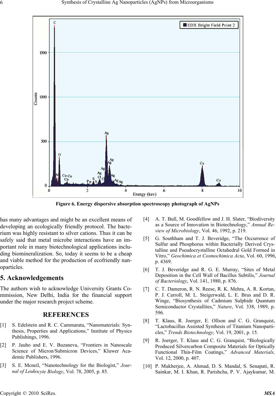 Synthesis of Crystalline Ag Nanoparticles (AgNPs) from Microorganisms 6 Figure 6. Energy dispersive absorption spectroscopy photograph of AgNPs has many advantages and might be an excellent means of developing an ecologically friendly protocol. The bacte- rium was highly resistant to silver cations. Thus it can be safely said that metal microbe interactions have an im- portant role in many biotechnological applications inclu- ding biomineralization. So, today it seems to be a cheap and viable method for the production of ecofriendly nan- oparticles. 5. Acknowledgements The authors wish to acknowledge University Grants Co- mmission, New Delhi, India for the financial support under the major research project scheme. REFERENCES [1] S. Edelstein and R. C. Cammarata, “Nanomaterials: Syn- thesis, Properties and Applications,” Institute of Physics Publishings, 1996. [2] P. Jauho and E. V. Buzaneva, “Frontiers in Nanoscale Science of Micron/Submicron Devices,” Kluwer Aca- demic Publishers, 1996. [3] S. E. Mcneil, “Nanotechnology for the Biologist,” Jour- nal of Leukocyte Biology, Vol. 78, 2005, p. 85. [4] A. T. Bull, M. Goodfellow and J. H. Slater, “Biodiversity as a Source of Innovation in Biotechnology,” Annual Re- view of Microbiology, Vol. 46, 1992, p. 219. [5] G. Southham and T. J. Beveridge, “The Occurrence of Sulfur and Phosphorus within Bacterially Derived Crys- talline and Pseudocrystalline Octahedral Gold Formed in Vitro,” Geochimica et Cosmochimica Acta, Vol. 60, 1996, p. 4369. [6] T. J. Beveridge and R. G. E. Murray, “Sites of Metal Deposition in the Cell Wall of Bacillus Subtilis,” Journal of Bacteriology, Vol. 141, 1980, p. 876. [7] C. T. Dameron, R. N. Reese, R. K. Mehra, A. R. Kortan, P. J. Carroll, M. L. Steigerwald, L. E. Brus and D. R. Winge, “Biosynthesis of Cadmium Sulphide Quantum Semiconductor Crystallites,” Nature, Vol. 338, 1989, p. 596. [8] T. Klaus, R. Joerger, E. Ollson and C. G. Granquist, “Lactobacillus Assisted Synthesis of Titanium Nanoparti- cles,” Trends Biotechnology, Vol. 19, 2001, p. 15. [9] R. Joerger, T. Klaus and C. G. Granquist, “Biologically Produced Silvercarbon Composite Materials for Optically Functional Thin-Film Coatings,” Advanced Materials, Vol. 12, 2000, p. 407. [10] P. Mukherjee, A. Ahmad, D. S. Mandal, S. Senapati, R. Sainkar, M. I. Khan, R. Parishcha, P. V. Ajaykumar, M. Copyright © 2010 SciRes. MSA 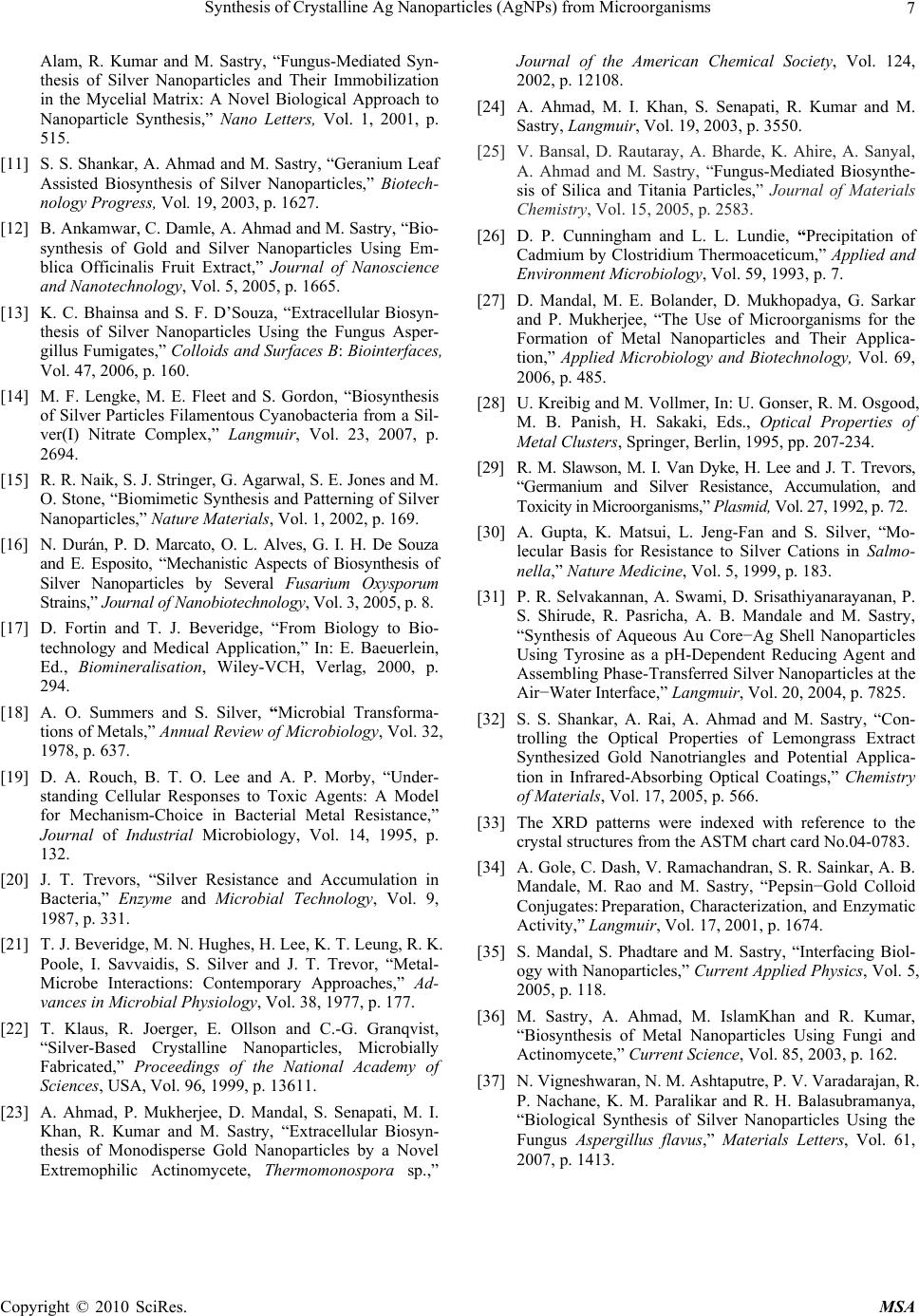 Synthesis of Crystalline Ag Nanoparticles (AgNPs) from Microorganisms7 Alam, R. Kumar and M. Sastry, “Fungus-Mediated Syn- thesis of Silver Nanoparticles and Their Immobilization in the Mycelial Matrix: A Novel Biological Approach to Nanoparticle Synthesis,” Nano Letters, Vol. 1, 2001, p. 515. [11] S. S. Shankar, A. Ahmad and M. Sastry, “Geranium Leaf Assisted Biosynthesis of Silver Nanoparticles,” Biotech- nology Progress, Vol. 19, 2003, p. 1627. [12] B. Ankamwar, C. Damle, A. Ahmad and M. Sastry, “Bio- synthesis of Gold and Silver Nanoparticles Using Em- blica Officinalis Fruit Extract,” Journal of Nanoscience and Nanotechnology, Vol. 5, 2005, p. 1665. [13] K. C. Bhainsa and S. F. D’Souza, “Extracellular Biosyn- thesis of Silver Nanoparticles Using the Fungus Asper- gillus Fumigates,” Colloids and Surfaces B: Biointerfaces, Vol. 47, 2006, p. 160. [14] M. F. Lengke, M. E. Fleet and S. Gordon, “Biosynthesis of Silver Particles Filamentous Cyanobacteria from a Sil- ver(I) Nitrate Complex,” Langmuir, Vol. 23, 2007, p. 2694. [15] R. R. Naik, S. J. Stringer, G. Agarwal, S. E. Jones and M. O. Stone, “Biomimetic Synthesis and Patterning of Silver Nanoparticles,” Nature Materials, Vol. 1, 2002, p. 169. [16] N. Durán, P. D. Marcato, O. L. Alves, G. I. H. De Souza and E. Esposito, “Mechanistic Aspects of Biosynthesis of Silver Nanoparticles by Several Fusarium Oxysporum Strains,” Journal of Nanobiotechnology, Vol. 3, 2005, p. 8. [17] D. Fortin and T. J. Beveridge, “From Biology to Bio- technology and Medical Application,” In: E. Baeuerlein, Ed., Biomineralisation, Wiley-VCH, Verlag, 2000, p. 294. [18] A. O. Summers and S. Silver, “Microbial Transforma- tions of Metals,” Annual Review of Microbiology, Vol. 32, 1978, p. 637. [19] D. A. Rouch, B. T. O. Lee and A. P. Morby, “Under- standing Cellular Responses to Toxic Agents: A Model for Mechanism-Choice in Bacterial Metal Resistance,” Journal of Industrial Microbiology, Vol. 14, 1995, p. 132. [20] J. T. Trevors, “Silver Resistance and Accumulation in Bacteria,” Enzyme and Microbial Technology, Vol. 9, 1987, p. 331. [21] T. J. Beveridge, M. N. Hughes, H. Lee, K. T. Leung, R. K. Poole, I. Savvaidis, S. Silver and J. T. Trevor, “Metal- Microbe Interactions: Contemporary Approaches,” Ad- vances in Microbial Physiology, Vol. 38, 1977, p. 177. [22] T. Klaus, R. Joerger, E. Ollson and C.-G. Granqvist, “Silver-Based Crystalline Nanoparticles, Microbially Fabricated,” Proceedings of the National Academy of Sciences, USA, Vol. 96, 1999, p. 13611. [23] A. Ahmad, P. Mukherjee, D. Mandal, S. Senapati, M. I. Khan, R. Kumar and M. Sastry, “Extracellular Biosyn- thesis of Monodisperse Gold Nanoparticles by a Novel Extremophilic Actinomycete, Thermomonospora sp.,” Journal of the American Chemical Society, Vol. 124, 2002, p. 12108. [24] A. Ahmad, M. I. Khan, S. Senapati, R. Kumar and M. Sastry, Langmuir, Vol. 19, 2003, p. 3550. [25] V. Bansal, D. Rautaray, A. Bharde, K. Ahire, A. Sanyal, A. Ahmad and M. Sastry,“Fungus-Mediated Biosynthe- sis of Silica and Titania Particles,” Journal of Materials Chemistry, Vol. 15, 2005, p. 2583. [26] D. P. Cunningham and L. L. Lundie, “Precipitation of Cadmium by Clostridium Thermoaceticum,” Applied and Environment Microbiology, Vol. 59, 1993, p. 7. [27] D. Mandal, M. E. Bolander, D. Mukhopadya, G. Sarkar and P. Mukherjee, “The Use of Microorganisms for the Formation of Metal Nanoparticles and Their Applica- tion,” Applied Microbiology and Biotechnology, Vol. 69, 2006, p. 485. [28] U. Kreibig and M. Vollmer, In: U. Gonser, R. M. Osgood, M. B. Panish, H. Sakaki, Eds., Optical Properties of Metal Clusters, Springer, Berlin, 1995, pp. 207-234. [29] R. M. Slawson, M. I. Van Dyke, H. Lee and J. T. Trevors, “Germanium and Silver Resistance, Accumulation, and Toxicity in Microorganisms,” Plasmid, Vol. 27, 1992, p. 72. [30] A. Gupta, K. Matsui, L. Jeng-Fan and S. Silver, “Mo- lecular Basis for Resistance to Silver Cations in Salmo- nella,” Nature Medicine, Vol. 5, 1999, p. 183. [31] P. R. Selvakannan, A. Swami, D. Srisathiyanarayanan, P. S. Shirude, R. Pasricha, A. B. Mandale and M. Sastry, “Synthesis of Aqueous Au Core−Ag Shell Nanoparticles Using Tyrosine as a pH-Dependent Reducing Agent and Assembling Phase-Transferred Silver Nanoparticles at the Air−Water Interface,” Langmuir, Vol. 20, 2004, p. 7825. [32] S. S. Shankar, A. Rai, A. Ahmad and M. Sastry, “Con- trolling the Optical Properties of Lemongrass Extract Synthesized Gold Nanotriangles and Potential Applica- tion in Infrared-Absorbing Optical Coatings,” Chemistry of Materials, Vol. 17, 2005, p. 566. [33] The XRD patterns were indexed with reference to the crystal structures from the ASTM chart card No.04-0783. [34] A. Gole, C. Dash, V. Ramachandran, S. R. Sainkar, A. B. Mandale, M. Rao and M. Sastry, “Pepsin−Gold Colloid Conjugates:Preparation, Characterization, and Enzymatic Activity,” Langmuir, Vol. 17, 2001, p. 1674. [35] S. Mandal, S. Phadtare and M. Sastry, “Interfacing Biol- ogy with Nanoparticles,” Current Applied Physics, Vol. 5, 2005, p. 118. [36] M. Sastry, A. Ahmad, M. IslamKhan and R. Kumar, “Biosynthesis of Metal Nanoparticles Using Fungi and Actinomycete,” Current Science, Vol. 85, 2003, p. 162. [37] N. Vigneshwaran, N. M. Ashtaputre, P. V. Varadarajan, R. P. Nachane, K. M. Paralikar and R. H. Balasubramanya, “Biological Synthesis of Silver Nanoparticles Using the Fungus Aspergillus flavus,” Materials Letters, Vol. 61, 2007, p. 1413. Copyright © 2010 SciRes. MSA |